. 2000 Sep;74(18):8277-85.
doi: 10.1128/jvi.74.18.8277-8285.2000.

In vivo genomic footprinting of the human T-cell leukemia virus type 1 (HTLV-1) long terminal repeat enhancer sequences in HTLV-1-infected human T-cell lines with different levels of Tax I activity

Affiliations

In vivo genomic footprinting of the human T-cell leukemia virus type 1 (HTLV-1) long terminal repeat enhancer sequences in HTLV-1-infected human T-cell lines with different levels of Tax I activity

S Datta et al. J Virol. 2000 Sep.

Abstract

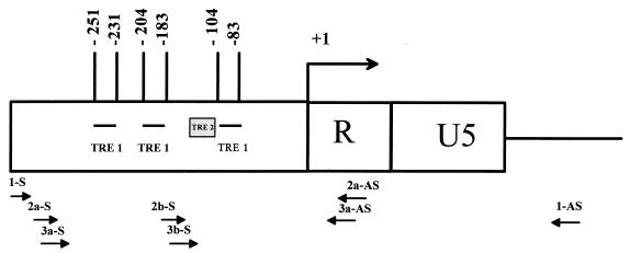
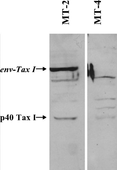
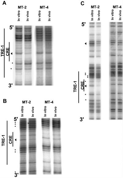
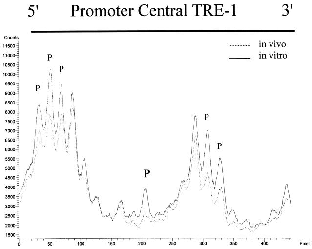
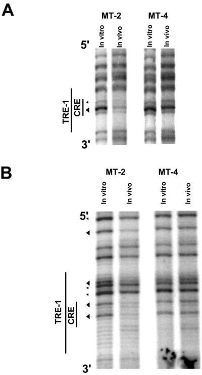
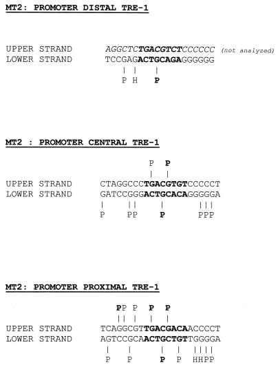
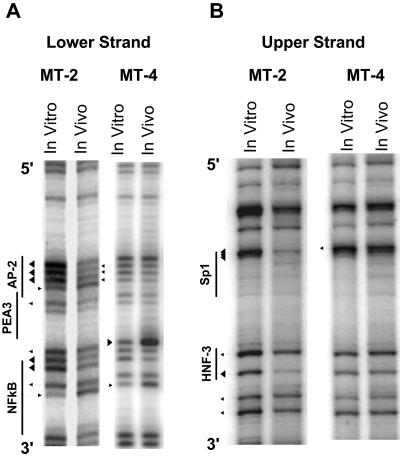
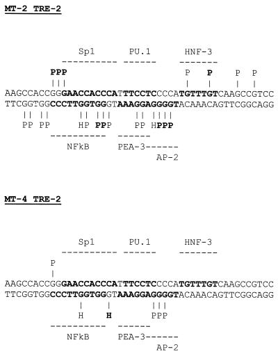
The Tax protein of human T-cell leukemia virus type 1 (HTLV-1) enhances viral gene expression through sequences in the U3 region of the viral long terminal repeat. These sequences consist of three imperfect 21-bp repeats (TRE-1s) and a region between the promoter-central and promoter-proximal 21-bp repeats (TRE-2). The TRE-1s contain a core cyclic AMP response element (CRE) motif and can be bound by CREB, ATF-1, ATF-2, and other members of the CREB-ATF superfamily of transcription factors. Tax enhances CREB binding to TRE-1 in vitro, and it promotes dimerization of CREB as well as other bZIP proteins. Using ligation-mediated PCR on in vivo dimethyl sulfate-treated HTLV-1-infected cell lines MT-2 and MT-4, we have compiled a profile of protein occupancy in the HTLV-1 enhancer sequences in the presence of high (MT-2) and low (MT-4) levels of biologically active Tax I. The in vivo footprinting showed that all three TRE-1s were bound by protein(s), but only in MT-2 cells. In MT-2 cells, all TRE-1s showed strong protection of the G residues in the central CRE, and the footprints extended to differing degrees into the GC-rich flanking sequences. This indicated Tax I-dependent loading of transcription factors onto the HTLV-1 TRE-1s in vivo. In vivo footprinting on TRE-2 indicated that this region was bound by proteins regardless of the Tax I status of the cell line. However, the presence of Tax I increased the extent and altered the profile of proteins binding TRE-2 in vivo.


Figures

FIG. 1
FIG. 1
Schematic of the HTLV-1 LTR and relative positions of oligonucleotides used for LMPCR. +1 signifies the start site of transcription. Relative positions of the HTLV-1-specific oligonucleotides used in LMPCR (see Materials and Methods for details) are also shown. Oligonucleotides ending in -S were used to analyze the lower strand, while those ending in -AS were used to analyze the upper strand.
FIG. 2
FIG. 2
Western blot analysis of Tax I levels in MT-2 and MT-4 cells. Whole-cell protein lysates from MT-2 and MT-4 cells were separated by SDS-PAGE and subjected to Western blot analysis using a polyclonal antibody to Tax I as described in Materials and Methods. The positions of authentic p40 Tax I as well as the Env-Tax I fusion protein made by MT-2 cells are indicated (see text for details). The band between p40 Tax I and Env-Tax I seen in both cell lines is a nonspecific band that is also observed in human T cells that do not express Tax I (not shown).
FIG. 3
FIG. 3
In vivo DMS footprinting of the lower strand of the TRE-1s. DNAs from in vivo DMS-treated MT-2 and MT-4 cells were piperidine cleaved and subjected to LMPCR as described in Materials and Methods. The reaction products were separated on polyacrylamide gels and exposed to film. Autoradiograms corresponding to the three TRE-1s are shown. (A) Promoter-distal TRE-1. A band is missing in the 5′ GC-rich flank most likely because the proviral DNA contains a base other than the predicted guanine residue at that position. (B) Promoter-central TRE-1. (C) Promoter-proximal TRE-1. Arrowheads pointing toward bands indicate hypersensitive residues; arrowheads pointing away from bands indicate protected residues. Larger arrowheads indicate major protections or hypersensitive sites.
FIG. 4
FIG. 4
PhosphorImager quantitation of the MT-2 lower-strand promoter-central TRE-1. The gel in Fig. 3B was exposed to a storage phosphor screen. The image was scanned, and the graph was generated by using ImageQuant 5.0 software. The protected and hypersensitive residues shown in Fig. 3B are indicated.
FIG. 5
FIG. 5
In vivo DMS footprinting of the upper strand of the promoter-central (A) and promoter-proximal (B) TRE-1s. In vivo DMS LMPCR was performed on MT-2 and MT-4 cells as described for Fig. 3.
FIG. 6
FIG. 6
Summary of TRE-1 footprints in the MT-2 cell line. The footprint data presented in Fig. 3 and 5 are summarized on the individual TRE-1 sequences. The CRE is in bold. “H” indicates hypersensitivity, and “P” indicates protection; bolded P's and H's reflect strong interactions. The upper strand of the promoter-distal TRE-1 is italicized to indicate that it was not analyzed.
FIG. 7
FIG. 7
In vivo DMS footprinting of TRE-2 in the MT-2 and MT-4 cell lines. In vivo DMS LMPCR was performed on MT-2 and MT-4 cells. (A) Lower strand of TRE-2 in MT-2 and MT-4 cells. The predicted binding sites for different transcription factors for this strand are also indicated. Some transcription factor binding sites overlap other binding sites on the opposite strand (Fig. 8). (B) Upper strand of TRE-2 in MT-2 and MT-4 cells.
FIG. 8
FIG. 8
Summary of TRE-2 footprints in the MT-2 and MT-4 cell lines. The footprint data presented in Fig. 6 are summarized on the TRE-2 sequence. Predicted transcription factor binding sites are shown in bold. Conventions are the same as for Fig. 6.

References

    1. Altman R, Harrich D, Garcia J A, Gaynor R B. Human T-cell leukemia virus types I and II exhibit different DNase I protection patterns. J Virol. 1988;62:1339–1346. - PMC - PubMed
    1. Ballard D W, Bohnlein E, Lowenthal J W, Wano Y, Franza B R, Greene W C. HTLV-1 tax induces cellular proteins that activate the kappa B element in the IL-2 receptor alpha gene. Science. 1988;241:1652–1655. - PubMed
    1. Baranger A M, Palmer C R, Hamm M K, Giebler H A, Brauweiler A, Nyborg J K, Schepartz A. Mechanism of DNA-binding enhancement by the human T-cell leukaemia virus transactivator Tax. Nature. 1995;376:606–608. - PubMed
    1. Brady J, Jeang K T, Duvall J, Khoury G. Identification of p40x-responsive regulatory sequences within the human T-cell leukemia virus type I long terminal repeat. J Virol. 1987;61:2175–2181. - PMC - PubMed
    1. Brauweiler A, Garl P, Franklin A A, Giebler H A, Nyborg J K. A molecular mechanism for human T-cell leukemia virus latency and Tax transactivation. J Biol Chem. 1995;270:12814–12822. - PubMed

Publication types

LinkOut - more resources