. 2000 Sep;74(18):8601-13.
doi: 10.1128/jvi.74.18.8601-8613.2000.

Phosphorylation of simian virus 40 T antigen on Thr 124 selectively promotes double-hexamer formation on subfragments of the viral core origin

Affiliations

Phosphorylation of simian virus 40 T antigen on Thr 124 selectively promotes double-hexamer formation on subfragments of the viral core origin

B A Barbaro et al. J Virol. 2000 Sep.

Abstract

Cell cycle-dependent phosphorylation of simian virus 40 (SV40) large tumor antigen (T-ag) on threonine 124 is essential for the initiation of viral DNA replication. A T-ag molecule containing a Thr-->Ala substitution at this position (T124A) was previously shown to bind to the SV40 core origin but to be defective in DNA unwinding and initiation of DNA replication. However, exactly what step in the initiation process is defective as a result of the T124A mutation has not been established. Therefore, to better understand the control of SV40 replication, we have reinvestigated the assembly of T124A molecules on the SV40 origin. Herein it is demonstrated that hexamer formation is unaffected by the phosphorylation state of Thr 124. In contrast, T124A molecules are defective in double-hexamer assembly on subfragments of the core origin containing single assembly units. We also report that T124A molecules are inhibitors of T-ag double hexamer formation. These and related studies indicate that phosphorylation of T-ag on Thr 124 is a necessary step for completing the assembly of functional double hexamers on the SV40 origin. The implications of these studies for the cell cycle control of SV40 DNA replication are discussed.


Figures

FIG. 1
FIG. 1
Sequences of representative oligonucleotides used to characterize the ability of T-ag and the T124A to form hexamers on subfragments of the core origin. Names of the oligonucleotides are given at the right. (A) Sequences present in the 64-bp core oligonucleotide. Locations of the AT-rich regions, site II, and the EP regions are depicted. SV40 sequences are numbered as described elsewhere (87). Arrows depict the four GAGGC pentanucleotides within site II that serve as binding sites for T-ag, numbered as previously described (41). (B) Diagram D1 provides the sequence of the 48-bp penta 1 + EP oligonucleotide, a derivative of the right-side assembly unit that supports only hexamer formation (81a). Although not depicted, we also synthesized the 48-bp penta 3 + EP oligonucleotide. Diagram D2 presents the sequence of the 48-bp penta 1 + EPm (mutant) oligonucleotide; in this molecule, the wild-type EP sequence was replaced by transition mutations. Although not shown, we also synthesized the 48-bp penta 3 + EPm oligonucleotide. Diagram D3 provides the sequence of the 47-bp penta 4 + AT oligonucleotide, a derivative of the left-side assembly unit that supports hexamer formation (81a). An additional member of this class of molecules, the 47-bp penta 2 + AT oligonucleotide, was also synthesized but is not depicted. Diagram D4 presents the sequence of the 47-bp penta 4 + ATm (mutant) oligonucleotide; in this molecule, transition mutations were used in place of the wild-type AT-rich region. We synthesized, but do not depict, an additional molecule in this class, the 47-bp penta 2 + ATm oligonucleotide. Diagram D5 depicts the sequence of the 47-bp control oligonucleotide, a molecule used to measure non-sequence-specific binding to DNA. Finally, lowercase boldface letters represent transition mutations in particular pentanucleotides or the AT or EP flanking regions.
FIG. 2
FIG. 2
Representative gel mobility shift assay used to assess the ability of T-ag or the T124A mutant to bind to single pentanucleotides in oligonucleotides derived from the penta 1,3 + EP assembly unit. (A) As positive controls, band shift reactions were conducted with the 64-bp core oligonucleotide and either T-ag (lane 2) or the T124A mutant (lane 3). Reaction products formed with the 48-bp penta 1 + EP oligonucleotide and either T-ag or the T124A mutation are shown in lane 5 or 6, respectively. The products formed with the 48-bp penta 1 + EPm oligonucleotide and either T-ag or the T124A mutant are shown in lane 8 or 9, respectively. Additional experiments were conducted with the 48-bp penta 3 + EP oligonucleotide and either T-ag (lane 11) or the T124A mutant (lane 12). Related experiments were conducted with the 48-bp penta 3 + EPm oligonucleotide and T-ag (lane 14) or the T124A mutant (lane 15). An additional set of reactions was performed with 47-bp control oligonucleotide and both T-ag and the T124A mutant (data not shown). Reactions in lanes 1, 4, 7, 10, and 13 were conducted in the absence of protein. All experiments were performed with AMP-PNP and 6 pmol of either T-ag or the T124A mutant. (B) The data presented in panel A and data from two additional experiments (data not shown) were quantitated with a Molecular Dynamics PhosphorImager, and the results are presented in a histogram. The percentage of input DNA shifted into hexamer, with 6 pmol of either T-ag or the T124A mutant, and AMP-PNP is shown on the ordinate.
FIG. 3
FIG. 3
Representative gel mobility shift assay used to assess the ability of T-ag or the T124A mutant to bind to single pentanucleotides in oligonucleotides derived from the penta 2,4 + AT assembly unit. (A) Control experiments were conducted with the 64-bp core oligonucleotide and either T-ag (lane 2) or the T124A mutant (lane 3). Reaction products formed with the 47-bp penta 4 + AT oligonucleotide and either T-ag or the T124A mutation are shown in lane 5 or 6, respectively. Lanes 8 and 9 present the products formed in reactions containing the 47-bp penta 4 + ATm oligonucleotide plus T-ag and the T124A mutant, respectively. An additional set of experiments were conducted with the 47-bp penta 2 + AT oligonucleotide and either T-ag (lane 11) or the T124A mutation (lane 12). To assess the contribution of the AT-rich region to binding, additional reactions were conducted with the 47-bp penta 2 + ATm oligonucleotide and either T-ag (lane 14) or the T124A mutation (lane 15). An additional set of experiments was performed with the 47-bp control oligonucleotide and either T-ag or the T124A mutant (data not shown). Reactions in lanes 1, 4, 7, 10, and 13 were conducted in the absence of protein. All experiments were performed with AMP-PNP and 6 pmol of either T-ag or the T124A mutant. (B) The data presented in panel A and data from two additional sets of experiments (data not shown) were quantitated with a Molecular Dynamics PhosphorImager. The percentage of input DNA shifted into hexamers, with 6 pmol of either T-ag or the T124A mutant, in the presence of AMP-PNP is shown on the ordinate. The percentage of input DNA shifted into hexamers is higher in panel B than in Fig. 2B. For unknown reasons, a relatively high percentage of the oligonucleotides derived from the penta 1,3 + EP assembly unit were trapped in the wells (Fig. 2A). Therefore, we are reluctant to make quantitative comparisons between the experiments in Fig. 2 and 3. However, the filter binding assays presented in Fig. 5 indicate that patterns of hexamer formation on the 48-bp penta 1 + EP and 47-bp penta 4 + AT oligonucleotides are roughly equivalent.
FIG. 4
FIG. 4
Sequences of representative oligonucleotides used to characterize the ability of T-ag and the T124A mutant to form double hexamers on subfragments of the core origin. Names of the oligonucleotides are presented at the right. Diagram D1 depicts sequences present in the 48-bp site II + EP oligonucleotide. Locations of the four GAGGC pentanucleotides in site II are depicted by arrows; the location of the EP is also indicated. Diagram D2 presents sequences present in the 48-bp penta 1,3 + EP oligonucleotide, a molecule containing an assembly unit for double hexamer formation that is located on the right side of the core origin (81a). Diagram D3 presents sequences comprising the 47-bp site II + AT oligonucleotide. Locations of the four pentanucleotides in site II and the AT-rich region are indicated. Diagram D4 presents sequences present in the 47-bp penta 2,4 + AT oligonucleotide, a molecule containing an assembly unit for double hexamer formation that is located on the left side of the core origin (81a). Lowercase boldface letters indicate transition mutations introduced at the indicated locations.
FIG. 5
FIG. 5
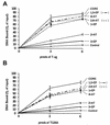
Filter binding assays to measure the relative abilities of T-ag and the T124A mutant to interact with subfragments of the core origin under replication conditions. The interaction of T-ag or the T124A mutant (0, 3, or 6 pmol) with 25 fmol of the indicated oligonucleotide was measured by nitrocellulose filter binding assays in the presence of AMP-PNP. The percentage of input DNA bound to a given filter was determined by scintillation counting. As a positive control, the interaction of T-ag and the T124A (0, 3, or 6 pmol) with the 64-bp core oligonucleotide was determined. As a negative control, identical reactions were conducted with the 47-bp control oligonucleotide.
FIG. 6
FIG. 6
Representative gel mobility shift assay used to assess the ability of T-ag or the T124A mutant to bind to oligonucleotides that support double hexamer formation. (A) Positive controls were conducted with the 64-bp core oligonucleotide and either T-ag (lane 2) or the T124A mutant (lane 3). Reaction products formed with the 48-bp site II + EP oligonucleotide plus T-ag and the T124A mutant are shown in lanes 5 and 6, respectively. Reactions conducted with the 47-bp site II + AT oligonucleotide plus T-ag and the T124A mutant are shown in lanes 8 and 9, respectively. To measure non-sequence-specific binding, additional reactions were conducted with the 47-bp control oligonucleotide (Ctrl.) and either T-ag (lane 11) or the T124A mutant (lane 12). Reactions were conducted with 6 pmol of either T-ag or the T124A mutant, in the presence of AMP-PNP; the reactions in lanes 1, 4, 7, and 10 were conducted in the absence of protein. (B) The data presented in panel A, and data from two additional sets of experiments (data not shown), were quantitated with a Molecular Dynamics PhosphorImager, and the results are presented in a histogram. The DH/H ratio was calculated as described in Materials and Methods.
FIG. 7
FIG. 7
Comparison of the abilities of T-ag and the T124A mutant to assemble on the penta 1,3 + EP assembly unit. (A) Results of experiments conducted with 25 fmol of the 48-bp penta 1,3 + EP oligonucleotide and different amounts of either T-ag or the T124A mutant (from 6 pmol of protein (240:1 protein-to-DNA ratio) to 0.75 pmol of protein (30:1 protein-to-DNA ratio) (lanes 5 to 12). As positive controls, reactions were conducted with the 64-bp core origin and either T-ag (lane 2) or the T124A mutant (lane 3) at a protein-to-DNA ratio of 240:1. The reactions in lanes 1 and 4 were conducted in the absence of protein. All reactions were performed in the presence of AMP-PNP. (B) The data in panel A were quantitated using a Molecular Dynamics PhosphorImager and used to calculate the DH/H ratio for both T-ag and the T124A mutant.
FIG. 8
FIG. 8
Comparison of the abilities of T-ag and the T124A mutant to assemble on the penta 2,4 + AT assembly unit. The experiments were conducted in the presence of AMP-PNP, with 25 fmol of the 47-bp penta 2,4 + AT oligonucleotide and different amounts of either T-ag or the T124A mutant (from 6 pmol of protein (240:1 protein to DNA ratio) to 0.75 pmol of protein (30:1 protein-to-DNA ratio) (lanes 5 to 12). As positive controls, reactions were conducted with the 64-bp core origin and either T-ag (lane 2) or the T124A mutant (lane 3) at a protein-to-DNA ratio of 240:1. The reactions in lanes 1 and 4 were conducted in the absence of protein. (B) The data in panel A were quantitated using a Molecular Dynamics PhosphorImager and used to calculate the DH/H ratio for both T-ag and the T124A mutant. Lower levels of double hexamer assembly on the penta 2,4 + AT assembly unit, relative to the penta 1,3 + EP assembly unit, are reflected in lower DH/H ratios (compare panel B with Fig. 7B).
FIG. 9
FIG. 9
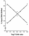
Double hexamer formation on the penta 1,3 + EP assembly unit is suppressed by T124A. Band shift reactions were conducted with 25 fmol of the 48-bp penta 1,3 + EP oligonucleotide, AMP-PNP, and 6 pmol of T-ag (left side of graph), 6 pmol of T124A (right side of graph), or 6 pmol of T-ag and T124A combined in the indicated ratios. The products of these reactions were loaded on 4 to 12% gradient polyacrylamide gels, and the percentages of input DNA shifted into hexamers (open squares) and double hexamers (filled diamonds) were determined by a PhosphorImager.
FIG. 10
FIG. 10
Model illustrating the role of Thr 124 phosphorylation during T-ag oligomerization on the penta 1,3 + EP assembly unit. The T-ag OBD is symbolized by the smaller ovoids, while the remaining regions of T-ag are symbolized by the larger ovoids (88). Pathways for T-ag and T124A oligomerization are shown on the left and right. The drawings on line 1 indicate that on a given assembly unit, T-ag and the T124A mutant prefer to bind to the pentanucleotides proximal to the flanking sequences (pentanucleotide 1 or 4; binding to pentanucleotide 1 is depicted). Upon monomer binding, a complicated set of protein-protein interactions (reviewed in reference 7) gives rise to hexamer formation; however, all steps required for hexamer formation are independent of the phosphorylation status of Thr 124. Phosphate residues on Thr 124 (P) are indicated. Depicted on line 2 is the observation that double hexamer formation on the penta 1,3 + EP assembly unit is highly dependent on phosphorylation of Thr 124. As a result, T-ag phosphorylated on Thr 124 is able to assemble a double hexamer, but the T124A molecule is blocked at the level of hexamer formation. Similar results were obtained with the penta 2,4 + AT assembly unit (see text). Although monomers of T-ag are depicted as being phosphorylated on Thr 124, it is possible that other oligomeric forms of T-ag are the actual substrates for phosphorylation. Finally, double hexamer formation is inhibited by phosphorylation of certain serine residues (reviewed in references to 26) (not illustrated).

References

    1. Adamczewski J P, Gannon J V, Hunt T. Simian virus 40 large T antigen associates with cyclin A and p33cdk2. J Virol. 1993;67:6551–6557. - PMC - PubMed
    1. Bell S P, Stillman B. ATP-dependent recognition of eukaryotic origins of DNA replication by a multiprotein complex. Nature. 1992;357:128–134. - PubMed
    1. Bhattacharyya S, Lorimer H E, Prives C. Murine polyomavirus and simian virus 40 large T antigens produce different structural alterations in viral origin DNA. J Virol. 1995;69:7579–7585. - PMC - PubMed
    1. Borowiec J A, Dean F B, Bullock P A, Hurwitz J. Binding and unwinding—how T antigen engages the SV40 origin of DNA replication. Cell. 1990;60:181–184. - PubMed
    1. Borowiec J A, Hurwitz J. ATP stimulates the binding of the simian virus 40 (SV40) large tumor antigen to the SV40 origin of replication. Proc Natl Acad Sci USA. 1988;85:64–68. - PMC - PubMed

Publication types

LinkOut - more resources