. 2000 Sep;74(18):8670-9.
doi: 10.1128/jvi.74.18.8670-8679.2000.

Localization of human immunodeficiency virus type 1 Gag and Env at the plasma membrane by confocal imaging

Affiliations

Localization of human immunodeficiency virus type 1 Gag and Env at the plasma membrane by confocal imaging

L Hermida-Matsumoto et al. J Virol. 2000 Sep.

Abstract

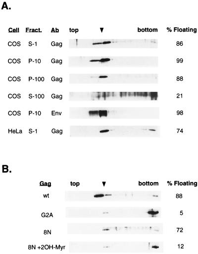
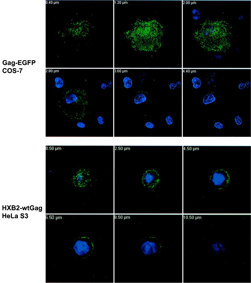
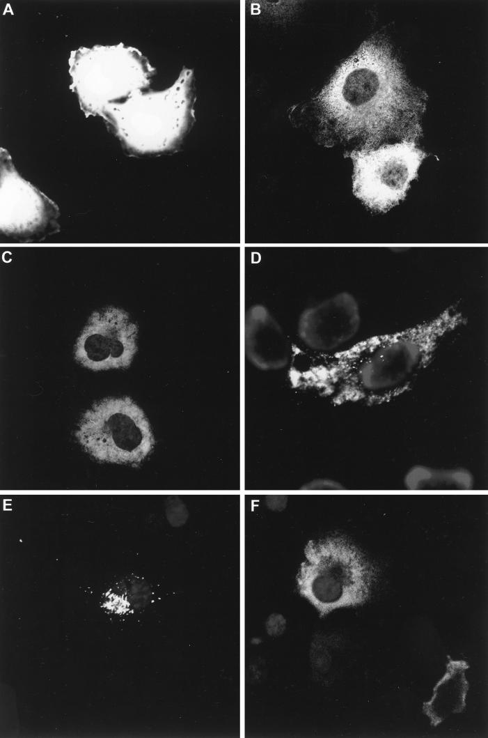
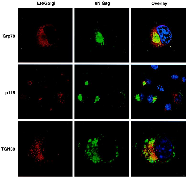
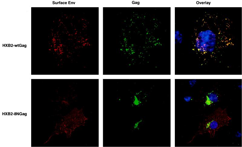
Budding of lentiviruses occurs at the plasma membrane, but the preceding steps involved in particle assembly are poorly understood. Since the Gag polyprotein mediates virion assembly and budding, studies on the localization of Gag within the cell should provide insight into the mechanism of particle assembly. Here, we utilize biochemical fractionation techniques as well as high-resolution confocal imaging of live cells to demonstrate that Gag is localized at the plasma membrane in a striking punctate pattern. Mutation of the N-terminal myristoylation site results in the formation of large cytosolic complexes, whereas mutation of the N-terminal basic residue cluster in the matrix domain redirects the Gag protein to a region partially overlapping the Golgi apparatus. In addition, we show that Gag and Env colocalize at the plasma membrane and that mistargeting of a mutant Gag to the Golgi apparatus alters the pattern of surface expression of Env.


Figures

FIG. 1
FIG. 1
Fractionation of wt Pr55Gag by sucrose flotation analysis. COS-1 cells transfected with pHXB2ΔBalID25S, which bears the gene encoding wt Pr55Gag, were harvested 48 h posttransfection. Cell homogenates were fractionated and subjected to sucrose flotation centrifugation as described in Materials and Methods. Fractions were collected from the top and analyzed by SDS-gel electrophoresis and Western blotting with an anti-p24CA antibody. (A) Fractionation of wt Pr55Gag S-1 cell homogenates and individual subcellular fractions. Blotting membranes containing wt Pr55Gag in S-1, P-10, and P-100 fractions were stripped and reprobed with a sheep anti-gp160/gp120Env polyclonal antibody (Env). Shown here is the Env fractionation profile for the P-10 fraction. The flotation profile of the HeLa S3 S-1 fraction is shown at the bottom. The distribution of Pr55Gag in the 10 to 65% sucrose interface (arrowhead) over the total protein is indicated at the right. The exposure time for the S-100 fraction was longer than that for the other fractions because very little wt Gag is present in this fraction (see Table 1). Ab, antibody. (B) Fractionation of wt Pr55Gag, G2A Gag, and 8N Gag by sucrose flotation analysis. Transfected COS cells were harvested 48 h posttransfection. Denucleated P-100 fractions were adjusted to 70% sucrose and analyzed by sucrose flotation as described above. For myristoylation inhibition, 8N Gag-transfected cells were incubated with 250 μM 2-OH-Myr for 5 h prior to sucrose flotation analysis.
FIG. 2
FIG. 2
Immunofluorescence of Pr55Gag in live and fixed COS-7 and HeLa S3 cells. (Top) Confocal microscopy analysis of live COS-7 cells transfected with wt Gag-EGFP and stained with Hoechst as described in Materials and Methods. A composite z stack corresponding to 6 confocal sections (from a total of 24) is shown. (Bottom) HeLa S3 cells were transfected with pHXB2ΔBalID25S expressing wt Pr55Gag. Cells were fixed and stained with an anti-p17MA polyclonal antibody and an FITC-conjugated secondary antibody. Nuclei were stained with Hoechst. A composite z stack of 6 confocal sections (out of 28) is shown.
FIG. 3
FIG. 3
Immunofluorescence of Pr55Gag and M domain Gag mutants. Single confocal sections of transfected COS-7 cells are shown. (A) Cells transfected with pEGFP; (B) cells transfected with pHXB2ΔBalID25S expressing G2A Gag; (C) cells transfected with pGag-EGFP and incubated overnight with 100 μM 2-OH-Myr; (D) cells transfected with pGag-EGFP and incubated overnight with 100 μM 2-OH-Myr followed by streptolysin O permeabilization; (E) cells transfected with pHXB2ΔBalID25S expressing 8N Gag; (F) cells transfected with pHXB2ΔBalID25S expressing 8N Gag and incubated overnight with 100 μM 2-OH-Myr to generate nonmyristoylated 8N Gag protein. EGFP confocal imaging was performed on live cells. G2A Gag and 8N Gag cells were fixed and stained as described in Materials and Methods. Cells were fixed and stained with anti-MA antibody and Hoechst as described above.
FIG. 4
FIG. 4
Intracellular localization of 8N Gag and costaining with ER and Golgi markers. Single confocal sections are shown for cells transfected with pHXB2 expressing and 8N Gag stained with cellular markers for ER (rhodamine) and Golgi and TGN (Cy 5) (left column), or anti-p17MA antibodies (FITC; middle column). The overlays are shown on the right. (Top) 8N Gag and Grp78 (ER). (Middle) 8N Gag and p115 (cis to medial Golgi marker). Notice the distinct morphologies of the 8N Gag fluorescence (FITC; center). (Bottom) 8N Gag and TGN38. Notice that the confocal section is focused on the TGN vesicles and that 8N Gag is out of focus.
FIG. 5
FIG. 5
wt Gag directs surface localization of Env to the sites of viral assembly. Cell surface staining of Env in COS cells transfected with pHXB2 expressing wt Gag (top) and 8N Gag (bottom) is shown. Transfected cells were fixed and stained for plasma membrane-associated Env as described in Materials and Methods. Left, Env staining (rhodamine); middle, Gag staining (FITC); right, Gag-Env overlay. Yellow areas indicate wt Gag and Env colocalization at the plasma membrane.

References

    1. Bryant M, Ratner L. Myristoylation-dependent replication and assembly of human immunodeficiency virus 1. Proc Natl Acad Sci USA. 1990;87:523–527. - PMC - PubMed
    1. Burniston M T, Cimarelli A, Colgan J, Curtis S P, Luban J. Human immunodeficiency virus type 1 Gag polyprotein multimerization requires the nucleocapsid domain and RNA and is promoted by the capsid-dimer interface and the basic region of matrix protein. J Virol. 1999;73:8527–8540. - PMC - PubMed
    1. Cosson P. Direct interaction between the envelope and matrix proteins of HIV-1. EMBO J. 1996;15:5783–5788. - PMC - PubMed
    1. Dorfman T, Luban J, Goff S P, Haseltine W A, Gottlinger H G. Mapping of functionally important residues of a cysteine-histidine box in the human immunodeficiency virus type 1 nucleocapsid protein. J Virol. 1993;67:6159–6169. - PMC - PubMed
    1. Facke M, Janetzko A, Shoeman R L, Krausslich H G. A large deletion in the matrix domain of the human immunodeficiency virus gag gene redirects virus particle assembly from the plasma membrane to the endoplasmic reticulum. J Virol. 1993;67:4972–4980. - PMC - PubMed

Publication types

MeSH terms

LinkOut - more resources