. 2000 Sep;74(18):8692-9.
doi: 10.1128/jvi.74.18.8692-8699.2000.

Membrane targeting properties of a herpesvirus tegument protein-retrovirus Gag chimera

Affiliations

Membrane targeting properties of a herpesvirus tegument protein-retrovirus Gag chimera

J B Bowzard et al. J Virol. 2000 Sep.

Abstract

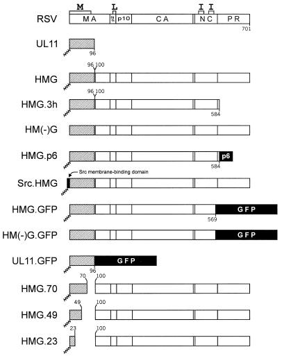
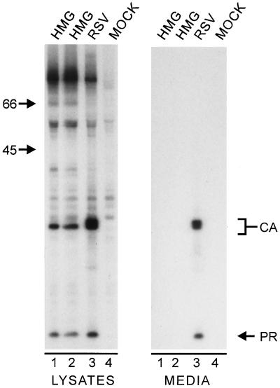
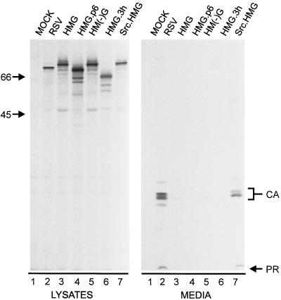
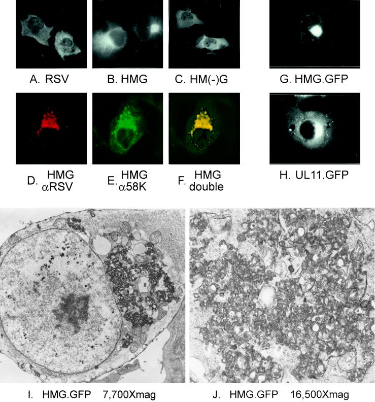
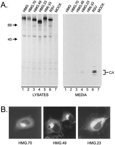
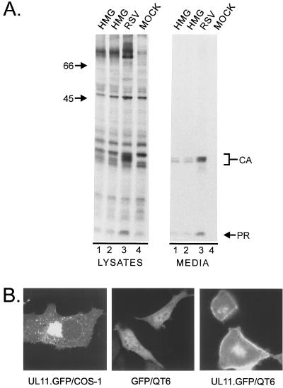
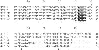
The retroviral Gag protein is capable of directing the production and release of virus-like particles in the absence of all other viral components. Budding normally occurs after Gag is transported to the plasma membrane by its membrane-targeting and -binding (M) domain. In the Rous sarcoma virus (RSV) Gag protein, the M domain is contained within the first 86 amino acids. When M is deleted, membrane association and budding fail to occur. Budding is restored when M is replaced with foreign membrane-binding sequences, such as that of the Src oncoprotein. Moreover, the RSV M domain is capable of targeting heterologous proteins to the plasma membrane. Although the solution structure of the RSV M domain has been determined, the mechanism by which M specifically targets Gag to the plasma membrane rather than to one or more of the large number of internal membrane surfaces (e.g., the Golgi apparatus, endoplasmic reticulum, and nuclear, mitochondrial, or lysosomal membranes) is unknown. To further investigate the requirements for targeting proteins to discrete cellular locations, we have replaced the M domain of RSV with the product of the unique long region 11 (U(L)11) gene of herpes simplex virus type 1. This 96-amino-acid myristylated protein is thought to be involved in virion transport and envelopment at internal membrane sites. When the first 100 amino acids of RSV Gag (including the M domain) were replaced by the entire UL11 sequence, the chimeric protein localized at and budded into the Golgi apparatus rather than being targeted to the plasma membrane. Myristate was found to be required for this specific targeting, as were the first 49 amino acids of UL11, which contain an acidic cluster motif. In addition to shedding new light on UL11, these experiments demonstrate that RSV Gag can be directed to internal cellular membranes and suggest that regions outside of the M domain do not contain a dominant plasma membrane-targeting motif.


Figures

FIG. 1
FIG. 1
UL11-Gag chimeras. The wild-type RSV Gag (unshaded) and UL11 (hatched) proteins are aligned at their N termini. The positions of the assembly domains are marked along the top of Gag. Sites cleaved by the viral protease are marked by vertical lines through Gag. N-terminal myristylation is indicated by a squiggled line. Numbers indicate the amino acids included in each construct. The p6 sequence of HIV, the first 10 amino acids of the Src oncoprotein, and the GFP sequences are indicated by black boxes.
FIG. 2
FIG. 2
Expression of HMG. COS-1 cells transfected with the indicated constructs were labeled for 2.5 h with l-[35S]methionine, and the Gag proteins from the media and cell lysates were immunoprecipitated with anti-RSV antibodies, separated by sodium dodecyl sulfate-polyacrylamide gel electrophoresis, and visualized by autoradiography. The numbers to the left are the positions (in kilodaltons) of molecular mass markers. The positions of the CA and PR Gag cleavage products are also indicated. Gag products in the medium are indicative of budding.
FIG. 3
FIG. 3
Release properties of modified forms of HMG. The indicated constructs were transfected into duplicate plates of COS-1 cells. The right panel (media) was from one set of plates that were treated as described in the legend to Fig. 2. Gag proteins in the left panel (lysates) were collected and visualized as for Fig. 2, but were from the second set of plates that had been labeled for only 5 min.
FIG. 4
FIG. 4
Microscopic analyses of HMG-expressing cells. COS-1 cells were transfected with the indicated constructs. Cells in panels A to F were analyzed by immunofluorescence. In panels A to C, antibodies specific for RSV Gag were used. Cells in panels D to F were double labeled with a mixture of rabbit antibodies against Gag and mouse antibodies against the Golgi 58K protein, which were detected by using a mixture of goat anti-rabbit antibodies conjugated to TRITC and goat anti-mouse antibodies conjugated to FITC. Panels D and E are the same field viewed by confocal microscopy with the appropriate wavelength to excite TRITC (D) or FITC (E) and were digitally combined to provide the image in panel F. Cells transfected with GFP constructs in panels G and H were viewed by light microscopy without fixing or staining. Cells in panels I and J were analyzed by standard electron microscopy.
FIG. 5
FIG. 5
Deletion analysis of UL11. (A) The level of particle release of the indicated mutants was measured by labeling the transfected cells as in Fig. 3. (B) COS-1 cells were transfected, fixed, and stained with RSV Gag-specific antibodies and visualized by confocal microscopy.
FIG. 6
FIG. 6
Expression of HMG in avian cells. The HMG and RSV Gag genes were transferred into the BHRCAN (12) vector for expression in avian cells. (A) Plasmids were transfected by the calcium-phosphate precipitation method into QT6 cells, and the proteins were labeled and collected as described in the legend to Fig. 2. (B) COS-1 or QT6 cells were transfected as for panel A with either GFP or UL11.GFP and viewed by confocal microscopy.
FIG. 7
FIG. 7
Alignment of UL11 homologs. The amino acid sequences of the UL11 homologs from several herpesviruses are shown. HHV, human herpesvirus. Identical amino acids are indicated by an asterisk, while similar amino acids are marked by a colon. A conserved acidic cluster motif is underlined.

References

    1. Baines J D, Jacob R J, Simmerman L, Roizman B. The herpes simplex virus 1 UL11 proteins are associated with cytoplasmic and nuclear membranes and with nuclear bodies of infected cells. J Virol. 1995;69:825–833. - PMC - PubMed
    1. Baines J D, Roizman B. The UL11 gene of herpes simplex virus 1 encodes a function that facilitates nucleocapsid envelopment and egress from cells. J Virol. 1992;66:5168–5174. - PMC - PubMed
    1. Bennett R P, Nelle T D, Wills J W. Functional chimeras of the Rous sarcoma virus and human immunodeficiency virus Gag proteins. J Virol. 1993;67:6487–6498. - PMC - PubMed
    1. Bennett R P, Rhee S, Craven R C, Hunter E, Wills J W. Amino acids encoded downstream of gag are not required by Rous sarcoma virus protease during Gag-mediated assembly. J Virol. 1991;65:272–280. - PMC - PubMed
    1. Bennett R P, Wills J W. Conditions for copackaging Rous sarcoma virus and murine leukemia virus Gag proteins during retroviral budding. J Virol. 1999;73:2045–2051. - PMC - PubMed

Publication types

MeSH terms

LinkOut - more resources