. 2000 Sep 1;28(17):3301-9.
doi: 10.1093/nar/28.17.3301.

Complex patterns of inheritance of an imprinted murine transgene suggest incomplete germline erasure

Affiliations

Complex patterns of inheritance of an imprinted murine transgene suggest incomplete germline erasure

M Kearns et al. Nucleic Acids Res. .

Abstract

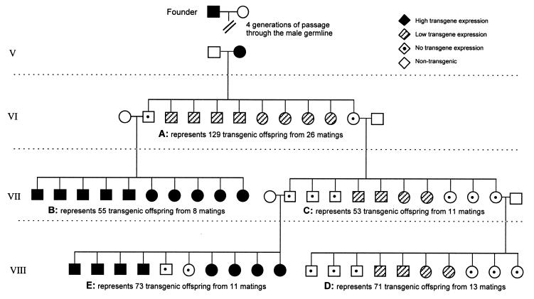
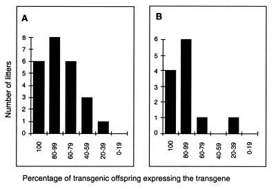
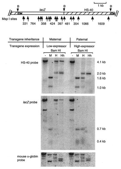
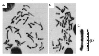
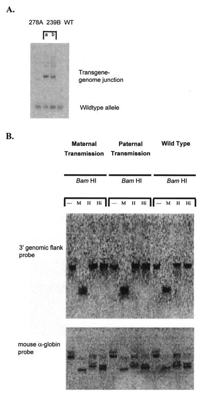
Here we report a transgenic mouse line that exhibits significant deviations from a classic pattern of parental imprinting. When the transgene is passed through the female germline, it is completely silenced in some offspring while in others expression is reduced. This variable expressivity does not appear to be the result of differences in the presence of unlinked modifiers. Female transmission of the transgene is associated with hypermethylation. The transgene is generally reactivated on passage through the male germline. Extended pedigrees reveal complex patterns of inheritance of the phenotype. The most likely explanation for this result is that the imprint is not completely erased and reset when passed through the germline of either sex. FISH analysis reveals that the transgene has integrated into chromosome 3 band E3, a region not known to carry imprinted genes, and the integration site shows no sign of allele-specific differential methylation. These findings, in conjunction with other recent work, raise the possibility that the introduction of foreign DNA into the mammalian genome, either through retrotransposition or transgenesis, may be associated with parental imprinting that is not always erased and reset during meiosis.


Figures

Figure 1
Figure 1
Representative pedigree of 239B transmission. The phenotypes depicted represent the percentages obtained for the total mice analysed for each of the five matings (A–E) adapted to a litter size of 10 pups. Genotyping was performed by Southern transfer and hybridisation using genomic mouse DNA probed with the lacZ gene and phenotypic analysis was determined by X-Gal staining of erythrocytes. All crosses were performed using P.O. heterozygote transgenic mice and P.O. wild-type mice of the opposite sex. Following convention, circles represent females and males are depicted by squares. It can be seen that the phenotype of the offspring depends not only on the phenotype of the transgenic parent but also grandparental effects are evident with male transmission. When the grandmother (female parent cross C) has a silenced transgene the silencing modification is not always reversed through the subsequent male germline (male parent cross E), resulting in some progeny with no transgene expression.
Figure 2
Figure 2
Distribution of litters based on the percentage of expressing offspring within a litter. Individual litters were grouped into categories depending on the percentage of expressing offspring within the litter. The numbers of litters obtained for each category are plotted on the y-axis. (A) Litters resulting from crosses between wild-type males and non-imprinted females. (B) Litters resulting from crosses between selected wild-type males and non-imprinted females. These selected wild-type males were generated by crossing two silenced parents.
Figure 3
Figure 3
Schematic representation of the transgene construct and the methylation state of the 239B transgene following male and female transmission. DNA was extracted from circulating erythrocytes collected from 12.5 d.p.c. embryos, then digested with either BamHI (B) alone or a combination of BamHI and either MspI (M), HpaII (H) or HhaI (Hh). Following separation on a 1.2% agarose gel, Southern blotting and hybridisation was performed using either a 4.1 kb HS-40 fragment or the lacZ gene. The membrane was also hybridised with a 1.4 kb fragment from the mouse α-globin gene to check for complete digestion of the enzymes. Not all band sizes are given as the difference in patterning is indicative of the differential methylation states of the transgene following male and female transmission.
Figure 4
Figure 4
Methylation state of the transgene array following two generations of imprinting. DNA was extracted from circulating erythrocytes and digested as described in Figure 3. Expressing and non-expressing individuals were collected from litters represented in Figure 1 (D, offspring with a non-expressing mother and grandmother; E, offspring with a non-expressing father and a non-expressing grandmother). A banding pattern, suggestive of extensive methylation, was obtained following female transmission (as seen in Fig. 3) and with the non-expressing individual resulting from inheritance through a non-expressing female then a non-expressing male.
Figure 5
Figure 5
(A and B) Representative metaphase cells of transgenic mouse line 239B showing a fluorescent signal (black arrowhead) on one mouse chromosome 3 band E3 after FISH with the DM2 transgene construct within p-127zetalacZHS-40. The chromosome 3 homologue is indicated with an open arrowhead. (C) Chromosome 3 from (A) aligned with the ideogram showing precise location of the probe signal in band E3. The signal has been painted white to increase clarity in black and white.
Figure 6
Figure 6
(A) The 3′ genomic flank sequence is unique in the genome. Southern blotting of DraI-digested genomic DNA and hybridisation with a 301 bp radiolabelled HindIII–EcoRI fragment of the 3′ genomic sequence from the site of integration (obtained by LMPCR) generated a single band in DNA samples from line 278A and wild-type (WT) mice and two bands in DNA from line 239B. The 239B DNA was obtained from a hemizygous mouse so that one allele generated a wild-type band and the other band represents the transgene–genome junction. Samples a and b are independent samples from line 239B. (B) Susceptibility of the 3′ site of integration of the transgene to digestion by methylation-sensitive restriction enzymes. Transgenic embryos were collected following either maternal or paternal transmission of the transgene as well as their wild-type littermates. DNA extracted from these 12.5 d.p.c. embryos was digested with BamHI alone or in combination with either MspI (M) (methylation-insensitive), HpaII (H) (methylation-sensitive) or HinP1I (Hi) (methylation-sensitive). The membrane was hybridised using a radiolabelled 301 bp HindIII–EcoRI fragment of the 3′ genomic DNA from the site of integration (obtained by LMPCR). The membrane was stripped and reprobed with the radiolabelled mouse α-globin gene to check for complete digestion of the DNA in all samples.

References

    1. Barlow D. (1995) Science, 270, 1610–1613. - PubMed
    1. Morison I.M. and Reeve,A.E. (1998) Hum. Mol. Genet., 7, 1599–1609. - PubMed
    1. Allen N.D., Norris,M.L. and Surani,M.A. (1990) Cell, 61, 853–861. - PubMed
    1. McGowan R., Campbell,R., Peterson,A. and Sapienza,C. (1989) Genes Dev., 3, 1669–1676. - PubMed
    1. Reik W., Howlett,S.K. and Surani,M.A. (1990) Development, (suppl.), 99–106.

Publication types

MeSH terms