. 2000 Sep 1;28(17):3370-8.
doi: 10.1093/nar/28.17.3370.

Differential effect of zinc finger deletions on the binding of CTCF to the promoter of the amyloid precursor protein gene

Affiliations

Differential effect of zinc finger deletions on the binding of CTCF to the promoter of the amyloid precursor protein gene

W W Quitschke et al. Nucleic Acids Res. .

Abstract

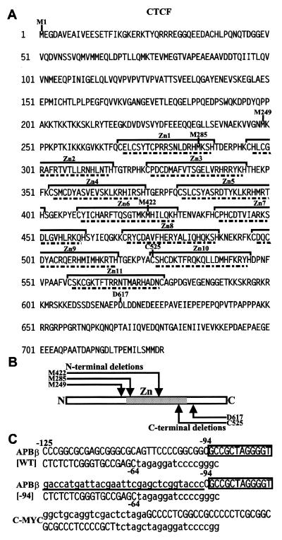
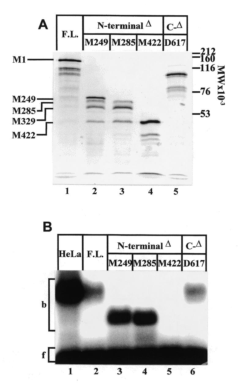
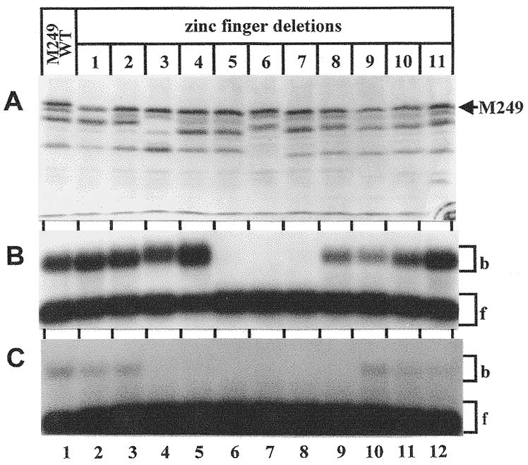
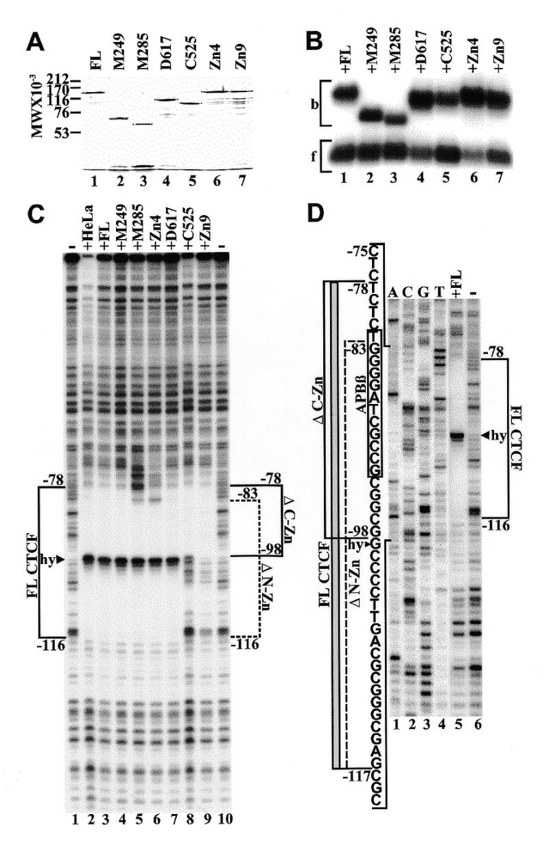
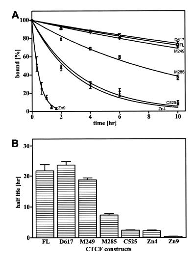
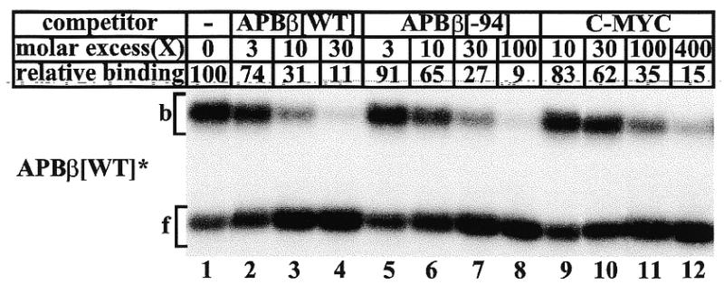
High levels of transcription from the amyloid precursor protein promoter are dependent on the binding of CTCF to the APBbeta core recognition sequence located between positions -82 and -93 upstream from the transcriptional start site. CTCF comprises 727 amino acids and contains 11 zinc finger motifs arranged in tandem that are flanked by 267 amino acids on the N-terminal side and 150 amino acids on the C-terminal side. Deletion of either the N- or the C-terminal regions outside of the zinc finger domain had no detrimental effect on the binding of CTCF to APBbeta. However, internal deletions of zinc fingers 5-7 completely abolished binding. The binding of full-length CTCF generated a DNase I protected domain extending from position -78 to -116, which was interrupted by a hypersensitive site at position -99. Selective deletions from the N- and C-terminal sides of the zinc finger domain showed that the N-terminal end of the zinc finger domain was aligned toward the transcriptional start site. Furthermore, deletions of zinc fingers peripheral to the essential zinc fingers 5-7 decreased the stability of the binding complex by interrupting sequence-specific interactions.


Figures

Figure 1
Figure 1
(A) Amino acid sequence of human CTCF. The first methionine is designated as M1. All other amino acids delineating selected deletions are indicated by the specific amino acid followed by its position in the sequence. The putative zinc fingers are indicated by brackets. Amino acids removed in individual zinc finger deletions are underlined. (B) Schematic representation of the CTCF molecule indicating its N-, C-terminal and zinc finger (Zn) domain. The approximate relative positions of N- and C-terminal deletions are indicated by arrows. (C) Sequence of the three oligonucleotides APBβ[WT], APBβ[–94] (derived from the APP promoter) and C-MYC used in their double-stranded form for mobility shift electrophoresis. Native APP and c-myc promoter sequences are written in capital letters and non-promoter sequences are written in lower case. The positions of nucleotides –125, –94 and –64 within the APP promoter are delineated in the APBβ oligonucleotides. In oligonucleotide APBβ[–94] the sequence upstream of position –94 is underlined, representing the exact reproduction of the sequence as it exists in expression vector APP[–94], which is derived from plasmid CAT2bGAL (26).
Figure 2
Figure 2
Analysis of N- and C-terminal deletions of CTCF. (A) Autoradiogram of [35S]methionine-labeled CTCF constructs. Full length CTCF (lane 1), successive N-terminal deletions at positions M249 (lane 2), M285 (lane 3), M422 (lane 4), and a C-terminal deletion at amino acid residue D617 (lane 5) were synthesized by in vitro transcription/translation and separated on a 7% SDS–polyacrylamide gel. The positions of methionine residues used as translational start sites are indicated. (B) Mobility shift electrophoresis of CTCF constructs bound to 32P-end-labeled oligonucleotide APBβ[WT] (Fig. 1C). In addition to CTCF partially purified from HeLa cell nuclear extract (lane 1), the same protein fractions as shown in (A), lanes 1–5, were used as binding factors (lanes 2–6).
Figure 3
Figure 3
Analysis of internal zinc finger deletions in CTCF. (A) Autoradiogram of [35S]methionine-labeled wild-type CTCF (lane 1) and successive zinc finger deletions (lanes 2–12). All zinc finger deletions were introduced into N-terminal deletion M249 by site-directed mutagenesis. Proteins were synthesized by in vitro transcription/translation and separated on a 7% SDS–polyacrylamide gel. The position of the protein initiating at methionine residue 249 (M249) is indicated. (B) Wild-type N-terminal deletion M249 (lane 1) and individual zinc finger deletions introduced into this construct (lanes 2–12) were bound to 32P-end-labeled APBβ[WT] (Fig. 1C) and separated by mobility shift electrophoresis. The same constructs as presented in (A) were used for the binding reaction. The bound (b) and free (f) oligonucleotides are indicated by brackets. (C) Same as in (B), except that the C-MYC oligonucleotide (Fig. 1C) was used as a labeled probe.
Figure 4
Figure 4
Binding of purified recombinant CTCF constructs to APP[WT]. (A) Coomassie blue stained SDS–polyacrylamide gel of full-length CTCF (lane 1), N-terminal deletions M249 (lane 2), M285 (lane 3), C-terminal deletions D617 (lane 4), C525 (lane 5), and internal zinc finger deletions Zn4 (lane 6) and Zn9 (lane 7). (B) Mobility shift electrophoresis of the same CTCF constructs shown in (A). All CTCF constructs were bound to radiolabeled oligonucleotide APBβ[WT] (Fig. 1C). Bound (b) and free (f) oligoncleotides are indicated by brackets. (C) DNase I footprinting of purified CTCF constructs bound to 5′-end-labeled wild-type APP fragment extending from position –193 to +100. Fragments were 5′-end-labeled at position –193 and digested with DNase I either in the absence of CTCF (lanes 1 and 10), or with full-length CTCF purified from HeLa cell nuclear extract (lane 2), full-length recombinant CTCF from P.pastoris (lane 3), N-terminal deletions M249 (lane 4) and M285 (lane 5), internal zinc finger deletion Zn4 (lane 6), C-terminal deletions D617 (lane 7) and C525 (lane 8), and internal zinc finger deletion Zn9 (lane 9). The DNase I footprint of full-length CTCF (FL CTCF) is delineated by a bracket from position –78 to –116 interrupted by a hypersensitive site (hy, arrowhead). Brackets also indicate the DNase I footprints generated by N-terminal (ΔN-Zn, position –83 to –116) and C-terminal (ΔC-Zn, position –98 to –78) zinc finger deletions. (D) DNase I footprinting of 5′-end-labeled wild-type APP fragment without CTCF (lane 1) or with bound full-length CTCF (lane 2). The DNase I footprinting reactions were co-electrophoresed with a dideoxy sequencing reaction obtained with the same 32P-labeled oligonucletide used for PCR amplification (lanes 3–6). The sequence within the bracketed region is provided together with the outlines of DNase I protected domains obtained with FL CTCF (gray bar). Brackets also indicate the outlines of the DNase I protected domains obtained with N- (ΔN-Zn) and C-terminal (ΔC-Zn) deletions of CTCF. The sequence of the APBβ binding sequence is boxed. The boundaries of the footprints are defined by the terminal nucleotides protected from DNase I digestion and their position should be considered accurate within 1 bp.
Figure 5
Figure 5
Dissociation of CTCF constructs bound to double-stranded oligonucleotide APBβ[WT]. (A) Deletions M249 (filled triangles), M285 (squares), C525 (filled diamonds), D617 (inverted triangles), Zn4 (circles), Zn9 (open triangles) and recombinant full-length CTCF (FL) (open diamonds) were bound to 5′-end-labeled APBβ[WT] and subsequently a 500-fold molar excess of unlabeled APBβ[WT] was added (time zero). The decreasing amount of radiolabeled binding complex as a function of time was monitored by mobility shift electrophoresis and the data were fitted to exponential decay curves. The relative amount of radiolabeled binding complex at time zero was assigned the value of 100% and all subsequent values are expressed as a fraction thereof. (B) From the exponential decay curves in (A), the half-lives of the CTCF binding complexes were calculated. The data points represent the averages of 2–4 independent determinations and error bars indicate the standard deviation.
Figure 6
Figure 6
Binding of recombinant CTCF constructs to APP[WT] and APP[–94]. (A) DNase I footprinting of fragments extending from position –193 to +100 in the wild-type APP promoter (lanes 1 and 2) or of fragments in which the region between positions –94 and –193 had been replaced with upstream vector sequences in plasmid APP[–94] (lanes 3–7). Fragments were 5′-end-labeled at position –193 and digested with DNase I in the absence of CTCF (lanes 1, 3 and 7), with full-length recombinant CTCF (lanes 2 and 4), or with internal zinc finger deletions Zn4 (lane 5) and Zn9 (lane 6). The protected domain of full-length CTCF bound to the wild-type APP sequence (FL CTCF[WT]) is indicated by a bracket between positions –78 and –116. Position –94, representing the point of divergence between the wild-type APP sequence (WT) and APP[–94] is indicated by an arrowhead. The protected domains of full-length CTCF and N-terminal zinc finger deletions bound to the APP[–94] sequence are indicated by brackets from position –78 to –98 (FL[–94])and –83 to –98 (ΔN-Zn), respectively. (B) DNase I footprinting of fragments extending from position –193 to +100 in plasmid APP[–94] in the absence of CTCF (lane 1) or with bound full-length CTCF (lane 2). The DNase I footprinting reactions were co-electrophoresed with a dideoxy sequencing ladder obtained with the same 32P-labeled oligonucletide used for PCR amplification (lanes 3–6). The sequence within the bracketed region is provided together with the outlines of DNase I protected domains obtained with full-length CTCF (FL[–94]) and internal deletion Zn4 (ΔN-Zn), The sequence of the APBβ binding sequence is boxed. The boundaries of the footprints are defined by the terminal nucleotides protected from DNase I digestion and their position should be considered accurate within 1 bp.
Figure 7
Figure 7
Mobility shift competition of CTCF binding to double-stranded oligonucleotide APBβ[WT]. Full-length recombinant CTCF was bound to 5′-end-labeled APBβ[WT] either without competitor (lane 1), or with a 3- (lane 2), 10- (lane 3), and 30-fold (lane 4) molar excess of unlabeled APBβ[WT], a 3- (lane 5), 10- (lane 6), 30- (lane 7), and 100-fold (lane 8) excess of unlabeled APBβ[–94], or a 10- (lane 9), 30- (lane 10) 100- (lane 11) and 400-fold (lane 12) excess of C-MYC (Fig. 4C). The amount of CTCF bound without competitor was assigned the value of 100, and all other binding activities are expressed as a fraction thereof.

References

    1. Glenner G.G. and Wong,C.W. (1984) Biochem. Biophys. Res. Commun., 122, 1131–1135. - PubMed
    1. Mann D.M., Jones,D., Prinja,D. and Purkiss,M.S. (1990) Acta Neuropathol., 80, 318–327. - PubMed
    1. Masters C.L., Simms,G., Weinman,N.A., Multhaup,G., McDonald,B.L. and Beyreuther,K. (1985) Proc. Natl Acad. Sci. USA, 82, 4245–4249. - PMC - PubMed
    1. Kang J., Lemaire,H.G., Unterbeck,A., Salbaum,J.M., Masters,C.L., Grzeschik,K.H., Multhaup,G., Beyreuther,K. and Müller-Hill,B. (1987) Nature, 325, 733–736. - PubMed
    1. Golde T.E., Estus,S., Usiak,M., Younkin,L.H. and Younkin,S.G. (1990) Neuron, 4, 253–267. - PubMed

Publication types

MeSH terms