. 2000 Aug 29;97(18):10044-9.
doi: 10.1073/pnas.160245097.

Conservation of early odontogenic signaling pathways in Aves

Affiliations

Conservation of early odontogenic signaling pathways in Aves

Y Chen et al. Proc Natl Acad Sci U S A. .

Abstract

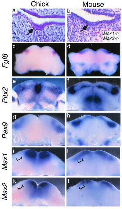
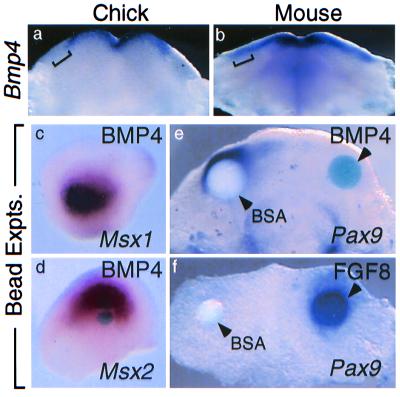
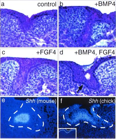
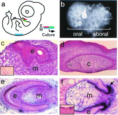
Teeth have been missing from birds (Aves) for at least 60 million years. However, in the chick oral cavity a rudiment forms that resembles the lamina stage of the mammalian molar tooth germ. We have addressed the molecular basis for this secondary loss of tooth formation in Aves by analyzing in chick embryos the status of molecular pathways known to regulate mouse tooth development. Similar to the mouse dental lamina, expression of Fgf8, Pitx2, Barx1, and Pax9 defines a potential chick odontogenic region. However, the expression of three molecules involved in tooth initiation, Bmp4, Msx1, and Msx2, are absent from the presumptive chick dental lamina. In chick mandibles, exogenous bone morphogenetic protein (BMP) induces Msx expression and together with fibroblast growth factor promotes the development of Sonic hedgehog expressing epithelial structures. Distinct epithelial appendages also were induced when chick mandibular epithelium was recombined with a tissue source of BMPs and fibroblast growth factors, chick skin mesenchyme. These results show that, although latent, the early signaling pathways involved in odontogenesis remain inducible in Aves and suggest that loss of odontogenic Bmp4 expression may be responsible for the early arrest of tooth development in living birds.


Figures

Figure 1
Figure 1
Phenotypic and molecular comparison between putative chick dental lamina and mouse dental lamina. (a) Transverse section through stage 27 chick mandible showing a representative epithelial thickening (arrow), proposed to represent a vestigial dental lamina (16). Not all embryos examined exhibited such structures. (b) Transverse section of E14.5 Msx1-Msx2 double mutant molar tooth germ arrested at the lamina stage; the stage shown is developmentally equivalent to E11.5 in wild-type embryos. [Reproduced with permission from ref. . (Copyright 1998, Company of Biologists LTD).] (cl) Molecular comparison between oral regions of stage 27 chick and E11.5 mouse mandibles. (c) Fgf8 expression in stage 27 chick mandibular epithelium. (d) Fgf8 expression in E11.5 mouse molar dental lamina. (e) Pitx2 expression in stage 27 chick mandibular epithelium. (f) Pitx2 expression in E11.5 mouse molar and incisor dental laminae. (g) Pax9 expression in stage 27 chick mandibular mesenchyme. (h) Pax9 expression in E11.5 mouse molar and incisor mesenchyme. (il) Differential Msx expression in chick and mouse mandibular mesenchyme. In chick, Msx1 (i) and Msx2 (k) expression is restricted to mesial mandibular mesenchyme and does not extend as far distally (bracketed). In mouse, Msx1 (j) and Msx2 (l) mesenchymal expression extends distally to the molar tooth-forming region (bracketed).
Figure 2
Figure 2
Expression of Bmp4 is absent from distal chick mandibular epithelium. (a) In the stage 27 chick mandible, Bmp4 is expressed in mesial oral epithelium and does not extend distally to the region of epithelial thickening (bracketed). (b) In contrast, in E11.5 mouse mandible, Bmp4 expression extends distally to the molar tooth-forming region (bracketed). (c and d) BMP4-soaked beads can induce Msx1 and Msx2 expression in stage 27 chick mandibular mesenchyme. BSA-soaked beads do not induce Msx1 or Msx2 expression (not shown). (e) BMP4 bead (Right) represses endogenous Pax9 expression in day 5 chick mandibular explants, whereas BSA bead (Left) does not. (f) FGF8 bead (Right) activates Pax9 expression in de-epithelialized day 4.5 chick mandibular explants, whereas BSA bead (Left) does not.
Figure 3
Figure 3
Induction of chick oral epithelial appendages by BMP and/or FGF. (a) Section through a control, untreated chick mandible after 6 days of culture showing region of thickened epithelium. (b and c) Bud-like structures induced in chick mandibles after 6 days of culture with 100 ng/ml of exogenous BMP4 (b) or FGF4 (c). (d) More advanced epithelial structure induced to form in chick mandibles after 6 days of culture with BMP4 and FGF4 (100 ng/ml each). Note convoluted epithelium (arrow). The clear space is a cyst. (e) Localization of Shh transcripts in the enamel knot of an E14.5 mouse molar tooth germ. (f) Shh expression induced in the central portion of the epithelial structure by addition of BMP4 and FGF4 to chick mandibles in explant culture. The dotted line in e and f indicates the location of the basal lamina separating epithelium and mesenchyme. (Inset) Shh is not expressed in control explants; the epithelium resides between the white lines.
Figure 4
Figure 4
Chick oral epithelial appendage structures induced in vitro by heterotypic, heterochronic recombination. (a) Scheme for recombinations in which a single piece of stage 27 chick mandibular epithelium, including both oral (red) and aboral (green) surfaces, was recombined with stage 34 chick dorsal skin mesenchyme (blue) and cultured for 5 days. (b) Whole mount showing that different epithelial appendages form from oral and aboral chick mandibular epithelia. Feather germs form from aboral epithelium (Right), whereas novel epithelial appendages form from oral epithelium (Left). (c) Histology of oral epithelial structures in the preceding experiment. A small cyst is present. (Inset) Whole-mount in situ demonstrating Bmp4 expression in stage 34 chick dorsal feather follicle mesenchyme. (d) Control recombination between stage 34 chick skin epithelium and stage 27 mandibular mensenchyme, which does not produce epithelial structures and in some cases (shown) results in cartilage formation (indicated by c). (e) Epithelial appendage structure formed after recombination of stage 27 chick mandibular epithelium with stage 35 chick skin mesenchyme after 2 days in organ culture and 6 days on CAM. Note condensation of mesenchymal cells within the epithelium. (f) AP activity detected (black, arrow) in condensed mesenchyme of a CAM-cultured chick mandibular epithelium-chick skin mesenchyme recombinant. (Inset) Absence of AP activity in naïve chick mandibular tissue. e, epithelium; m, mesenchyme.

References

    1. Maas R, Bei M. Crit Rev Oral Biol Med. 1997;8:4–39. - PubMed
    1. Thesleff I, Sharpe P. Mech Dev. 1997;67:11–23. - PubMed
    1. Mina M, Kollar E J. Arch Oral Biol. 1987;32:123–127. - PubMed
    1. Lumsden A G S. Development (Cambridge, UK) 1988;103,Suppl.:155–169. - PubMed
    1. Kettunen P, Thesleff I. Dev Dyn. 1998;211:256–268. - PubMed

Publication types

MeSH terms

LinkOut - more resources