. 2000 Aug 29;97(18):9931-6.
doi: 10.1073/pnas.170153097.

Hexose permeation pathways in Plasmodium falciparum-infected erythrocytes

Affiliations

Hexose permeation pathways in Plasmodium falciparum-infected erythrocytes

C J Woodrow et al. Proc Natl Acad Sci U S A. .

Abstract

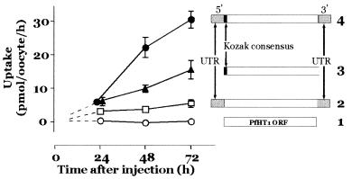
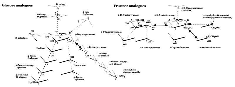
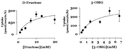
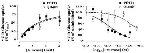
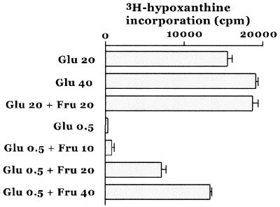
Plasmodium falciparum requires glucose as its energy source to multiply within erythrocytes but is separated from plasma by multiple membrane systems. The mechanism of delivery of substrates such as glucose to intraerythrocytic parasites is unclear. We have developed a system for robust functional expression in Xenopus oocytes of the P. falciparum asexual stage hexose permease, PfHT1, and have analyzed substrate specificities of PfHT1. We show that PfHT1 (a high-affinity glucose transporter, K(m) approximately 1.0 mM) also transports fructose (K(m) approximately 11.5 mM). Fructose can replace glucose as an energy source for intraerythrocytic parasites. PfHT1 binds fructose in a furanose conformation and glucose in a pyranose form. Fructose transport by PfHT1 is ablated by mutation of a single glutamine residue, Q169, which is predicted to lie within helix 5 of the hexose permeation pathway. Glucose transport in the Q169N mutant is preserved. Comparison in oocytes of transport properties of PfHT1 and human facilitative glucose transporter (GLUT)1, an archetypal mammalian hexose transporter, combined with studies on cultured P. falciparum, has clarified hexose permeation pathways in infected erythrocytes. Glucose and fructose enter erythrocytes through separate permeation pathways. Our studies suggest that both substrates enter parasites via PfHT1.


Figures

Figure 1
Figure 1
Influence of untranslated sequences on expression of PfHT1 in Xenopus oocytes. Sequence modifications outside the ORF for PfHT1 are shown next to induced glucose uptake. Oocytes from a single toad were injected with RNA (5 ng) transcribed from different plasmid constructs and assayed for glucose uptake at the times indicated (six to eight oocytes per uptake; data displayed are mean ± SE). Constructs are: (1) PfHT1 ORF (open rectangle); (2) PfHT1 ORF with 5′ and 3′ UTRs from Xenopus β-globin (shaded); (3) PfHT1 ORF with Kozak consensus (filled); and (4) PfHT1 ORF with both UTRs and Kozak consensus. Data represent one of two independent experiments.
Figure 2
Figure 2
Analogues used in studies on PfHT1 and GLUT1. Structures are represented as chair conformations. For glucose and fructose, solid arrows indicate interconversions occurring in solution. For analogues, a single common form is shown for simplicity. Percentages of analogues in each conformation at equilibrium in solution are shown in Table 2.
Figure 3
Figure 3
Transport of fructose and 3-OMG by PfHT1. (Left) Initial uptake rates of d-fructose (representative of three independent experiments; see Table 2) and (Right) 3-OMG (representative of three independent experiments; see Table 1) are plotted against concentration of substrate (eight oocytes per concentration; mean ± SE). In this experiment, d-fructose Km = 6.8 mM; for 3-OMG Km = 0.9 mM.
Figure 4
Figure 4
Transport properties of Q169N mutant PfHT1. (Left) Initial mean d-glucose uptake rates for PfHT1 and Q169N mutant (one of three independent experiments in each case; eight oocytes per concentration; mean ± SE). For PfHT1 in this experiment, Km = 1.6 mM, Vmax = 302 pmol/oocyte/h; for Q169N Km = 1.4 mM, Vmax = 650 pmol/oocyte/h. (Right) d-fructose inhibition of d-[14C]glucose uptake in PfHT1 and Q169N mutant. Uptakes (eight oocytes per concentration; mean ± SE) are expressed relative to d-glucose uptake in uncompeted oocytes.
Figure 5
Figure 5
In vitro studies on P. falciparum by using hexose analogues. Standard culture medium contains ≈20 mM d-glucose (Glu 20). The effects of supplementing this medium on [3H]hypoxanthine incorporation with glucose (Glu 40; final concentration 40 mM d-glucose); fructose (Fru 20; d-fructose 20 mM) are shown (Upper). The effects of culture in 0.5 mM glucose (Glu 0.5; containing 10% human serum and glucose-free RPMI 1640), and of increasing concentrations of d-fructose supplements (Fru 10, 20, and 40; respectively 10 mM, 20 mM, and 40 mM fructose) are shown (Lower). Representative of three independent experiments.

References

    1. Kirk K, Horner H A, Kirk J. Mol Biochem Parasitol. 1996;82:195–205. - PubMed
    1. Woodrow C J, Penny J I, Krishna S. J Biol Chem. 1999;274:7272–7277. - PubMed
    1. Pardridge W M, Boado R J. In: The Blood–Brain Barrier: Cellular and Molecular Biology. Pardridge W M, editor. New York: Raven; 1993. pp. 395–440.
    1. Krishna S, Woodrow C J. In: Transport and Trafficking in the Malaria-Infected Erythrocyte. Cardew G, editor. Vol. 226. London: Wiley; 1999. pp. 126–144.
    1. Kaback H R. In: Transport Processes in Eukaryotic and Prokaryotic Organisms. Konings W N, Kaback H R, Lolkema J S, editors. Vol. 2. Amsterdam: Elsevier; 1996. pp. 203–227.

Publication types

MeSH terms

LinkOut - more resources