

doi: 10.1073/pnas.170207097.
Human mesothelial cells are unusually susceptible to simian virus 40-mediated transformation and asbestos cocarcinogenicity
Affiliations
- PMID: 10954737
- PMCID: PMC27818
- DOI: 10.1073/pnas.170207097
Item in Clipboard
Human mesothelial cells are unusually susceptible to simian virus 40-mediated transformation and asbestos cocarcinogenicity
Proc Natl Acad Sci U S A.
.
Abstract
Mesothelioma, a malignancy associated with asbestos, has been recently linked to simian virus 40 (SV40). We found that infection of human mesothelial cells by SV40 is very different from the semipermissive infection thought to be characteristic of human cells. Mesothelial cells are uniformly infected but not lysed by SV40, a mechanism related to p53, and undergo cell transformation at an extremely high rate. Exposure of mesothelial cells to asbestos complemented SV40 mutants in transformation. Our data provide a mechanistic explanation for the ability of SV40 to transform mesothelial cells preferentially and indicate that asbestos and SV40 may be cocarcinogens.
Figures
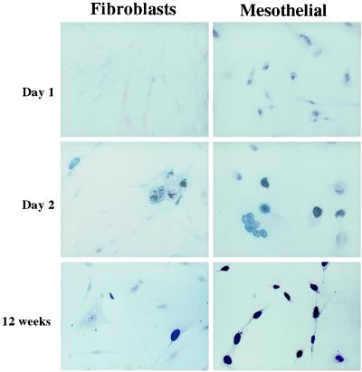
Tag immunostaining. WI38 HF (Left) and HM3 (Right), at the indicated time after infection. Similar results were obtained with the other cells. No substantial differences were observed among HM1–3. In HM, the nuclear staining was punctate at 24 h, then it became granular, with the formation of intranuclear bodies, and finally obscured the entire nucleus. In HF, only a fraction of cells expressed Tag. These cells formed ill-looking cell clumps and giant cells, with clear evidence of cytopathic effects, such as vacuolization and lysis. (Original magnification, ×400.)
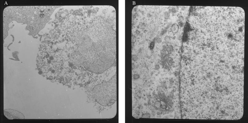
EM of WI38 HF (A) and HM3 (B) infected with SV40 72 h earlier. Note that infected HF are lysed and full of viral particles (the individual viral particles are not clearly visible at this magnification). HM, instead, have intact nuclear membrane, and viral particles (round structures) are seen only in the nucleus (right side of the photograph). (Original magnifications: A, ×4, 400; B, ×20,000.) The same results were obtained when HM2 were used; HM1 were not tested.
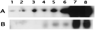
Southern blot for SV40 DNA in HM3 and WI38. (A) SV40 DNA extracted from the cells. (B) SV40 DNA recovered in the tissue culture medium. Lanes: 1 and 2, HM 48 h after infection; 3 and 4, HF 48 h after infection; 5 and 6, HM 72 h after infection; 7 and 8, HF 72 h after infection. Each lane represents an independent infection experiment. Almost identical results were obtained with HM2 and MRC-5 HF. HM1 and CCD1069Sk were not tested. The different amounts of DNA (see text) were determined by Cherenkov counting of the individual lanes. DNAs were normalized for number of Tag-positive cells.
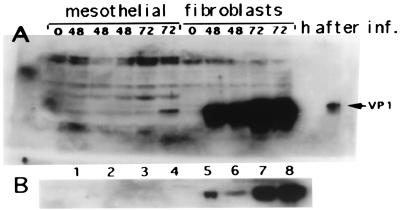
Western blot for SV40 VP1 in HM3 and WI38 after infection with SV40. Blots were developed by enhanced chemiluminescence. (A) VP-1 in the total cell extracts (100 μg per lane). (B) VP1 in the tissue culture medium (samples were normalized for number of Tag-positive cells). Lanes: 1 and 2, HM 48 h after infection; 3 and 4, HM 72 h after infection; 5 and 6, HF 48 h after infection; 7 and 8, HF 72 h after infection. Each lane represents an independent infection experiment. Almost identical results were obtained with all of the other HM and HF. Cell extracts were normalized for number of Tag-positive cells.
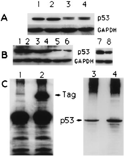
(A) p53 expression in HM and HF. One hundred micrograms of total protein extracts was loaded into each lane. GAPDH, glyceraldehyde-3-phosphate dehydrogenase. Lanes: 1, HM2; 2, HM3; 3, WI38; 4, CCD1069Sk [HM1 and MRC5 (not shown) produced almost identical results]. (B) Lanes 1–6, p53 expression in untreated HM (lanes 1 and 2), HM treated with 5 μM scrambled oligo (lanes 3 and 4), and HM treated with 5 μM antisense p53 oligo (lanes 5 and 6). Cells were harvested and lysed 48 h after the onset of treatment; 100 μg of total protein extracts was loaded per lane (each lane represent an independent experiment). Lanes 7 and 8, p53 expression in HM 5 days after treatment (and 72 h after SV40 infection) with scrambled oligos (lane 7) and antisense p53 (lane 8). (C) Lanes 1 and 2, Tag immunoprecipitation in SV40-infected HM. Lane 1, scrambled control oligo-treated HM; lane 2, antisense p53-treated HM. Tag was precipitated with the monoclonal anti-Tag AB-1 (Tag amino terminus). The membrane was probed with the monoclonal anti-Tag AB-2 (Tag carboxyl terminus), followed by monoclonal anti-mouse IgG conjugated with horseradish peroxidase. Lanes 3 and 4, Tag/p53 coimmunoprecipitation. The membrane shown on the left was stripped of antibodies and probed with the monoclonal anti-p53 DO-1 directly conjugated with horseradish peroxidase. Lane 3, control oligo-treated HM; lane 4, antisense p53-treated HM.
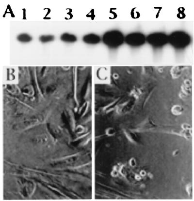
(A) Southern blot analysis of SV40 DNA synthesis in SV40-infected HM3 cells treated with a control scrambled oligo (5 μM) (lanes 1–4) and treated with antisense p53 (5 μM) (lanes 5–8). Almost identical results were obtained with HM2; HM1 were not tested. (B and C) HM2 treated with control scrambled oligo (B; 25 μM); or antisense p53 (C; 25 μM) and infected 48 h later with SV40. Cells are shown 72 h after infection. In C, note empty spaces because of lysis in cells treated with antisense p53. Little or no cell lysis was observed in cells treated with control oligos (B). (Original magnification, ×400.)
Comment in
-
Simian virus 40 and the human mesothelium.Proc Natl Acad Sci U S A. 2000 Aug 29;97(18):9830-1. doi: 10.1073/pnas.190325697. Proc Natl Acad Sci U S A. 2000. PMID: 10954758 Free PMC article. No abstract available.
References
-
- Testa J R, Pass H I, Carbone M. In: Principles and Practice of Oncology. 6th Ed. De Vita V, Hellman S, Rosenberg S, editors. Williams & Wilkins, Philadelphia: Lippincott; 2000. , in press.
-
- Robledo R, Mossman B T. J Cell Physiol. 1999;180:158–166. - PubMed
-
- Rosenthal G J, Simeonova P, Corsini E. Rev Environ Health. 1999;14:11–19. - PubMed
-
- Butel J, Lednicky J. J Natl Cancer Inst. 1999;91:119–134. - PubMed
Publication types
MeSH terms
Substances
Grants and funding
LinkOut - more resources
Full Text Sources
Other Literature Sources
Medical
Research Materials
Miscellaneous
