. 2000 Sep;20(18):6646-58.
doi: 10.1128/MCB.20.18.6646-6658.2000.

Meiotic segregation, synapsis, and recombination checkpoint functions require physical interaction between the chromosomal proteins Red1p and Hop1p

Affiliations

Meiotic segregation, synapsis, and recombination checkpoint functions require physical interaction between the chromosomal proteins Red1p and Hop1p

D Woltering et al. Mol Cell Biol. 2000 Sep.

Abstract

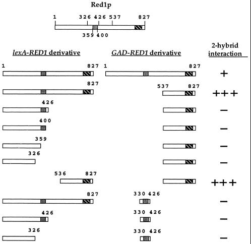
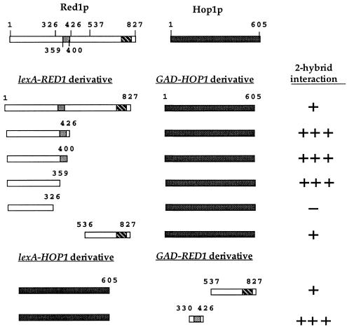
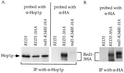
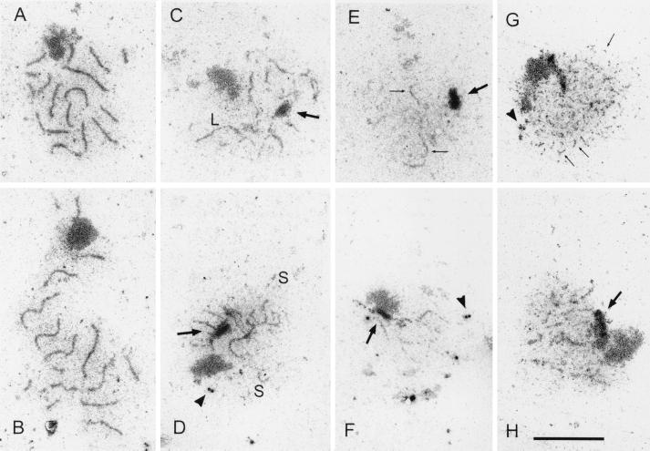
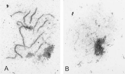
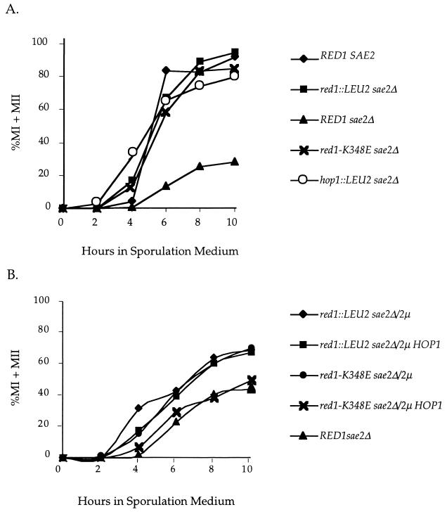
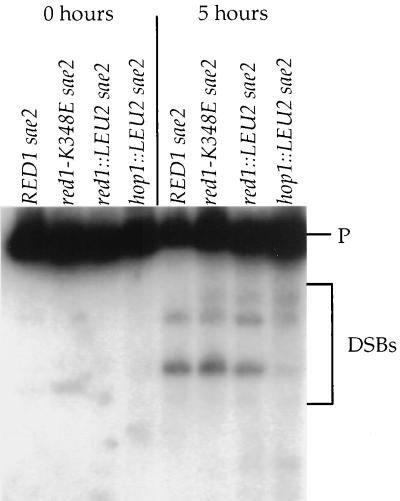
In yeast, HOP1 and RED1 are required during meiosis for proper chromosome segregation and the consequent formation of viable spores. Mutations in either HOP1 or RED1 create unique as well as overlapping phenotypes, indicating that the two proteins act alone as well as in concert with each other. To understand which meiotic processes specifically require Red1p-Hop1p hetero-oligomers, a novel genetic screen was used to identify a single-point mutation of RED1, red1-K348E, that separates Hop1p binding from Red1p homo-oligomerization. The Red1-K348E protein is stable, phosphorylated in a manner equivalent to Red1p, and undergoes efficient homo-oligomerization; however, its ability to interact with Hop1p both by two-hybrid and coimmunoprecipitation assays is greatly reduced. Overexpression of HOP1 specifically suppresses red1-K348E, supporting the idea that the only defect in the protein is a reduced affinity for Hop1p. red1-K348E mutants exhibit reduced levels of crossing over and spore viability and fail to undergo chromosome synapsis, thereby implicating a role for Red1p-Hop1p hetero-oligomers in these processes. Furthermore, red1-K348E suppresses the sae2/com1 defects in meiotic progression and sporulation, indicating a previously unknown role for HOP1 in the meiotic recombination checkpoint.


Figures

FIG. 1
FIG. 1
Two-hybrid analysis of various RED1 constructs. The two boxes present in Red1p represent regions of predicted coiled-coil structure. Protein-protein interactions were assayed by β-galactosidase filter assays in strain L40. Plasmids used: pNH223 (lexA-RED11–827); pDW35 (lexA-RED11-326); pDW61 (lexA-RED11–359); pDW62 (lexA-RED11-400); pDW28 (lexA-RED11–426); pDW58 (lexA-RED1536–827); pJ63 (GAD-RED11–827); pGAD-RED1537–827 and pDW54 (GAD-RED1330–426). One plus sign indicates that the cells turned blue in 90 min at 30°C; three plus signs indicates the cells turned blue by 15 min; a minus sign indicates a failure to turn blue by 4 h.
FIG. 2
FIG. 2
Two-hybrid analysis of various RED1 constructs with HOP1. Protein-protein interactions were assayed in L40 by β-galactosidase filter assays in strain L40. Plasmids used: pNH223 (lexA-RED11–827); pDW35 (lexA-RED11–326); pDW61 (lexA-RED11–359); pDW62 (lexA-RED11–400); pDW28 (lexA-RED11–426); pDW58 (lexA-RED1536–827); pLP27 (lexA-HOP1); pJ63 (GAD-RED11–827); pGAD-RED1537–827; pDW54 (GAD-RED1330–426) and pNH108 (GAD-HOP1). One plus sign indicates that the cells turned blue in 90 min at 30°C; three plus signs indicates the cells turned blue by 15 min; a minus sign indicates a failure to turn blue by 4 h.
FIG. 3
FIG. 3
Co-IP of different Red1-3HA proteins with Hop1p from meiotic extracts of SK1 diploids. Cells from YTS3 transformed with either p1b-1 (RED1), pNH212 (RED1-3HA), or pNH212-K348E (red1-K348E-3HA) were sporulated for 3 h. Hop1p and Red1-3HAp were immunoprecipitated from soluble extracts prepared from equivalent numbers of cells using α-Hop1p (A) or α-HA (B) antibody. The Hop1p IPs were first probed with α-HA to detect Red1-3HAp. The blot was then stripped and reprobed with α-Hop1p antibodies to detect Hop1p. The α-HA IPs were probed with α-HA antibodies.
FIG. 4
FIG. 4
SC formation in various wild-type and mutant SK1 strains. Chromosome spreads were prepared for electron microscopy as described in Materials and Methods. (A and B) YTS3::pSB3 (RED1); (C and D) YTS3::pSB3-K348E (red1-K348E); (E and F) YTS3::pRS306 (red1::LEU2); (G and H) DW10 (hop1::LEU2). The arrows point to polycomplexes, and the arrowheads point to unduplicated spindle pole bodies. The bar corresponds to 5 μm. S and L denote short and long regions of dense structures, respectively.
FIG. 5
FIG. 5
SC formation in SK1 red1 diploids overexpressing HOP1. Chromosome spreads were prepared for electron microscopy as described in Materials and Methods. (A) NH305::pBB14 (red1-K348E)/2μm HOP1 (pNH83); (B) NH305::pRS402 (red1::LEU2)/2μm HOP1 (pNH83).
FIG. 6
FIG. 6
Meiotic progression in various red1 sae2Δ SK1 diploids. Cells were fixed at different times after transfer to sporulation medium and stained with DAPI. The numbers of bi- and tetranucleate cells (indicative of passage through MI and MII, respectively) were determined by epifluorescence microscopy. Three independent cultures were examined for each strain. The graph shows the mean value for each diploid. (A) RED SAE2, NH305::pBB16; red1::LEU2 sae2Δ, NH306::pRS402; RED1 sae2Δ, NH306::pBB16; red1-K348E sae2Δ, NH306::pBB14; hop1::LEU2 sae2Δ, NH311::pRS402. (B) red1::LEU2 sae2Δ/2μm, NH306/pRS422; red1::LEU2 sae2Δ/2μm HOP1, NH306/pDW72; red1-K348E sae2Δ/2μm, NH306::pBB19/pRS422; red1-K348E sae2Δ/2μm HOP1, NH306::pBB19/pDW72; RED1 sae2Δ, NH306::pBB16.
FIG. 7
FIG. 7
DSBs analysis in SK1 diploids. DNA was isolated from vegetative cells (0 h) or cells 5 h after transfer to sporulation medium (5 h) as described in Materials and Methods. After digestion with BglII, the DNA was probed with a fragment to detect DSBs occurring near THR4 (48). P represents the 10-kb parental fragment. The DSB fragments are indicated by the bracket. Strains used: NH306::pBB16 (RED1 sae2Δ); NH306::pBB14 (red1-K384E sae2Δ); NH306::pRS402 (red1::LEU2 sae2Δ); NH311::pRS402 (hop1::LEU2 sae2Δ).

References

    1. Alani E, Padmore R, Kleckner N. Analysis of wildtype and rad50 mutants of yeast suggests an intimate relationship between meiotic chromosome synapsis and recombination. Cell. 1990;61:419–436. - PubMed
    1. Bai C, Elledge S J. Gene identification using the yeast two-hybrid system. Methods Enzymol. 1997;283:141–156. - PubMed
    1. Bailis J M, Roeder G S. Pachytene exit controlled by reversal of Mek1-dependent phosphorylation. Cell. 2000;101:211–221. - PubMed
    1. Bailis J M, Roeder G S. Synaptonemal complex morphogenesis and sister-chromatid cohesion require Mek1-dependent phosphorylation of a meiotic chromosomal protein. Genes Dev. 1998;12:3551–3563. - PMC - PubMed
    1. Bascom-Slack C A, Ross L O, Dawson D S. Chiasmata, crossovers and meiotic chromosome segregation. Adv Genet. 1997;35:253–284. - PubMed

Publication types

MeSH terms

LinkOut - more resources