. 2000 Sep;20(18):6704-11.
doi: 10.1128/MCB.20.18.6704-6711.2000.

Characterization of the role of AMP-activated protein kinase in the regulation of glucose-activated gene expression using constitutively active and dominant negative forms of the kinase

Affiliations

Characterization of the role of AMP-activated protein kinase in the regulation of glucose-activated gene expression using constitutively active and dominant negative forms of the kinase

A Woods et al. Mol Cell Biol. 2000 Sep.

Abstract

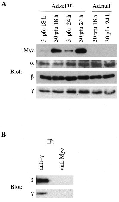
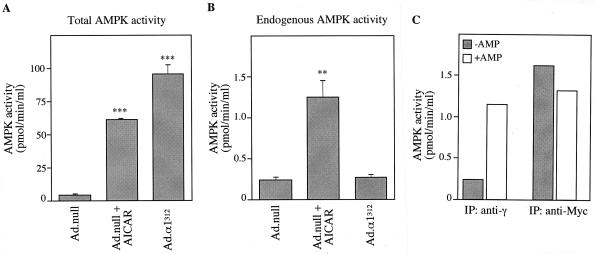
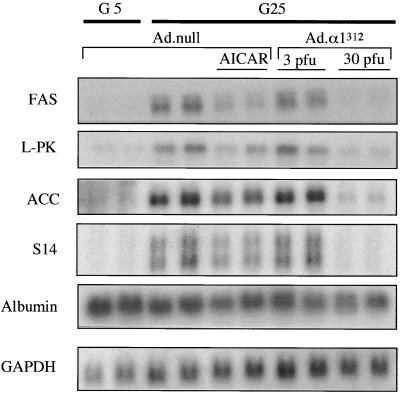
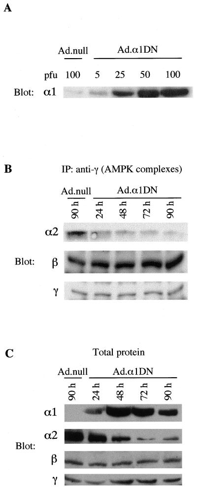
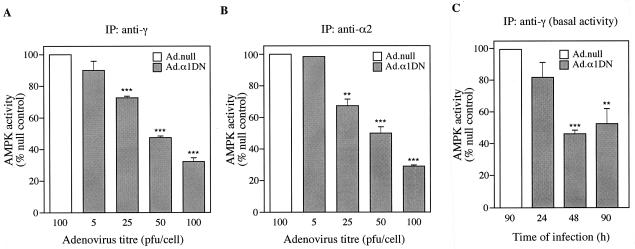
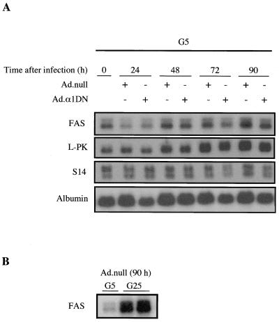
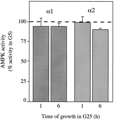
In the liver, glucose induces the expression of a number of genes involved in glucose and lipid metabolism, e.g., those encoding L-type pyruvate kinase and fatty acid synthase. Recent evidence has indicated a role for the AMP-activated protein kinase (AMPK) in the inhibition of glucose-activated gene expression in hepatocytes. It remains unclear, however, whether AMPK is involved in the glucose induction of these genes. In order to study further the role of AMPK in regulating gene expression, we have generated two mutant forms of AMPK. One of these (alpha1(312)) acts as a constitutively active kinase, while the other (alpha1DN) acts as a dominant negative inhibitor of endogenous AMPK. We have used adenovirus-mediated gene transfer to express these mutants in primary rat hepatocytes in culture in order to determine their effect on AMPK activity and the transcription of glucose-activated genes. Expression of alpha1(312) increased AMPK activity in hepatocytes and blocked completely the induction of a number of glucose-activated genes in response to 25 mM glucose. This effect is similar to that observed following activation of AMPK by 5-amino-imidazolecarboxamide riboside. Expression of alpha1DN markedly inhibited both basal and stimulated activity of endogenous AMPK but had no effect on the transcription of glucose-activated genes. Our results suggest that AMPK is involved in the inhibition of glucose-activated gene expression but not in the induction pathway. This study demonstrates that the two mutants we have described will provide valuable tools for studying the wider physiological role of AMPK.


Figures

FIG. 1
FIG. 1
Expression of α1312 in hepatocytes. (A) Primary rat hepatocytes in culture were infected with Ad.α1312 or Ad.null at either 3 or 30 PFU/cell. Cell lysates from hepatocytes harvested 18 or 24 h postinfection were analyzed by Western blotting for expression of the recombinant α1312 protein (using an anti-Myc antibody) or the endogenous AMPK subunits (using either an anti-α antibody which recognizes both α1 and α2 isoforms, but not the truncated α1312 protein, or an anti-β or anti-γ antibody). (B) Cell lysates from hepatocytes infected with Ad.α1312 (30 PFU/cell for 24 h) were immunoprecipitated (IP) with either an anti-γ antibody or an anti-Myc antibody. Immune complexes were resolved by SDS-PAGE, and the presence of the β and γ subunits was determined by Western blotting. In each case, a representative blot from two independent experiments is shown.
FIG. 2
FIG. 2
Activity of α1312 in hepatocytes. Hepatocytes were grown in the presence of 25 mM glucose and 100 nM insulin and infected with either Ad.null or Ad.α1312 (30 PFU/cell). Twenty-four hours postinfection, hepatocytes were incubated in the presence or absence of 250 μM AICA riboside (AICAR) for 1 h before harvesting. (A) Total activity (endogenous AMPK and expressed α1312) was measured in cell lysates, without prior immunoprecipitation, using the SAMS peptide assay. (B) Endogenous AMPK activity was measured in immune complexes isolated by immunoprecipitation using an anti-γ antibody bound to protein G-Sepharose. (C) AMPK activity present in either anti-γ (endogenous) or anti-Myc (expressed α1312) immunoprecipitates was measured in the absence (shaded bars) or presence (open bars) of 0.2 mM AMP. Activities shown are the mean ± the SEM from four experiments (A and B) or the average of two independent experiments assayed in duplicate (C). ∗∗∗ denotes a significant difference from the Ad.null value (P < 0.0005), and ∗∗ indicates that P was <0.005. Activities are plotted as picomoles of 32P incorporation/minutes per milliliter of lysate.
FIG. 3
FIG. 3
Expression of α1312 inhibits the transcription of glucose-activated genes in cultured hepatocytes. Hepatocytes were grown in medium containing either 5 mM glucose and 100 nM insulin (G5) or 25 mM glucose and 100 nM insulin (G25) and were infected with Ad.α1312 (3 or 30 PFU/cell) or Ad.null (30 PFU/cell) and incubated for 18 h. At the end of this period, hepatocytes were treated with or without AICA riboside (AICAR) (250 μM) and incubated for 4 h. Total RNA was extracted, electrophoresed on a 1% agarose gel, and subjected to Northern blot analysis using cDNA probes encoding either FAS, L-PK, ACC, S14, albumin, or GAPDH. Blots were washed extensively and exposed to autoradiographic film at −70°C for 1 to 2 days. The blot shown is representative of two independent experiments.
FIG. 4
FIG. 4
Expression of α1DN in hepatocytes. (A) Hepatocytes in culture were infected with varying titers of Ad.α1DN or Ad.null. Twenty-four hours after infection, cell lysates were resolved by SDS-PAGE, transferred to a PVDF membrane, and probed with an anti-α1 antibody. The α1-specific antibody used for detection recognizes both the endogenous α1 subunit and the recombinant α1 mutant protein. (B) Hepatocytes were infected with Ad.α1DN (10 PFU/cell), and at varying times after infection, AMPK complexes were isolated by immunoprecipitation (IP) with an anti-γ antibody. Proteins within the immune complex were resolved by SDS-PAGE, transferred to a PVDF membrane, and probed with either α2-, β-, or γ-specific antibodies. A control lane shows the expression of the subunits in an immune complex isolated from hepatocytes infected for 90 h with Ad.null (10 PFU/cell). (C) Hepatocytes were infected with either Ad.α1DN (10 PFU/cell) or Ad.null (10 PFU/cell). At various times after infection, total protein in cell lysates was analyzed for expression of α1 and the endogenous α2, β, and γ subunits, using subunit-specific AMPK antibodies. In each case, a representative blot from two independent experiments is shown.
FIG. 5
FIG. 5
Expression of α1DN inhibits AMPK activity. (A and B) Hepatocytes were infected with varying titers of Ad.α1DN or Ad.null. Forty-four hours after infection, AICA riboside (500 μM) was added to the culture medium, and the hepatocytes were incubated for a further 1 h. AMPK complexes were isolated from cell lysates by immunoprecipitation (IP) with either an anti-γ antibody (A) or an anti-α2 antibody (B). Activity present in the immune complexes was measured by phosphorylation of the SAMS peptide. For panel C, hepatocytes were infected with either Ad.α1DN (10 PFU/cell) or Ad.null (10 PFU/cell) in the absence of AICA riboside. At various times postinfection, AMPK activity present in immune complexes isolated using an anti-γ antibody was determined as described above. In each case, results are plotted as a percentage of the activity present in hepatocytes infected with Ad.null and are the mean ± the SEM of four independent experiments. ∗∗∗ denotes a significant difference from the Ad.null value (P < 0.0005), and ∗∗ indicates that P was <0.005.
FIG. 6
FIG. 6
Overexpression of α1DN has no effect on the transcription of glucose-activated genes. (A) Hepatocytes were incubated in the presence of 5 mM glucose and 100 nM insulin (G5) for 18 h before infection with either Ad.α1DN (10 PFU/cell) or Ad.null (10 PFU/cell). At various times after infection, total RNA was extracted from the cells and subjected to Northern blotting with cDNA probes encoding either FAS, L-PK, ACC, S14, or albumin. Blots were washed extensively and exposed to autoradiographic film at −70°C for 1 to 2 days. (B) Northern blot analysis of RNA isolated from hepatocytes 90 h after infection with Ad.null (10 PFU/cell) cultured in medium containing 100 nM insulin and either 5 mM glucose (G5) or 25 mM glucose (G25). In each case, a representative blot from at least four independent experiments is shown.
FIG. 7
FIG. 7
Effect of extracellular glucose concentration on AMPK activity in hepatocytes. Hepatocytes were cultured overnight in medium containing 5 mM glucose and 100 nM insulin (G5). Medium was removed, and fresh G5 medium or medium containing 25 mM glucose and 100 nM insulin (G25) was added. Hepatocytes were harvested following incubation for a further 1 or 6 h. AMPK activity present in immune complexes isolated using either an anti-α1 (shaded bars) or anti-α2 (hatched bars) antibody was determined by phosphorylation of the SAMS peptide. Activities are plotted as a percentage of AMPK activity present in hepatocytes maintained in G5 and are the mean ± the SEM of three experiments. The 100% value for the activity in G5 is indicated by the dashed line.

References

    1. Carling D, Aguan K, Woods A, Verhoeven A J M, Beri R K, Brennan C H, Sidebottom C, Davison M D, Scott J. Mammalian AMP-activated protein kinase is homologous to yeast and plant protein kinases involved in the regulation of carbon metabolism. J Biol Chem. 1994;269:11442–11448. - PubMed
    1. Celenza J L, Carlson M. Mutational analysis of the Saccharomyces cerevisiae SNF1 protein kinase and evidence for functional interaction with the SNF4 protein. Mol Cell Biol. 1989;9:5034–5044. - PMC - PubMed
    1. Celenza J L, Carlson M. A yeast gene that is essential for release from glucose repression encodes a protein kinase. Science. 1986;233:1175–1180. - PubMed
    1. Cheung P C F, Salt I P, Davies S P, Hardie D G, Carling D. Characterization of AMP-activated protein kinase γ-subunit isoforms and their role in AMP binding. Biochem J. 2000;346:659–669. - PMC - PubMed
    1. Chomczynski P, Sacchi N. Single-step method of RNA isolation by acid guanidinium thiocyanate-phenol-chloroform extraction. Anal Biochem. 1987;162:156–159. - PubMed

Publication types

MeSH terms

LinkOut - more resources