. 2000 Sep;20(18):6882-90.
doi: 10.1128/MCB.20.18.6882-6890.2000.

Beta-catenin-histone deacetylase interactions regulate the transition of LEF1 from a transcriptional repressor to an activator

Affiliations

Beta-catenin-histone deacetylase interactions regulate the transition of LEF1 from a transcriptional repressor to an activator

A N Billin et al. Mol Cell Biol. 2000 Sep.

Abstract

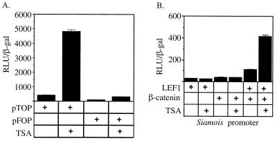
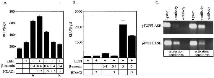
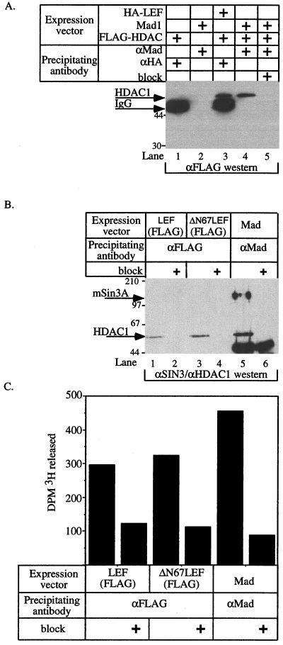
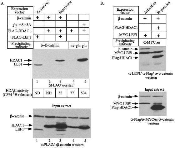
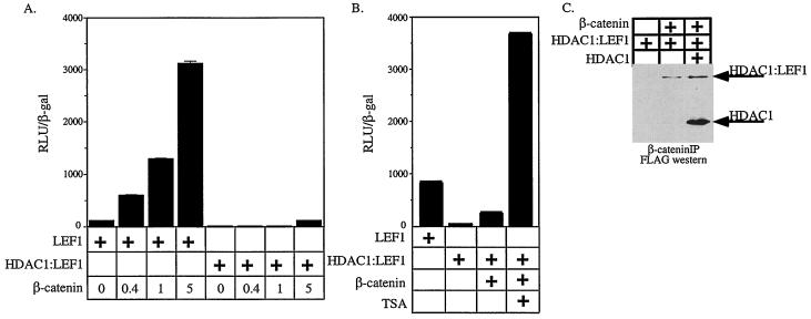
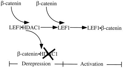
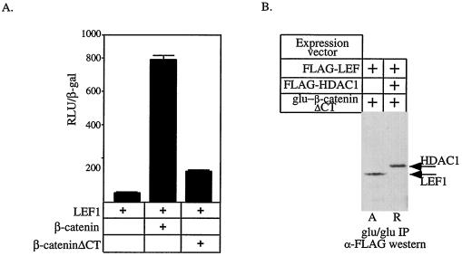
Recent evidence suggests that certain LEF/TCF family members act as repressors in the absence of Wnt signaling. We show here that repression by LEF1 requires histone deacetylase (HDAC) activity. Further, LEF1 associates in vivo with HDAC1, and transcription of a model LEF1-dependent target gene is modulated by the ratio of HDAC1 to beta-catenin, implying that repression by LEF1 is mediated by promoter-targeted HDAC. Consistent with this hypothesis, under repression conditions the promoter region of a LEF1 target gene is hypoacetylated. By contrast, when the reporter is activated, its promoter becomes hyperacetylated. Coexpression of beta-catenin with LEF1 and HDAC1 results in the formation of a beta-catenin/HDAC1 complex. Surprisingly, the enzymatic activity of HDAC1 associated with beta-catenin is attenuated. Together, these findings imply that activation of LEF1-dependent genes by beta-catenin involves a two-step mechanism. First, HDAC1 is dissociated from LEF1 and its enzymatic activity is attenuated. This first step yields a promoter that is inactive but poised for activation. Second, once HDAC1-dependent repression has been overridden, beta-catenin binds LEF1 and the beta-catenin-LEF1 complex is competent to activate the expression of downstream target genes.


Figures

FIG. 1
FIG. 1
LEF-dependent reporters are derepressed by deacetylase inhibitors. Luciferase activity was measured from cells either untreated or following an 8-h treatment with the specific HDAC inhibitor TSA. (A) pTOPFLASH reporter constructs containing binding sites for LEF/TCF family members or the pFOP-FLASH reporter lacking LEF/TCF binding sites. (B) Expression vectors encoding LEF1 and β-catenin were cotransfected with the siamois promoter in the combinations indicated by the plus signs.
FIG. 2
FIG. 2
Reciprocal regulation of LEF-dependent reporters by HDAC1 and β-catenin. (A and B) Expression from the pTOPFLASH promoter was tested in the presence of different combinations of expression vectors encoding LEF1, β-catenin, and HDAC1 as indicated by the plus signs. (A) Cells were transfected with the amount (in micrograms) of HDAC expression plasmid indicated. A and R denote the combinations and amounts of expression vectors referred to as activation and repression conditions, respectively. (B) Cells were transfected with 0.4 or 5 μg of β-catenin expression plasmid in the presence or absence of 3 μg of HDAC1 expression plasmid as indicated. (C) Chromatin was immunoprecipitated using antiserum specific for acetylated amino-terminal tails of histone H4 from cells transfected under repression conditions and activation conditions. The pTOPFLASH and pFOPFLASH DNAs were detected by PCR using primers (see Materials and Methods) that spanned the wild-type or mutant LEF binding sites. “Lysate” represents 1:50 of the amount of lysate used in the immunoprecipitations and controls for DNA recovery. The data shown are representative of 4 independent experiments.
FIG. 3
FIG. 3
LEF1 and HDAC1 associate in vivo. (A and B) 293 cells were transfected with expression vectors encoding the indicated proteins in the combinations denoted by plus signs. After 24 h, cell extracts were prepared and proteins were precipitated with the indicated antisera. “block” indicates incubation of the antiserum with cognate peptide. Proteins associated with LEF1 were detected by Western blotting using antiserum specific for the FLAG epitope (A) or antisera specific for mSin3A and HDAC1 (B). Arrows mark HDAC1 and mSin3A. (C) The deacetylase activity associated with LEF1, ΔN67LEF1, or Mad1. ΔN67LEF lacks its amino-terminal β-catenin-binding domain. Western blotting of cell extracts prepared from the various transfected cell populations indicated abundant and equivalent expression from the transfected cDNAs (data not shown).
FIG. 4
FIG. 4
β-Catenin can interact with HDAC1 and attenuate HDAC activity. (A) 293 cells were transfected with expression vectors encoding the indicated cDNAs. The amount of FLAG-tagged LEF1 or FLAG-tagged HDAC1 associated with β-catenin or mSin3A in each extract was detected by Western blotting for the FLAG epitope following immunoprecipitation with antisera specific for β-catenin or the Glu-Glu epitope on mSin3A (top panel). The values at the bottom of the top panel are the HDAC activities determined for each immunoprecipitation. ND, not determined. Western blotting of whole-cell extracts shows that the transfected HDAC1, LEF1, and β-catenin proteins are expressed to similar levels (bottom panel). The top and bottom panels are aligned so that the IPs shown in a given lane were performed from the cell extracts shown in the lane directly below. (B) 293 cells were transfected with expression vectors encoding the indicated cDNAs. The proteins associated with MYC-LEF1 were detected with a mixture of antibodies specific for the FLAG epitope, LEF1, and β-catenin (top panel). Western blotting of whole-cell extracts shows that the transfected HDAC1, LEF1, and β-catenin proteins are expressed to similar levels (bottom panel). The panels are aligned as in panel A. In both panels, “Activation” and “Repression” mark the lanes showing experiments performed under activation and repression conditions, respectively.
FIG. 5
FIG. 5
HDAC1-LEF1 chimeras cannot be activated by β-catenin. (A and B) 293 cells were transfected with expression constructs encoding the proteins as indicated and the pTOPFLASH reporter. The amount of β-catenin transfected, in micrograms, is indicated. In panel B, cells transfected with HDAC1-LEF1 and β-catenin were analyzed following an 8-h treatment with TSA. (C) Cells were transfected with the indicated expression constructs. Immunoprecipitation were performed from nuclear extracts with anti-β-catenin and HDAC1, and HDAC1-LEF1 was detected by Western blotting.
FIG. 6
FIG. 6
Model for β-catenin-dependent gene activation. See the text for details.
FIG. 7
FIG. 7
β-Catenin activates transcription in two steps. (A) The activity of a pTOPFLASH was determined by transfecting 293 cells with expression vectors encoding LEF1, β-catenin, and β-cateninΔCT in the indicated combinations. (B) 293 cells were transfected with the indicated expression constructs. Proteins complexed to β-cateninΔCT were identified by immunoprecipitation using a monoclonal antibody that recognizes the Glu-Glu epitope on the plasmid-expressed β-catenin protein. Associated LEF1 or HDAC1 was detected by probing Western blots for their FLAG epitopes. In each cell extract, FLAG-LEF1 was expressed to similar levels (data not shown). A and R mark the lanes showing experiments performed under activation and repression conditions, respectively.

References

    1. Ayer D E. Histone deacetylases: transcriptional repression with SINers and NuRDs. Trends Cell Biol. 1999;9:193–198. - PubMed
    1. Ayer D E, Eisenman R N. A switch from Myc:Max to Mad:Max heterocomplexes accompanies monocyte/macrophage differentiation. Genes Dev. 1993;7:2110–2119. - PubMed
    1. Behrens J, von Kries J P, Kuhl M, Bruhn L, Wedlich D, Grosschedl R, Birchmeier W. Functional interaction of beta-catenin with the transcription factor LEF-1. Nature. 1996;382:638–642. - PubMed
    1. Bienz M. TCF: transcriptional activator or repressor? Curr Opin Cell Biol. 1998;10:366–372. - PubMed
    1. Boyes J, Byfield P, Nakatani Y, Ogryzko V. Regulation of activity of the transcription factor GATA-1 by acetylation. Nature. 1998;396:594–598. - PubMed

Publication types

MeSH terms

LinkOut - more resources