. 2000 Sep;20(18):6996-7006.
doi: 10.1128/MCB.20.18.6996-7006.2000.

A human condensin complex containing hCAP-C-hCAP-E and CNAP1, a homolog of Xenopus XCAP-D2, colocalizes with phosphorylated histone H3 during the early stage of mitotic chromosome condensation

Affiliations

A human condensin complex containing hCAP-C-hCAP-E and CNAP1, a homolog of Xenopus XCAP-D2, colocalizes with phosphorylated histone H3 during the early stage of mitotic chromosome condensation

J A Schmiesing et al. Mol Cell Biol. 2000 Sep.

Abstract

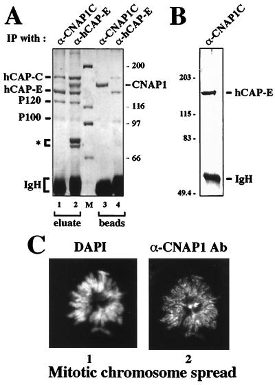
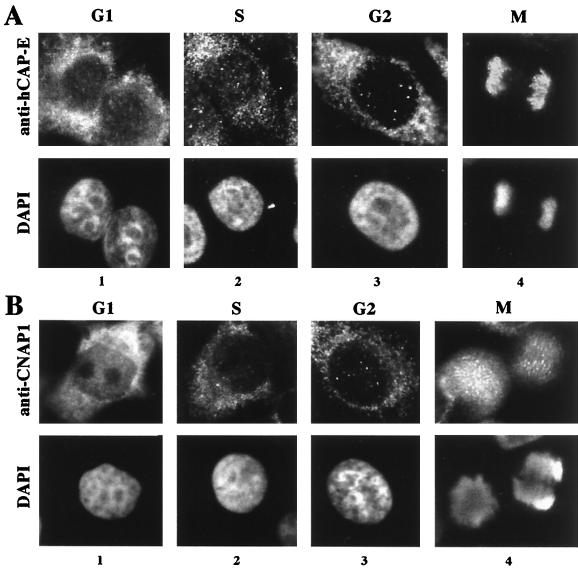
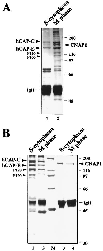
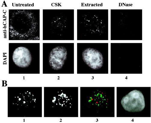
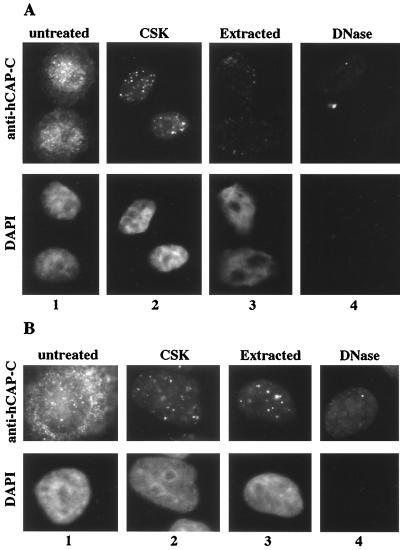
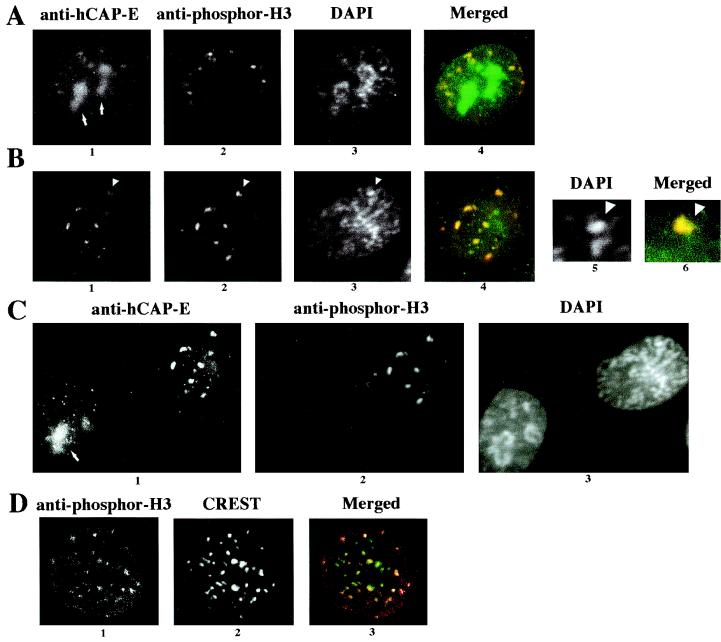
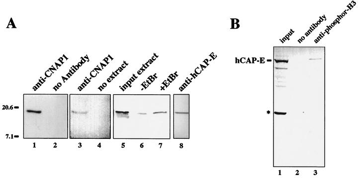
Structural maintenance of chromosomes (SMC) family proteins play critical roles in structural changes of chromosomes. Previously, we identified two human SMC family proteins, hCAP-C and hCAP-E, which form a heterodimeric complex (hCAP-C-hCAP-E) in the cell. Based on the sequence conservation and mitotic chromosome localization, hCAP-C-hCAP-E was determined to be the human ortholog of the Xenopus SMC complex, XCAP-C-XCAP-E. XCAP-C-XCAP-E is a component of the multiprotein complex termed condensin, required for mitotic chromosome condensation in vitro. However, presence of such a complex has not been demonstrated in mammalian cells. Coimmunoprecipitation of the endogenous hCAP-C-hCAP-E complex from HeLa extracts identified a 155-kDa protein interacting with hCAP-C-hCAP-E, termed condensation-related SMC-associated protein 1 (CNAP1). CNAP1 associates with mitotic chromosomes and is homologous to Xenopus condensin component XCAP-D2, indicating the presence of a condensin complex in human cells. Chromosome association of human condensin is mitosis specific, and the majority of condensin dissociates from chromosomes and is sequestered in the cytoplasm throughout interphase. However, a subpopulation of the complex was found to remain on chromosomes as foci in the interphase nucleus. During late G(2)/early prophase, the larger nuclear condensin foci colocalize with phosphorylated histone H3 clusters on partially condensed regions of chromosomes. These results suggest that mitosis-specific function of human condensin may be regulated by cell cycle-specific subcellular localization of the complex, and the nuclear condensin that associates with interphase chromosomes is involved in the reinitiation of mitotic chromosome condensation in conjunction with phosphorylation of histone H3.


Figures

FIG. 1
FIG. 1
Identification of a 155-kDa polypeptide as CNAP1. (A) Silver staining of the immunoprecipitated proteins from HeLa nuclear extracts using anti-hCAP-E antibody. The arrows indicate hCAP-C and hCAP-E. The open arrowhead indicates a 155-kDa protein (p155) corresponding to CNAP1. Positions of size markers (M) in panels A, C, and D are indicated in kilodaltons. IgH, immunoglobulin heavy chain (B) Peptide sequence analysis of p155. Numbers represent the amino acid residues in the CNAP1 protein. Black bars below the schematic diagram of CNAP1 protein indicate the positions of identified peptide sequences. The open boxes labeled CNAP1N and CNAP1C represent recombinant proteins corresponding to the N- and C-terminal regions of CNAP1 used to generate antibodies. (C) Western blot analysis of recombinant CNAP1 fusion proteins with antibodies specific for N- and C-terminal domains of CNAP1. Antibodies specific for CNAP1N and CNAP1C were used to test cross-reactivity of the two antibodies with GST-CNAP1N (lanes 1 and 3) and GST-CNAP1C (lanes 2 and 4). Lanes 2 and 3 demonstrate the lack of cross-reactivity between the two antibodies. Specific signals corresponding to the GST fusion proteins are indicated with arrowheads. (D) Detection of the endogenous CNAP1 by Western blot analysis of crude HeLa nuclear extracts using anti-CNAP1C antibody (Ab) (lanes 1 and 2). Lane 1 was further probed with anti-hCAP-E antibody to confirm the size of CNAP1 relative to that of hCAP-E.
FIG. 2
FIG. 2
Coimmunoprecipitation of the hCAP-C–hCAP-E complex with anti-CNAP1C antibody using mitotic HeLa extracts. (A) Silver staining of reciprocal coimmunoprecipitation. A pattern of immunoprecipitated (IP) proteins by anti-CNAP1C (lanes 1 and 3) is compared to that by anti-hCAP-E (lanes 2 and 4). Eluate, proteins that were eluted from beads with 2 M guanidine (see Materials and Methods); beads, proteins that remained on the beads after the elution; IgH, immunoglobulin heavy chain. Three polypeptides indicated by an asterisk are unique to anti-hCAP-E immunoprecipitation. Sizes of markers are indicated in kilodaltons. (B) Western blot analysis of the proteins immunoprecipitated with anti-CNAP1C antibody probed with anti-hCAP-E. (C) CNAP1 localization on mitotic chromosomes. Immunofluorescent staining of a HeLa mitotic chromosome spread using DAPI (panel 1) to visualize DNA and anti-CNAP1C antibody (Ab) to detect CNAP1 protein (panel 2). The bright spots in the center correspond to centrosomes.
FIG. 3
FIG. 3
Analysis of the subcellular localization of the hCAP-C–hCAP-E complex and CNAP1 in HeLa cells at different cell cycle stages, as indicated at the top. The proteins are detected by immunofluorescent staining with antibody specific for hCAP-E, representing hCAP-C–hCAP-E (A) and CNAP1 (B). The top panels show immunofluorescent staining, and the bottom panels represent DAPI staining.
FIG. 4
FIG. 4
Coimmunoprecipitation of human condensin complex from S-phase cytoplasmic and mitotic HeLa extracts. (A) Coimmunoprecipitation of the complex with anti-hCAP-E middle domain antibody from S-phase cytoplasmic (lane 1) and mitotic (lane 2) extracts. hCAP-C, hCAP-E, CNAP1, P120, and P100 are indicated. IgH, immunoglobulin heavy chain. (B) Coimmunoprecipitation of the complex with anti-CNAP1 antibody using S-phase cytoplasmic and mitotic extracts. Extracts used are indicated at the top. CNAP1 remained on beads after guanidine elution (lanes 3 and 4). In both panels, sizes are indicated in kilodaltons.
FIG. 5
FIG. 5
Nuclear foci of the human condensin complex. (A) Immunofluorescent staining of the hCAP-C–hCAP-E complex in G2-synchronized HeLa cells with in situ extraction and DNA digestion. The top panels show immunofluorescent staining of the hCAP-C–hCAP-E complex using anti-hCAP-C antibody; the bottom panel indicates DNA visualized by DAPI staining. The extraction steps are indicated at the top (see Materials and Methods). (B) Immunofluorescent costaining of the CSK-extracted cell with anti-hCAP-C and CREST antibodies. Panel 1, anti-hCAP-C; panel 2, CREST antibody; panel 3, merged image (red [anti-hCAP-C] and green [CREST]); panel 4, DAPI.
FIG. 6
FIG. 6
Immunofluorescent staining of the hCAP-C–hCAP-E complex in G1 (A)- and S (B)-phase-synchronized HeLa cells with in situ extraction and DNA digestion. The top panels show immunofluorescent staining with anti-hCAP-C antibody; the bottom panels indicate DNA visualized by DAPI staining. Each extraction step is indicated at the top.
FIG. 7
FIG. 7
Colocalization analysis of the condensin complex with phosphor-H3. HeLa cells were treated with CSK buffer and costained with anti-hCAP-E, anti-phosphor-H3, and DAPI as indicated at the top. Colocalization of anti-hCAP-E (green) and anti-phosphor-H3 staining (red) is shown as a merged image (colocalization is shown in yellow). (A) Late-G2-phase cell. Nucleolus staining with hCAP-E is indicated by an arrows. (B) Early-prophase cell. The arrowheads in panels 1, 2, and 3 identify a site of locally condensed DNA, which is enlarged to show DAPI (panel 5) and the merged image of hCAP-E (green) and phosphor-H3 (red) (panel 6). (C) Comparison of early prophase (same as in panel B) and interphase cells. Staining is indicated at the top, and the nucleolus staining of anti-hCAP-E is indicated with an arrow. (D) Colocalization of phosphor-H3 and centromeric regions. The CSK-extracted cell was stained with anti-phosphor-H3 (panel 1) and CREST (panel 2); the merged image is shown in panel 3 (red [phosphor-H3] and green [CREST]). These images were captured with confocal microscopy.
FIG. 8
FIG. 8
Coimmunoprecipitation of phosphor-H3 with the condensin complex. (A) Mitotic HeLa extracts were used for coimmunoprecipitation using anti-CNAP1 antibody (lanes 1, 3, 6, and 7). The immunoprecipitated materials eluted from antibody beads with 1 M salt were probed with anti-phosphor-H3 antibody in a Western blot analysis. Controls include protein A beads alone incubated with extract (lane 2) and protein A beads bound by antibody without extract (lane 4). In lane 7, EtBr was added to remove residual DNA contaminants from the extract prior to immunoprecipitation, which was compared to the sample without EtBr (lane 6) and the input extracts (lane 5). Lane 8 shows coimmunoprecipitation with anti-hCAP-E antibody probed with anti-phosphor-H3 antibody. (B) Reciprocal immunoprecipitation with anti-phosphor-H3 antibody probed with anti-hCAP-E antibody. The full-length hCAP-E is indicated. Input extract in lane 1 also shows the smaller degradation product of hCAP-E indicated by an asterisk, which was not coprecipitated with anti-phosphor-H3 (lane 3). Protein A beads alone did not coprecipitate hCAP-E from the extract (lane 2). In both panels, sizes are indicated in kilodaltons.

References

    1. Bootsma D, Budke L, Vos O. Studies on synchronous division of tissue culture cells initiated by excess thymidine. Exp Cell Res. 1964;33:301–309. - PubMed
    1. Brenner S, Pepper D, Berns M W, Tan E, Brinkley B R. Kinetochore structure, duplication, and distribution in mammalian cells: analysis by human autoantibodies from scleroderma patients. J Cell Biol. 1981;91:95–102. - PMC - PubMed
    1. Cubizolles F, Legagneux V, Le Guellec R, Chartrain I, Uzbekov R, Ford C, Le Guellec K. pEg7, a new Xenopus protein required for mitotic chromosome condensation in egg extracts. J Cell Biol. 1998;143:1437–1446. - PMC - PubMed
    1. de Belle I, Cai S, Kohwi-Shigematsu T. The genomic sequences bound to special AT-rich sequence-binding protein 1 (SATB1) in vivo in Jurkat T cells are tightly associated with the nuclear matrix at the bases of the chromatin loops. J Cell Biol. 1998;141:335–348. - PMC - PubMed
    1. Earnshaw W C, Rothfield N. Identification of a family of human centromere proteins using autoimmune sera from patients with scleroderma. Chromosoma. 1985;91:313–321. - PubMed

Publication types