. 2000 Sep;20(18):7037-48.
doi: 10.1128/MCB.20.18.7037-7048.2000.

The N terminus of the centromere H3-like protein Cse4p performs an essential function distinct from that of the histone fold domain

Affiliations

The N terminus of the centromere H3-like protein Cse4p performs an essential function distinct from that of the histone fold domain

Y Chen et al. Mol Cell Biol. 2000 Sep.

Abstract

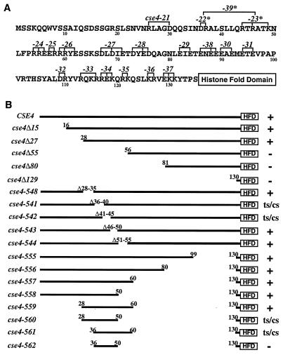
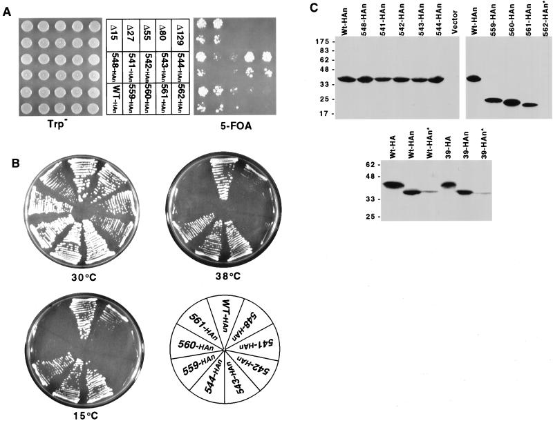
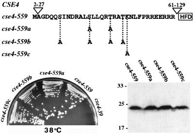
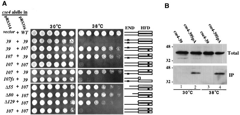
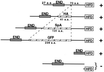
Cse4p is an evolutionarily conserved histone H3-like protein that is thought to replace H3 in a specialized nucleosome at the yeast (Saccharomyces cerevisiae) centromere. All known yeast, worm, fly, and human centromere H3-like proteins have highly conserved C-terminal histone fold domains (HFD) but very different N termini. We have carried out a comprehensive and systematic mutagenesis of the Cse4p N terminus to analyze its function. Surprisingly, only a 33-amino-acid domain within the 130-amino-acid-long N terminus is required for Cse4p N-terminal function. The spacing of the essential N-terminal domain (END) relative to the HFD can be changed significantly without an apparent effect on Cse4p function. The END appears to be important for interactions between Cse4p and known kinetochore components, including the Ctf19p/Mcm21p/Okp1p complex. Genetic and biochemical evidence shows that Cse4p proteins interact with each other in vivo and that nonfunctional cse4 END and HFD mutant proteins can form functional mixed complexes. These results support different roles for the Cse4p N terminus and the HFD in centromere function and are consistent with the proposed Cse4p nucleosome model. The structure-function characteristics of the Cse4p N terminus are relevant to understanding how other H3-like proteins, such as the human homolog CENP-A, function in kinetochore assembly and chromosome segregation.


Figures

FIG. 1
FIG. 1
Mutagenesis of the Cse4p N terminus. (A) Alanine scanning mutations. The N-terminal 135 amino acids of wild-type Cse4p are shown. Numbers identifying each cse4 mutation (cse4-21 to -39) are above the sequence at the positions where the charged residues (R, D, K, and E) indicated by brackets were changed to alanine for each allele. Mutant alleles that exhibit growth and/or chromosome loss phenotypes are denoted by asterisks. (B) N-terminal deletions. The N terminus of Cse4p is depicted as a thick line terminating at an open box representing the HFD, which starts at amino acid 135. Numbers above the lines indicate the residues remaining in the construct, except for alleles 541 to 548, where the residues deleted are indicated. The cse4-encoded mutant proteins initiate with methionine and continue with the amino acids indicated. The viability and growth phenotypes of the mutants are given on the right: +, growth at all of the temperatures tested (30, 15, and 38°C); ts, no growth at 38°C; cs, no growth at 15°C; −, inviable (unable to complement the cse4 null mutation).
FIG. 2
FIG. 2
Phenotypes of Cse4p N-terminal mutants. (A) Plasmid shuffle test (see Materials and Methods) showing the ability of various cse4 alleles (middle) to complement the cse4 null lethality. HAn indicates HA epitope-tagged alleles. The bottom of each pair of spots is a 10-fold dilution of the cell suspension used for the top spots. Only yeast strains carrying nonlethal cse4 mutations can grow on 5-FOA medium (right side). (B) Viable cse4 alleles were tested for conditional growth phenotypes. Plates were photographed after 3 days at 30°C, 5 days at 38°C, and 14 days at 15°C, respectively. (C) Immunoblot analysis of cse4 mutant proteins. Immunoblots of representative viable and lethal cse4 mutants are shown in the top two blots. The positions of protein molecular size markers (kilodaltons) are indicated at the left. Proteins were extracted from the cse4 mutant strains indicated above the lanes. The Vector lane contains proteins from cells carrying a vector without a cse4 insert. Asterisks denote cases in which the cells carried an untagged wild-type (Wt) CSE4 allele in addition to the tagged cse4 mutant allele. The HA- and HAn-tagged alleles differ in the location of the HA tag, either at amino acid 83 (Cse4HA) or at the extreme N terminus (Cse4HAn).
FIG. 3
FIG. 3
Mutational analysis of possible posttranslational modification sites in the Cse4p END. (Top) The initiating methionine and the 33-amino-acid END sequence in the N terminus of Cse4-559p. Potential phosphorylation (S or T) or acetylation (K) sites were changed to alanine codons, generating the three new alleles shown. (Bottom left) Plate showing growth phenotypes at 38°C (5 days) of strains carrying the indicated HAn-tagged cse4 mutant alleles as their sole source of Cse4p. All strains grew well at 30°C (data not shown). (Bottom right) Immunoblot showing the expression of cse4-encoded mutant proteins. The values on the left are molecular sizes in kilodaltons.
FIG. 4
FIG. 4
Genetic and biochemical evidence for Cse4p protein-protein interactions. (A) Interallelic complementation between cse4 END and HFD mutant alleles. The cse4 mutant alleles indicated at the left were expressed from low-copy-number pRS314 and pRS316 vectors and tested for interallelic complementation as described in Materials and Methods. Threefold serial cell dilutions were spotted left to right. The cse4-107fs frameshift mutation terminates translation of the cse4-107-encoded protein between the N terminus and the HFD. Cse4p dimers possibly formed are schematically shown at the right, with × denoting the END mutations in Cse4-39p and ■ indicating the HFD mutation in Cse4-107p. WT, wild type. (B) Coprecipitation of cse4-encoded mutant proteins. Extracts were prepared from cells coexpressing cse4-107HA and either protein A-tagged (lanes 2 and 4) or untagged (lanes 1 and 3) cse4-39. Protein A-tagged protein complexes were precipitated using IgG-Sepharose beads. Approximately 0.5% of the total extracts (Total) and 10 to 15% of the precipitated proteins (IP) were subjected to SDS-PAGE and immunoblot analysis using anti-HA antibodies. The values to the left are molecular sizes in kilodaltons.
FIG. 5
FIG. 5
Localization of GFP-tagged Cse4p proteins. Cells were fixed and prepared for microscopy as described in Materials and Methods. The top panels show GFP fluorescence, the middle panels show DAPI staining, and the bottom panels show the overlaid GFP and DAPI images. (Left) Yeast cells (KC100) carrying only wild-type Cse4GFP were grown at 30°C and shifted to 37°C for 5 h (three to four doublings) before fixation. (Middle) KC100 cells expressing only Cse4-107GFP grown at the permissive temperature (30°C). (Right) KC100 cells carrying cse4-107HA and cse4Δ55GFP on separate URA3 and TRP1 vectors, respectively, were streaked and grown at 38°C, the restrictive temperature for both mutations. A single colony was picked, inoculated into prewarmed medium, and grown for an additional 15 h (three to four doublings) at 38°C before fixation and microscopy.
FIG. 6
FIG. 6
CHIP of Cse4Δ55GFP. Cse4-107HA cells were transformed with plasmids carrying cse4GFP or cse4Δ55GFP. Transformants were grown at 30 or 38°C, and CHIP was performed as described in Materials and Methods. The ethidium bromide-stained gel shows the products of PCR using primers for CEN3 (CEN3, 243 bp), DNA 177 kbp to the right of CEN3 (R, 278 bp), and DNA 4 kbp to the left of CEN3 (L, 213 bp). The PCR template was total chromatin (T) or chromatin immunoprecipitated by α-GFP or α-HA antibody (PGFP and PHA, respectively).
FIG. 7
FIG. 7
Cse4p N terminus interaction with components of the Mcm21p-Ctf19p-Okp1p complex. (A) Suppression of phenotypes due to cse4-39 by high-copy MCM21. The cse4-39 mutant yeast strain was transformed with a high-copy-number plasmid carrying MCM21 and tested for growth at 30, 38, and 15°C. (B) Synthetic lethality between mcm21Δ and cse4-39. Cells from strain YC220 carrying cse4-39 and a disrupted mcm21 gene (mcm21Δ::HIS3) depend on a CSE4-URA3 plasmid for viability and therefore failed to grow on 5-FOA medium (middle sector). The 5-FOA sensitivity was relieved by introduction of either an MCM21 plasmid (right sector) or a CSE4 plasmid (left sector). (C) Two-hybrid analysis of Cse4p-Ctf19p interactions. Two-hybrid reporter cells (PJ69-4A) were cotransformed with plasmids expressing all or part of Cse4p fused to the GAL4 AD and a plasmid carrying full-length Ctf19p fused to the GAL4 BD. Different AD fusion constructs were derived from the cse4 alleles shown at the left and tested with BD-Ctf19p. The designation Vector indicates that no CSE4 or CTF19 sequences are present in those clones. Transformants were plated in a series of fivefold dilutions spotted on the media indicated. Interaction between AD and BD fusion proteins allowed growth on 3-aminotriazole medium.
FIG. 8
FIG. 8
Flexible spacing between Cse4p END and HFD. The Cse4p END is spaced 70 amino acids (a.a.) away from the Cse4p HFD in the wild-type protein. This spacing can vary from zero (Cse4-559p) to 309 amino acids (Cse4GFP) without appreciably compromising Cse4p function (indicated by a plus sign); however, Cse4p function was not restored when the END and HFD peptides were expressed separately (indicated by a minus sign).

References

    1. Anderson M T, Tjioe I M, Lorincz M C, Parks D R, Herzenberg L A, Nolan G P. Simultaneous fluorescence-activated cell sorter analysis of two distinct transcriptional elements within a single cell using engineered green fluorescent proteins. Proc Natl Acad Sci USA. 1996;93:8508–8511. - PMC - PubMed
    1. Baker R E, Fitzgerald-Hayes M, O'Brien T C. Purification of the yeast centromere binding protein CP1 and a mutational analysis of its binding site. J Biol Chem. 1989;264:10843–10850. - PubMed
    1. Baker R E, Harris K, Zhang K. Mutations synthetically lethal with cep1 target S. cerevisiae kinetochore components. Genetics. 1998;149:73–85. - PMC - PubMed
    1. Baker R E, Masison D C. Isolation of the gene encoding the Saccharomyces cerevisiae centromere-binding protein CP1. Mol Cell Biol. 1990;10:2458–2467. - PMC - PubMed
    1. Basrai M A, Kingsbury J, Koshland D, Spencer F, Hieter P. Faithful chromosome transmission requires Spt4p, a putative regulator of chromatin structure in Saccharomyces cerevisiae. Mol Cell Biol. 1996;16:2838–2847. - PMC - PubMed

Publication types

MeSH terms