Case Reports
. 2000 Oct;67(4):967-75.
doi: 10.1086/303071. Epub 2000 Aug 24.

Defective peroxisome membrane synthesis due to mutations in human PEX3 causes Zellweger syndrome, complementation group G

Affiliations
Case Reports

Defective peroxisome membrane synthesis due to mutations in human PEX3 causes Zellweger syndrome, complementation group G

A C Muntau et al. Am J Hum Genet. 2000 Oct.

Abstract

Zellweger cerebro-hepato-renal syndrome is a severe congenital disorder associated with defective peroxisomal biogenesis. At least 23 PEX genes have been reported to be essential for peroxisome biogenesis in various species, indicating the complexity of peroxisomal assembly. Cells from patients with peroxisomal biogenesis disorders have previously been shown to segregate into >/=12 complementation groups. Two patients assigned to complementation group G who had not been linked previously to a specific gene defect were confirmed as displaying a cellular phenotype characterized by a lack of even residual peroxisomal membrane structures. Here we demonstrate that this complementation group is associated with mutations in the PEX3 gene, encoding an integral peroxisomal membrane protein. Homozygous PEX3 mutations, each leading to C-terminal truncation of PEX3, were identified in the two patients, who both suffered from a severe Zellweger syndrome phenotype. One of the mutations involved a single-nucleotide insertion in exon 7, whereas the other was a single-nucleotide substitution eight nucleotides from the normal splice site in the 3' acceptor site of intron 10. Expression of wild-type PEX3 in the mutant cell lines restored peroxisomal biogenesis, whereas transfection of mutated PEX3 cDNA did not. This confirmed that the causative gene had been identified. The observation of peroxisomal formation in the absence of morphologically recognizable peroxisomal membranes challenges the theory that peroxisomes arise exclusively by growth and division from preexisting peroxisomes and establishes PEX3 as a key factor in early human peroxisome synthesis.


Figures
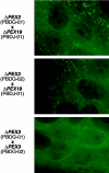
Figure  1
Figure 1
Somatic cell–fusion experiments. Fusion of cocultivated fibroblasts was performed using methods described elsewhere (Roscher et al. 1989). Fusion of fibroblasts from patients PBDG-01 and PBDG-02 with fibroblasts from a patient carrying a PEX19 mutation (PBDJ-01 [Matsuzono et al. 1999]) restored peroxisomes in the majority of multinucleated cells, as demonstrated by a punctate immunofluorescent pattern after staining for catalase. Somatic cell fusion of fibroblasts from patient PBDG-01 with fibroblasts from patient PBDG-02 revealed only diffuse cytosolic staining with anti-catalase antibodies.
Figure  2
Figure 2
Intracellular localization of PMPs in CG-G cell lines. Indirect immunofluorescence microscopy was performed with fibroblasts from a healthy control (A, E, I, and M), from a patient with Zellweger syndrome who was assigned to CG-E (B, F, J, and N), and from the patients PBDG-01 (C, G, K, and O), and PBDG-02 (D, H, L, and P). The cells were stained using rhodamine-labeled antibodies to ALDP, a PMP (A–D); fluorescein-labeled antibodies to catalase, a peroxisomal matrix protein (E–H); fluorescein-labeled antibodies to PEX14, a PMP (I–L), or the mitochondrial marker MitoTracker (M–P). In normal fibroblasts, staining with antibodies to ALDP (A), catalase (E), and PEX14 (I) all yielded a punctate pattern, indicating the presence of peroxisomal membranes and intact peroxisomes. Fibroblasts from a patient with Zellweger syndrome who was assigned to CG-E (PBDE) were stained with antibodies to the PMPs ALDP (B) and PEX14 (J). Note the colocalization of the punctate patterns, indicating the presence of peroxisomal membranes. These membranes presumably are nonfunctional peroxisomal ghosts. Incubation with anti-catalase antibodies (F) resulted in a diffuse cytosolic localization, consistent with the absence of intact peroxisomes. In cells of PBDG-01 (C) and PBDG-02 (D), no ALDP positive particles were visible, indicating the complete absence of morphologically detectable peroxisomal membranes. Incubation with anti-catalase antibodies (G and H) exhibited a diffuse cytosolic pattern, consistent with the absence of intact peroxisomes. Staining the cells by use of an antibody specific for PEX14 (K and L) reveals a mitochondrial pattern, as demonstrated by colocalization of PEX14 with MitoTracker (O and P).
Figure  3
Figure 3
Mutation analyses. A, Genomic organization of the human PEX3 gene. Exons are indicated as boxes. B, Exon 7 sequencing results and predicted protein sequences of a wild-type control (wt) and patient PBDG-01. Note the homozygous thymine insertion (arrow). The frameshift causes a premature stop two codons after the insertion. C, Genomic sequencing of the exon-intron boundary of a control individual (wt) and patient PBDG-02. Note the homozygous substitution of a thymine by a guanine in the 3′ splice site of intron 10, at position −8 (arrow). D, PCR amplification of PEX3 fragments from total RNA of PBDG-02 after synthesis of first-strand cDNA by reverse transcription using primer 1255R (for primer sequences, see table 1). Amplification of a fragment containing exons 5–10 by primers 404F and 957R did not reveal any difference between the control individual (wt) and the patient (PBDG-02). The fragment containing exons 5–11 amplified by primer 404F and the exon 11–specific primer 1062R could not be amplified from the patient’s cDNA. Amplification of exons 5–12 by primers 404F and 1120R produced an aberrantly sized PCR product from the patient’s cDNA. E, Sequence analysis of the PCR fragment containing exons 5–12 show that the PEX3 cDNA of patient PBDG-02 is missing 97 bp, which were present in wt PEX3 cDNA. The 97-bp deletion corresponds to the sequence of exon 11 and leads to direct fusion of exons 10–12. Premature termination of the protein is predicted from the frameshift.
Figure  4
Figure 4
Functional complementation of patient fibroblasts. In untransfected cells from PBDG-01 (A, B) and PBDG-02 (G and H), no ALDP positive particles were visible (A and G). Incubation with anti-catalase antibodies (B and H) showed a diffuse cytosolic localization. Five days after transfection with a human PEX3 cDNA vector (pcDNA3.1/Pex3-Myc-His [Kammerer et al. 1998]), punctate patterns for ALDP (C and I) and catalase (D and J) indicated that these proteins were colocalized either at or within restored peroxisomes. To generate the mutated PEX3 expression vectors pcDNA3-PBDG-01 and pcDNA3-PBDG-02, RT-PCR was performed using the primers 5′F3 and 1255R, and the resulting fragments were cloned into pcDNA3. Transfection of the mutant PEX3 cDNA constructs pcDNA3-PBDG-01 and pcDNA3-PBDG-02 into cells from PBDG-01 (E and F) and PBDG-02 (K and L), respectively, did not lead to restoration of a peroxisomal punctate pattern with either ALDP (E and K) or catalase (F and L) immunofluorescence. In an additional experiment, transfection efficiency was verified by cotransfection of pcDNA3-PBDG-01 and pcDNA3-PBDG-02 constructs, with the green fluorescent protein-expression vector pEGFP-N1 (Clontech) as a reporter plasmid (data not shown).
Figure  5
Figure 5
PEX3 in vitro binding assays. A, Schematic diagram of wild-type PEX3 and truncated PEX3 of patient PBDG-01. The mutation in PBDG-01 results in a C-terminally truncated PEX3 protein, whereas predicted transmembrane regions (TM) remain unaffected. B, Pull-down assays of in vitro–translated PEX3, with immobilized GST-PEX19, performed as described elsewhere (Glöckner et al. 2000). The left-hand gel was loaded with 10% of the [35S]-methionine-labeled PEX3 translation products used as input for the binding assays. To ensure that even weak binding of the mutated PEX3 would be detected, the amount of mutated PEX3 was chosen such that it clearly exceeded the amount of wild-type PEX3 (wt), which was bound by GST-PEX19, whereas no detectable signal was obtained when the interaction of the mutated PEX3 protein (PBDG-01) with PEX19 was analyzed (central gel). GST alone did not bind to either protein (right-hand gel). Several repetitions of the assay yielded similar results. C, Construct lacking the first start codon and the following 63 bp of the PEX3 gene (66 bp).We generated this construct to demonstrate that the molecular weight of the lower band in the PEX3 wild-type lanes (wt) is a result of the utilization of the internal start codon at position 199 of the coding region. In vitro translation of this construct by the internal start codon yielded a protein corresponding to the lower band of wild-type PEX3.

References

Electronic-Database Information

    1. Online Mendelian Inheritance in Man (OMIM), http://www.ncbi.nlm.nih.gov/Omim (for PBD [MIM 601539], RCDP [MIM 215100], and PEX3 [MIM 603164])

References

    1. Arias JA, Moser AB, Goldfischer SL (1985) Ultrastructural and cytochemical demonstration of peroxisomes in cultured fibroblasts from patients with peroxisomal deficiency disorders. J Cell Biol 100:1789–1792 - PMC - PubMed
    1. Baerends RJ, Rasmussen SW, Hilbrands RE, van der Heide M, Faber KN, Reuvekamp PT, Kiel JA, Cregg JM, van der Klei IJ, Veenhuis M (1996) The Hansenula polymorpha PER9 gene encodes a peroxisomal membrane protein essential for peroxisome assembly and integrity. J Biol Chem 271:8887–8894 - PubMed
    1. Beldjord C, Lapoumeroulie C, Pagnier J, Benabadji M, Krishnamoorthy R, Labie D, Bank A (1988) A novel beta thalassemia gene with a single base mutation in the conserved polypyrimidine sequence at the 3′ end of IVS 2. Nucleic Acids Res 16:4927–4935 - PMC - PubMed
    1. Glöckner CJ, Mayerhofer PU, Landgraf P, Muntau AC, Holzinger A, Gerber JK, Kammerer S, Adamski J, Roscher AA (2000) Human adrenoleukodystrophy protein and related peroxisomal ABC transporters interact with the peroxisomal assembly protein PEX19p. Biochem Biophys Res Commun 271:144–150 - PubMed
    1. Götte K, Girzalsky W, Linkert M, Baumgart E, Kammerer S, Kunau WH, Erdmann R (1998) Pex19p, a farnesylated protein essential for peroxisome biogenesis. Mol Cell Biol 18:616–628 - PMC - PubMed

Publication types

MeSH terms