

ADP is not an agonist at P2X(1) receptors: evidence for separate receptors stimulated by ATP and ADP on human platelets
- PMID: 10960076
- PMCID: PMC1572284
- DOI: 10.1038/sj.bjp.0703517
ADP is not an agonist at P2X(1) receptors: evidence for separate receptors stimulated by ATP and ADP on human platelets
Abstract
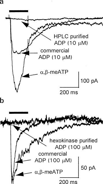
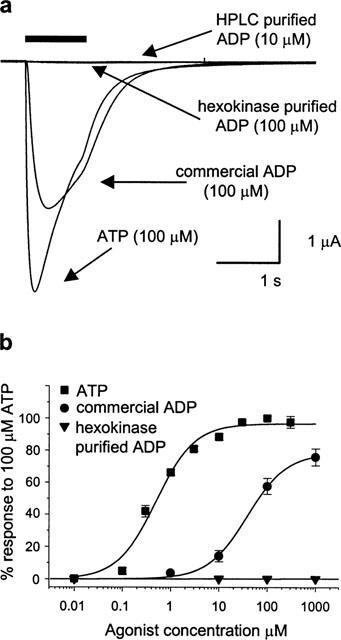
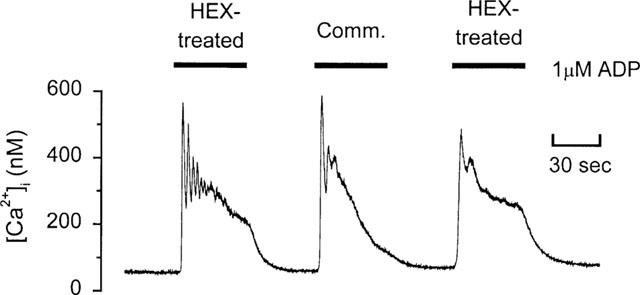
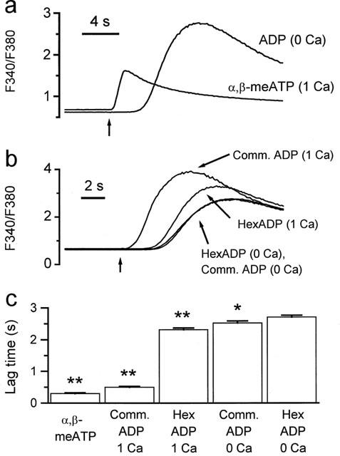
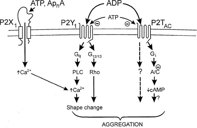
ADP, an important agonist in thrombosis and haemostasis, has been reported to activate platelets via three receptors, P2X(1), P2Y(1) and P2T(AC). Given the low potency of ADP at P2X(1) receptors and recognized contamination of commercial samples of adenosine nucleotides, we have re-examined the activation of P2X(1) receptors by ADP following HPLC and enzymatic purification. Native P2X(1) receptor currents in megakaryocytes were activated by alpha, beta-meATP (10 microM) and commercial samples of ADP (10 microM), but not by purified ADP (10 - 100 microM). Purified ADP (up to 1 mM) was also inactive at recombinant human P2X(1) receptors expressed in Xenopus oocytes. Purification did not modify the ability of ADP to activate P2Y receptors coupled to Ca(2+) mobilization in rat megakaryocytes. In human platelets, P2X(1) and P2Y receptor-mediated [Ca(2+)](i) responses were distinguished by their different kinetics at 13 degrees C. In 1 mM Ca(2+) saline, alpha,beta-meATP (10 microM) and commercial ADP (40 microM) activated a rapid [Ca(2+)](i) increase (lag time < or =0.5 s) through the activation of P2X(1) receptors. Hexokinase treatment of ADP shifted the lag time by approximately 2 s, indicating loss of the P2X(1) receptor-mediated response. A revised scheme is proposed for physiological activation of P2 receptors in human platelets. ATP stimulates P2X(1) receptors, whereas ADP is a selective agonist at metabotropic (P2Y(1) and P2T(AC)) receptors.
Figures
References
-
- BEZPROZVANNY I., EHRLICH B.E. The inositol 1,4,5-trisphosphate (InsP3) receptor. J. Memb. Biol. 1995;145:205–216. - PubMed
-
- BOARDER M.R., HOURANI S.M.O. The regulation of vascular function by P2 receptors: multiple sites and multiple receptors. TIPS. 1998;19:99–107. - PubMed
-
- BOEYNAEMS J.-M., COMMUNI D., SAVI P., HERBERT J.M. P2Y receptors: in the middle of the road. TIPS. 2000;21:1–3. - PubMed
-
- BOEYNAEMS J.-M., PEARSON J.D. P2 purinoceptors on vascular endothelial cells: physiological significance and transduction mechanisms. TIPS. 1990;11:34–37. - PubMed
-
- BORN G.V.R. Aggregation of blood platelets by adenosine diphosphate and its reversal. Nature. 1962;194:927–929. - PubMed
Publication types
MeSH terms
Substances
LinkOut - more resources
Full Text Sources
Other Literature Sources
Miscellaneous
