

Characterization of vibrio cholerae O1 antigen as the bacteriophage K139 receptor and identification of IS1004 insertions aborting O1 antigen biosynthesis
- PMID: 10960093
- PMCID: PMC94657
- DOI: 10.1128/JB.182.18.5097-5104.2000
Characterization of vibrio cholerae O1 antigen as the bacteriophage K139 receptor and identification of IS1004 insertions aborting O1 antigen biosynthesis
Abstract
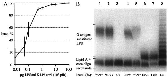
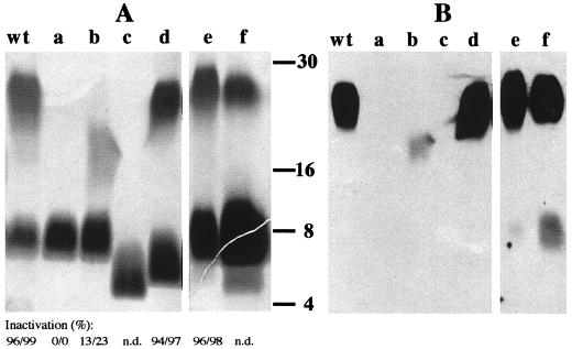
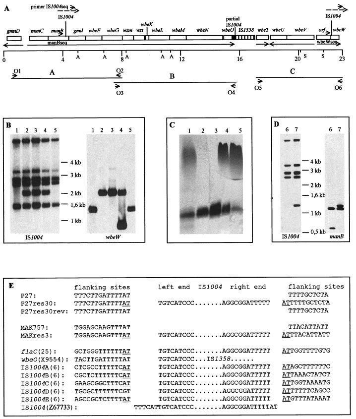
Bacteriophage K139 was recently characterized as a temperate phage of O1 Vibrio cholerae. In this study we have determined the phage adsorption site on the bacterial cell surface. Phage-binding studies with purified lipopolysaccharide (LPS) of different O1 serotypes and biotypes revealed that the O1 antigen serves as the phage receptor. In addition, phage-resistant O1 El Tor strains were screened by using a virulent isolate of phage K139. Analysis of the LPS of such spontaneous phage-resistant mutants revealed that most of them synthesize incomplete LPS molecules, composed of either defective O1 antigen or core oligosaccharide. By applying phage-binding studies, it was possible to distinguish between receptor mutants and mutations which probably caused abortion of later steps of phage infection. Furthermore, we investigated the genetic nature of O1-negative strains by Southern hybridization with probes specific for the O antigen biosynthesis cluster (rfb region). Two of the investigated O1 antigen-negative mutants revealed insertions of element IS1004 into the rfb gene cluster. Treating one wbeW::IS1004 serum-sensitive mutant with normal human serum, we found that several survivors showed precise excision of IS1004, restoring O antigen biosynthesis and serum resistance. Investigation of clinical isolates by screening for phage resistance and performing LPS analysis of nonlysogenic strains led to the identification of a strain with decreased O1 antigen presentation. This strain had a significant reduction in its ability to colonize the mouse small intestine.
Figures
Similar articles
-
O antigen is the receptor of Vibrio cholerae serogroup O1 El Tor typing phage VP4.J Bacteriol. 2013 Feb;195(4):798-806. doi: 10.1128/JB.01770-12. Epub 2012 Dec 7. J Bacteriol. 2013. PMID: 23222721 Free PMC article.
-
Characterization of Vibrio cholerae bacteriophage K139 and use of a novel mini-transposon to identify a phage-encoded virulence factor.Mol Microbiol. 1995 Nov;18(4):685-701. doi: 10.1111/j.1365-2958.1995.mmi_18040685.x. Mol Microbiol. 1995. PMID: 8817491
-
Characterization of Vibrio cholerae O1 El tor galU and galE mutants: influence on lipopolysaccharide structure, colonization, and biofilm formation.Infect Immun. 2001 Jan;69(1):435-45. doi: 10.1128/IAI.69.1.435-445.2001. Infect Immun. 2001. PMID: 11119535 Free PMC article.
-
DNA fingerprinting of Vibrio cholerae strains with a novel insertion sequence element: a tool to identify epidemic strains.J Clin Microbiol. 1996 Jun;34(6):1453-61. doi: 10.1128/jcm.34.6.1453-1461.1996. J Clin Microbiol. 1996. PMID: 8735097 Free PMC article.
-
Genetic organization of the regions associated with surface polysaccharide synthesis in Vibrio cholerae O1, O139 and Vibrio anguillarum O1 and O2: a review.Gene. 1998 Nov 26;223(1-2):269-82. doi: 10.1016/s0378-1119(98)00407-7. Gene. 1998. PMID: 9858748 Review.
Cited by
-
Use of bacteriophage Ba1 to identify properties associated with Bordetella avium virulence.Infect Immun. 2002 Mar;70(3):1219-24. doi: 10.1128/IAI.70.3.1219-1224.2002. Infect Immun. 2002. PMID: 11854203 Free PMC article.
-
Outer membrane vesicles mediate transport of biologically active Vibrio cholerae cytolysin (VCC) from V. cholerae strains.PLoS One. 2014 Sep 4;9(9):e106731. doi: 10.1371/journal.pone.0106731. eCollection 2014. PLoS One. 2014. PMID: 25187967 Free PMC article.
-
O antigen is the receptor of Vibrio cholerae serogroup O1 El Tor typing phage VP4.J Bacteriol. 2013 Feb;195(4):798-806. doi: 10.1128/JB.01770-12. Epub 2012 Dec 7. J Bacteriol. 2013. PMID: 23222721 Free PMC article.
-
Core lipopolysaccharide-specific phage SSU5 as an Auxiliary Component of a Phage Cocktail for Salmonella biocontrol.Appl Environ Microbiol. 2014 Feb;80(3):1026-34. doi: 10.1128/AEM.03494-13. Epub 2013 Nov 22. Appl Environ Microbiol. 2014. PMID: 24271179 Free PMC article.
-
Transmission of Vibrio cholerae is antagonized by lytic phage and entry into the aquatic environment.PLoS Pathog. 2008 Oct;4(10):e1000187. doi: 10.1371/journal.ppat.1000187. Epub 2008 Oct 24. PLoS Pathog. 2008. PMID: 18949027 Free PMC article.
References
-
- Berche P, Poyart C, Abachin E, Lelievre H, Vandepitte J, Dodin A, Fournier J M. The novel epidemic strain O139 is closely related to the pandemic strain O1 of Vibrio cholerae. J Infect Dis. 1994;170:701–704. - PubMed
-
- Bik E M, Bunschoten A E, Willems R J L, Chang A C Y, Mooi F R. Genetic organization and functional analysis of the otn DNA essential for cell-wall polysaccharide synthesis in Vibrio cholerae O139. Mol Microbiol. 1996;20:799–811. - PubMed
Publication types
MeSH terms
Substances
Grants and funding
LinkOut - more resources
Full Text Sources
Other Literature Sources
