. 2000 Sep;182(18):5114-20.
doi: 10.1128/JB.182.18.5114-5120.2000.

Bacteriophage phiYeO3-12, specific for Yersinia enterocolitica serotype O:3, is related to coliphages T3 and T7

Affiliations

Bacteriophage phiYeO3-12, specific for Yersinia enterocolitica serotype O:3, is related to coliphages T3 and T7

M Pajunen et al. J Bacteriol. 2000 Sep.

Abstract

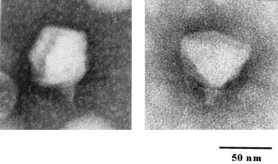
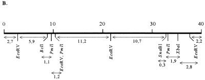
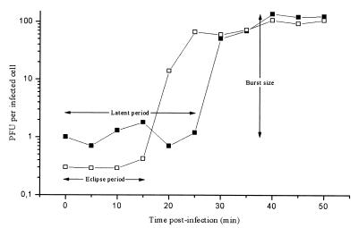
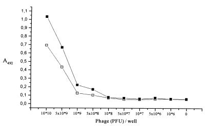
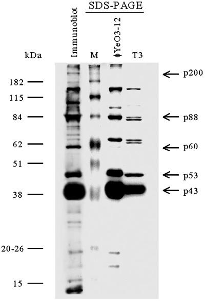
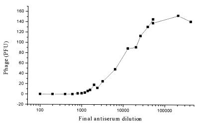
Bacteriophage phiYeO3-12 is a lytic phage of Yersinia enterocolitica serotype O:3. The phage receptor is the lipopolysaccharide O chain of this serotype that consists of the rare sugar 6-deoxy-L-altropyranose. A one-step growth curve of phiYeO3-12 revealed eclipse and latent periods of 15 and 25 min, respectively, with a burst size of about 120 PFU per infected cell. In electron microscopy phiYeO3-12 virions showed pentagonal outlines, indicating their icosahedral nature. The phage capsid was shown to be composed of at least 10 structural proteins, of which a protein of 43 kDa was predominant. N-terminal sequences of three structural proteins were determined, two of them showing strong homology to structural proteins of coliphages T3 and T7. The phage genome was found to consist of a double-stranded DNA molecule of 40 kb without cohesive ends. A physical map of the phage DNA was constructed using five restriction enzymes. The phage infection could be effectively neutralized using serum from a rabbit immunized with whole phiYeO3-12 particles. The antiserum also neutralized T3 infection, although not as efficiently as that of phiYeO3-12. phiYeO3-12 was found to share, in addition to the N-terminal sequence homology, several common features with T3, including morphology and nonsubjectibility to F exclusion. The evidence conclusively indicated that phiYeO3-12 is the first close relative of phage T3 to be described.


Figures

FIG. 1
FIG. 1
Electron micrograph of negatively stained φYeO3-12 virions.
FIG. 2
FIG. 2
Characterization of the φYeO3-12 genome. (A) Agarose gel electrophoresis of φYeO3-12 DNA after single and double digestions with restriction enzymes BclI, EcoRV, PmlI, SnaBI, and XbaI. (B) Restriction map of phage φYeO3-12 DNA (scale in kilobases) constructed on the basis of the data in panel A. The restriction fragments are indicated by double-headed arrows, below which the fragment sizes in kilobases are given.
FIG. 2
FIG. 2
Characterization of the φYeO3-12 genome. (A) Agarose gel electrophoresis of φYeO3-12 DNA after single and double digestions with restriction enzymes BclI, EcoRV, PmlI, SnaBI, and XbaI. (B) Restriction map of phage φYeO3-12 DNA (scale in kilobases) constructed on the basis of the data in panel A. The restriction fragments are indicated by double-headed arrows, below which the fragment sizes in kilobases are given.
FIG. 3
FIG. 3
One-step growth curve of bacteriophage φYeO3-12 on Y. enterocolitica serotype O:3 strain 6471/76-c at RT. Shown are the PFU per infected cell in chloroform-treated cultures (□) and in untreated cultures (■) at different time points. Each data point is a mean from three experiments.
FIG. 4
FIG. 4
Characterization of the rabbit antiserum specific for φYeO3-12 particles. The sensitivity of the antiserum in detection of immobilized native phage particles was assessed using EIA. Shown are the absorbance values obtained using antiserum dilutions of 1:10,000 (□) and 1:2,000 (■). Each data point is a mean from three experiments.
FIG. 5
FIG. 5
SDS-PAGE of the φYeO3-12 and T3 particles and immunoblot of the φYeO3-12 proteins developed with rabbit antiserum. The phage proteins discussed in the text are indicated by arrows at the right. Protein molecular size markers are shown at the left.
FIG. 6
FIG. 6
Neutralization efficiency of the rabbit antiserum specific for φYeO3-12 particles. Shown are obtained PFU values of φYeO3-12 at different antiserum dilutions. Each data point is a mean from one to five experiments.

Similar articles

Cited by

References

    1. Ackermann H-W, DuBow M S. Family Podoviridae. In: Ackermann H-W, DuBow M S, editors. Viruses of prokaryotes. II. Natural groups of bacteriophages. Boca Raton, Fla: CRC Press; 1987. pp. 85–100.
    1. Ackermann H-W, DuBow M S, Gershman M, Karska-Wysocki B, Kasatiya S S, Loessner M J, Mamet-Bratley M D, Regué M. Taxonomic changes in tailed phages of enterobacteria. Arch Virol. 1997;142:1381–1390. - PubMed
    1. Adams M H, Wade E. Classification of bacterial viruses: the relationship of two Serratia phages to coli-dysentery phages T3, T7, and D44. J Bacteriol. 1954;68:320. - PMC - PubMed
    1. Al-Hendy A, Toivanen P, Skurnik M. Expression cloning of the Yersinia enterocolitica O:3 rfb gene cluster in Escherichia coli K12. Microb Pathog. 1991;10:47–59. - PubMed
    1. Al-Hendy A, Toivanen P, Skurnik M. Lipopolysaccharide O side chain of Yersinia enterocolitica O:3 is an essential virulence factor in an orally infected murine model. Infect Immun. 1992;60:870–875. - PMC - PubMed

Publication types

MeSH terms

LinkOut - more resources