. 2000 Sep;182(18):5153-66.
doi: 10.1128/JB.182.18.5153-5166.2000.

ZipA-induced bundling of FtsZ polymers mediated by an interaction between C-terminal domains

Affiliations

ZipA-induced bundling of FtsZ polymers mediated by an interaction between C-terminal domains

C A Hale et al. J Bacteriol. 2000 Sep.

Abstract

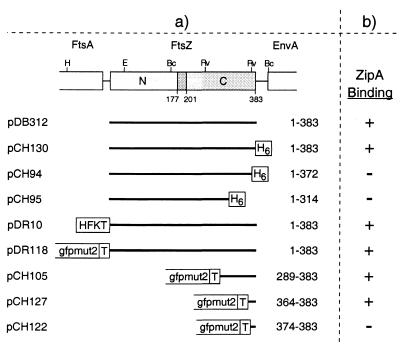
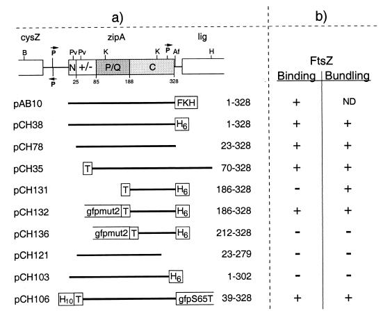
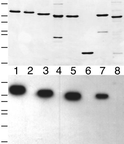
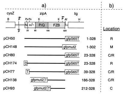
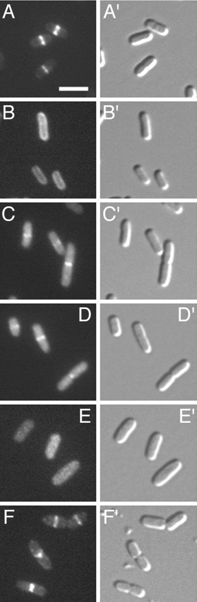
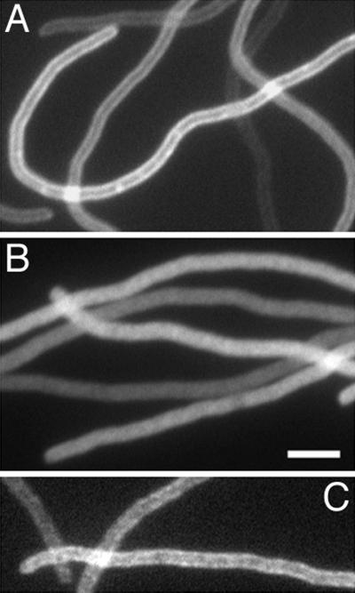
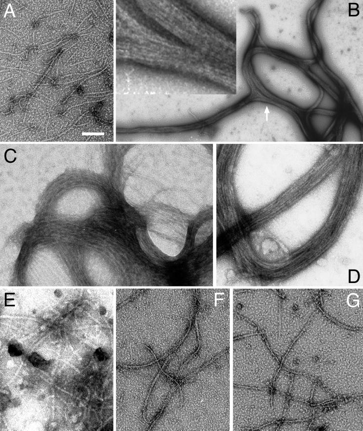
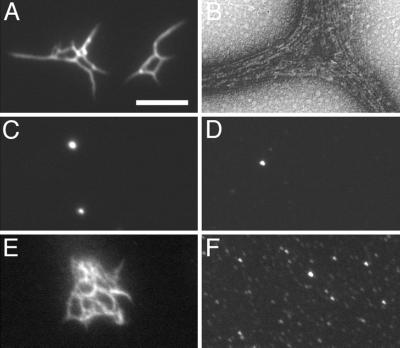
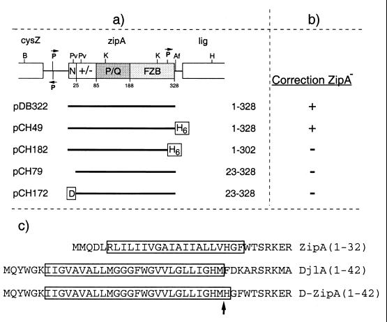
FtsZ and ZipA are essential components of the septal ring apparatus, which mediates cell division in Escherichia coli. FtsZ is a cytoplasmic tubulin-like GTPase that forms protofilament-like homopolymers in vitro. In the cell, the protein assembles into a ring structure at the prospective division site early in the division cycle, and this marks the first recognized event in the assembly of the septal ring. ZipA is an inner membrane protein which is recruited to the nascent septal ring at a very early stage through a direct interaction with FtsZ. Using affinity blotting and protein localization techniques, we have determined which domain on each protein is both sufficient and required for the interaction between the two proteins in vitro as well as in vivo. The results show that ZipA binds to residues confined to the 20 C-terminal amino acids of FtsZ. The FtsZ binding (FZB) domain of ZipA is significantly larger and encompasses the C-terminal 143 residues of ZipA. Significantly, we find that the FZB domain of ZipA is also required and sufficient to induce dramatic bundling of FtsZ protofilaments in vitro. Consistent with the notion that the ability to bind and bundle FtsZ polymers is essential to the function of ZipA, we find that ZipA derivatives lacking an intact FZB domain fail to support cell division in cells depleted for the native protein. Interestingly, ZipA derivatives which do contain an intact FZB domain but which lack the N-terminal membrane anchor or in which this anchor is replaced with the heterologous anchor of the DjlA protein also fail to rescue ZipA(-) cells. Thus, in addition to the C-terminal FZB domain, the N-terminal domain of ZipA is required for ZipA function. Furthermore, the essential properties of the N domain may be more specific than merely acting as a membrane anchor.


Figures

FIG. 1
FIG. 1
FtsZ plasmids used to define the ZipA-binding domain. (a) The physical map of ftsZ and portions of flanking genes in E. coli are shown at the top of the panel. The positions of BclI (Bc), EcoRI (E), EcoRV (Rv), and HindIII (H) restriction sites are indicated. The positions in the E. coli FtsZ polypeptide of two domains (N and C) and a connecting core helix (residues ca. 177 to 201) were inferred from the crystal structure of FtsZ from Methanococcus jannaschii (14, 15). Inserts of plasmids are presented below the map, and the FtsZ residues they encode are given at the right of each insert. All plasmids were derivatives of pET21, such that transcription of inserts was under control of the T7lac promoter (Novagen). Plasmid pDB312 encodes native FtsZ. All others encode either full-length or portions of FtsZ fused to various tags, as indicated. H6, stretch of six histidine residues; HFKT, combination tag peptide which includes a stretch of 10 histidine residues and a substrate site for heart muscle kinase (10); T, T7.tag peptide. (b) The ability of these FtsZ derivatives to bind radiolabeled ZipA-FKH was determined by affinity blotting. +, protein binds ZipA; −, protein does not bind ZipA.
FIG. 2
FIG. 2
Binding of radiolabeled ZipA to a C-terminal domain of FtsZ. Purified proteins (lanes 1 to 4) and whole-cell extracts (lanes 5 to 8) were separated on two identical SDS-PAGE gels. One gel was stained with Coomassie brilliant blue to visualize protein bands (top), and proteins in the other gel were blotted to a nitrocellulose filter which was subsequently incubated with radiolabeled ZipA-FKH (bottom). Lanes 1 to 4 contain 50 pmol of native FtsZ(1–383) (lane 1), FtsZ(1–383)-H (lane 2), FtsZ(1–372)-H (lane 3), or FtsZ(1–314)-H (lane 4). Lanes 5 to 8 contain 10 μl of extract of cells overexpressing Gfp-T-FtsZ(1–383) (lane 5), Gfp-T-FtsZ(289–383) (lane 6), Gfp-T-FtsZ(364–383) (lane 7), or Gfp-T-FtsZ(374–383) (lane 8). Extracts were prepared from cells of strain BL21(λDE3)/plysS containing the appropriate plasmid (Fig. 1) after growth in the presence of IPTG and by resuspension of cells in SDS-PAGE lysis buffer to the equivalent of 20.0 OD600 units. Bands corresponding to the overexpressed proteins are indicated by an asterisk in the upper panel. The positions of molecular mass standards (66, 45, 36, 29, and 24 kDa [top to bottom]) are indicated on the left of the panels.
FIG. 3
FIG. 3
Protease inaccessibility of ZipA in intact spheroplasts. Spheroplasts of PB103 cells were either left untreated (lanes 2) or treated with protease (lanes 1 and 3) and/or detergent (lanes 3 and 4). Samples were used to prepare three identical Western blots. TonB (top) and FtsZ (bottom) were detected with specific antibodies; ZipA (middle) was detected by incubation of the blot with radiolabeled HFKT-FtsZ.
FIG. 4
FIG. 4
ZipA plasmids used to define the FtsZ-binding domain. (a) The physical map of zipA and portions of flanking genes in E. coli are shown at the top of the panel. The positions of AflII (Af), BamHI (B), HindIII (H), KpnI (K), and PvuII (Pv) restriction sites are indicated. The four domains of the ZipA polypeptide we previously proposed are denoted N (N-terminal membrane anchor), +/− (highly charged domain), P/Q (proline- and glutamine-rich domain), and C (C-terminal domain). The +/− domain includes the MAP-Tau repeat-like sequence proposed by RayChaudhuri to mediate binding to and bundling of FtsZ polymers (30). Inserts of plasmids are presented below the map, and the ZipA residues they encode are given at the right of each insert. All plasmids were derivatives of pET21, such that transcription of inserts was under the control of the T7lac promoter (Novagen). As indicated, inserts encode either full-length or portions of ZipA fused to various tags. H10, stretch of 10 histidine residues; FKH, combination tag which includes a stretch of six histidine residues and a substrate site for heart muscle kinase. See also the legend to Fig. 1. (b) The ability of these ZipA derivatives to bind radiolabeled HFKT-FtsZ was determined by affinity blotting. +, protein binds FtsZ; −, protein does not bind FtsZ. The ability of proteins to bundle FtsZ polymers was assessed by electron microscopy in all cases. The results obtained with the three Gfp-tagged derivatives were confirmed by fluorescence microscopy. +, numerous bundles and bundle networks observed. −, no bundles observed.
FIG. 5
FIG. 5
Binding of radiolabeled FtsZ to a C-terminal domain of ZipA. Purified proteins (50 pmol/lane) were separated on two identical SDS-PAGE gels. One gel was stained with Coomassie brilliant blue to visualize protein bands (top), and proteins in the other gel were blotted to a nitrocellulose filter which was subsequently incubated with radiolabeled HFKT-FtsZ (bottom). Lanes contained ZipA(1–328)-H (lane 1), ZipA(1–302)-H (lane 2), ZipA(23–328) (lane 3), ZipA(23–279) (lane 4), T-ZipA(70–328) (lane 5), T-ZipA(186–328)-H (lane 6), Gfp-T-ZipA(186–328)-H (lane 7), or Gfp-T-ZipA(212–328)-H (lane 8). The positions of molecular mass standards (66, 45, 36, 29, 24, 20, and 14 kDa [top to bottom]) are indicated on the left of the panels.
FIG. 6
FIG. 6
Plasmids used to sublocalize ZipA derivatives. (a) The C-domain of ZipA is renamed FZB (FtsZ-binding domain) to indicate that this domain coincides with the portion of ZipA found here to be required and sufficient for binding FtsZ (see the text). Inserts were cloned into the vector pMLB1113 such that expression of the fusion proteins is under control of the lac promoter and lacIq. D, transmembrane domain corresponding to residues 1 to 32 of DjlA. See also the legend to Fig. 1. (b) Cellular location of fusion proteins in strain PB103. R, virtually all fluorescence associated with the septal ring; M, fluorescence evenly distributed along the entire cell membrane; C, fluorescence evenly distributed throughout the cytoplasm; C/R, a significant portion of total fluorescence throughout the cytoplasm and the rest associated with the septal ring.
FIG. 7
FIG. 7
Localization of Gfp-tagged ZipA derivatives in wild-type cells. Cells were chemically fixed and observed under fluorescence (A to F) and differential interference contrast (A′ to F′) optics. Images faithfully reflected the distributions of fluorescence seen prior to fixation. Panels show cells of strain PB103 (wild type) expressing ZipA derivatives from plasmids pCH50 [Plac::zipA(1–328)- gfp] (A), pCH148 [Plac::zipA(1–302)-gfp] (B), pCH80 [Plac::zipA(23–328)-gfp] (C), pCH138 [Plac::gfp-t-zipA(186–328)] (D), pCH93 [Plac::gfp-t-zipA(212–328)] (E), or pCH174 [Plac::djlA(1–32)-zipA(23–328)-gfp] (F). Cells were grown in the presence of 5 μM (A), 10 μM (B), 25 μM (C), or 100 μM (D to F) IPTG. None of the plasmids interfered noticeably with the normal division phenotype under these conditions. Bar, 3 μm.
FIG. 8
FIG. 8
Localization of Gfp-tagged ZipA derivatives in FtsZ cells. Filaments of the FtsZ CID strain PB143/pDB346 (ftsZ0/cI857 PλR::ftsZ) carrying pCH50 [Plac::zipA(1–328)-gfp] (A), pCH80 [Plac::zipA(23–328)-gfp] (B), or pCH174 [Plac::djlA(1–32)-zipA(23–328)-gfp] (C) are shown. Cells were grown at 30°C (leading to depletion of FtsZ) in the presence of 5 μM (A and B) or 100 μM (C) IPTG. The resulting filaments were chemically fixed and observed under fluorescence optics. Images faithfully reflected the distributions of fluorescence seen prior to fixation. Bar, 3 μm.
FIG. 9
FIG. 9
ZipA-induced bundling of FtsZ polymer filaments. Purified native FtsZ (A to E) and FtsZ(1–314)-H (F and G) was incubated in the presence of 1.0 mM GTP and in the presence of either 10 mM Mg2+ (A, B, and D to G) or 2 mM EDTA (C). After 5 min, buffer (A and F), ZipA(23–328) (B, C, and G), Gfp-T-ZipA(186–328)-H (D), or Gfp-T-ZipA(212–328)-H (E) was added. After an additional 10 min, samples were applied to a microscope grid, stained with uranyl acetate, and examined under an electron microscope. Each protein in the reactions was present at 5 μM. The inset in panel B represents a portion (arrow) of the bundle network in more detail, emphasizing the side-by-side arrangement of polymers within the bundles. Polymers or polymer bundles were completely absent in control reactions in which GTP or FtsZ were omitted (data not shown). Bar, 76 nm (C), 100 nm (A, E to G, and inset in B), 125 nm (D), or 600 nm (B).
FIG. 10
FIG. 10
Detection of polymer bundles by fluorescence microscopy. Purified native FtsZ (6.0 μM) was incubated in the presence of GTP (1 mM) and Mg2+ (10 mM) at 30°C. After 5 min, purified H-T-ZipA(39–328)-Gfp was added to 0.6 μM. After an additional 10 min, one sample was applied to a microscope slide and observed immediately by fluorescence microscopy (A) and another was used for observation by EM (B). The fluorescent samples shown in panels C to F were prepared identically, except that GTP was replaced with GDP (C), FtsZ was replaced with buffer (D), or H-T-ZipA(39–328)-Gfp was replaced with either Gfp-T-ZipA(186–328)-H (E) or Gfp-T-ZipA(212–328)-H (F). EM grids prepared from the reactions shown in panels C to F showed extensive bundle networks as in panel A (E), no bundles but many individual FtsZ protofilaments (F), or no individual protofilaments or polymer bundles (C and D) (data not shown). Bar, represents 0.1 μm (B) or 3.4 μm (A and C to F).
FIG. 11
FIG. 11
Plasmids used to test correction of ZipA by ZipA derivatives. (a) Inserts were cloned into the vector pMLB1113 such that expression of the proteins is under control of the lac promoter and lacIq. See also the legends to Fig. 1 and 6. (b) The ability of the plasmids to correct a ZipA phenotype was determined as described in the text. +, correction; −, no correction. (c) Shown are the N-terminal domains of ZipA, DjlA, and DjlA(1–32)-ZipA(23–328). Transmembrane segments as predicted by the Dense Alignment Surface method (4) (http://www.biokemi.su.se/∼server/DAS/) are boxed. Residue numbers are indicated in parentheses. The arrow marks the junction between DjlA and ZipA residues in the DjlA-ZipA fusion.

Similar articles

Cited by

References

    1. Blanar M A, Rutter W J. Interaction cloning: identification of a helix-loop-helix zipper protein that interacts with c-Fos. Science. 1992;256:1014–1018. - PubMed
    1. Broome-Smith J K, Spratt B G. A vector for the construction of translational fusions to TEM β-lactamase and the analysis of protein export signals and membrane protein topology. Gene. 1986;49:341–349. - PubMed
    1. Clarke D J, Jacq A, Holland I B. A novel DnaJ-like protein in Escherichia coli inserts into the cytoplasmic membrane with a type III topology. Mol Microbiol. 1996;20:1273–1286. - PubMed
    1. Cserzo M, Wallin E, Simon I, von Heijne G, Elofsson A. Prediction of transmembrane alpha-helices in prokaryotic membrane proteins: the dense alignment surface method. Protein Eng. 1997;10:673–676. - PubMed
    1. de Boer P, Crossley R, Rothfield L. The essential bacterial cell-division protein FtsZ is a GTPase. Nature. 1992;359:254–256. - PubMed

Publication types

MeSH terms

LinkOut - more resources