

The amino terminus of Pseudomonas aeruginosa outer membrane protein OprF forms channels in lipid bilayer membranes: correlation with a three-dimensional model
- PMID: 10960112
- PMCID: PMC94676
- DOI: 10.1128/JB.182.18.5251-5255.2000
The amino terminus of Pseudomonas aeruginosa outer membrane protein OprF forms channels in lipid bilayer membranes: correlation with a three-dimensional model
Abstract
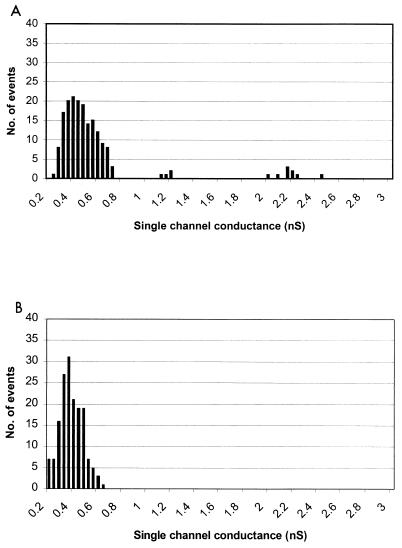
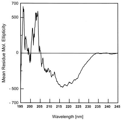
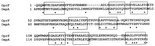
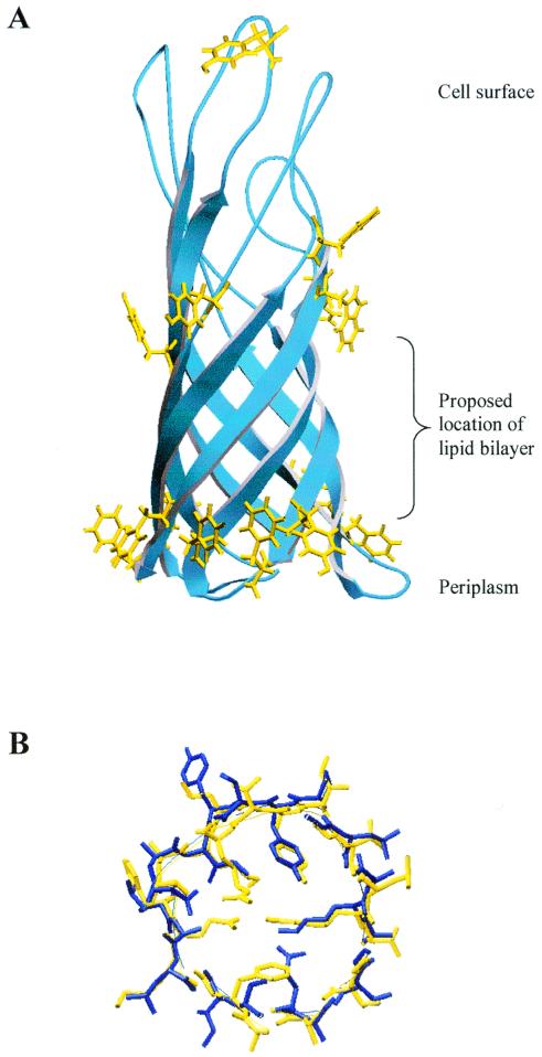
Pseudomonas aeruginosa OprF forms 0.36-nS channels and, rarely, 2- to 5-nS channels in lipid bilayer membranes. We show that a protein comprising only the N-terminal 162-amino-acid domain of OprF formed the smaller, but not the larger, channels in lipid bilayers. Circular dichroism spectroscopy indicated that this protein folds into a beta-sheet-rich structure, and three-dimensional comparative modeling revealed that it shares significant structural similarity with the amino terminus of the orthologous protein Escherichia coli OmpA, which has been shown to form a beta-barrel. OprF and OmpA share only 15% identity in this domain, yet these results support the utility of modeling such widely divergent beta-barrel domains in three dimensions in order to reveal similarities not readily apparent through primary sequence comparisons. The model is used to further hypothesize why porin activity differs for the N-terminal domains of OprF and OmpA.
Figures
References
-
- Arora A, Rinehart D, Szabo G, Tamm L K. Refolded outer membrane protein A of Escherichia coli forms ion channels with two conductance states in planar lipid bilayers. J Biol Chem. 2000;275:1594–1600. - PubMed
-
- Benz R, Hancock R E W. Properties of the large ion-permeable pores formed from protein F of Pseudomonas aeruginosa in lipid bilayer membranes. Biochim Biophys Acta. 1981;646:298–308. - PubMed
-
- Brahms S, Brahms J. Determination of protein secondary structure in solution by vacuum ultraviolet circular dichroism. J Mol Biol. 1980;138:149–178. - PubMed
Publication types
MeSH terms
Substances
LinkOut - more resources
Full Text Sources
Other Literature Sources
Miscellaneous
