

Human renal cell carcinoma expresses distinct binding sites for growth hormone-releasing hormone
- PMID: 10962030
- PMCID: PMC27063
- DOI: 10.1073/pnas.180313097
Human renal cell carcinoma expresses distinct binding sites for growth hormone-releasing hormone
Abstract
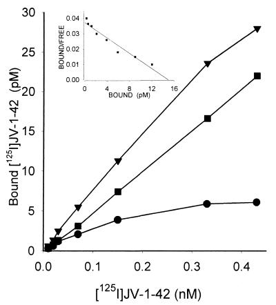
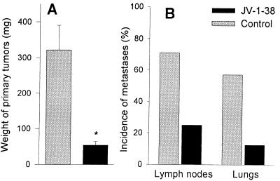
Antagonists of growth hormone-releasing hormone (GHRH) inhibit the proliferation of various human cancers in vitro and in vivo by mechanisms that include apparent direct effects through specific binding sites expressed on tumors and that differ from pituitary human GHRH (hGHRH) receptors. In this study, GHRH antagonist JV-1-38 (20 microgram/day per animal s.c.) inhibited the growth of orthotopic CAKI-1 human renal cell carcinoma (RCC) by 83% and inhibited the development of metastases to lung and lymph nodes. Using ligand competition assays with (125)I-labeled GHRH antagonist JV-1-42, we demonstrated the presence of specific high-affinity (K(d) = 0.25 +/- 0.03 nM) binding sites for GHRH with a maximal binding capacity (B(max)) of 70.2 +/- 4.1 fmol/mg of membrane protein in CAKI-1 tumors. These receptors bind GHRH antagonists preferentially and display a lower affinity for hGHRH. The binding of (125)I-JV-1-42 is not inhibited by vasoactive intestinal peptide (VIP)-related peptides sharing structural homology with hGHRH. The receptors for GHRH antagonists on CAKI-1 tumors are distinct from binding sites detected with (125)I-VIP (K(d) = 0.89 +/- 0.14 nM; B(max) = 183.5 +/- 2.6 fmol/mg of protein) and also have different characteristics from GHRH receptors on rat pituitary as documented by the insignificant binding of [His(1),(125)I-Tyr(10), Nle(27)]hGHRH(1-32)NH(2). Reverse transcription-PCR revealed the expression of splice variants of hGHRH receptor in CAKI-1 RCC. Biodistribution studies demonstrate an in vivo uptake of (125)I-JV-1-42 by the RCC tumor tissue. The presence of specific receptor proteins that bind GHRH antagonists in CAKI-1 RCC supports the view that distinct binding sites that mediate the inhibitory effect of GHRH antagonists are present on various human cancers.
Figures
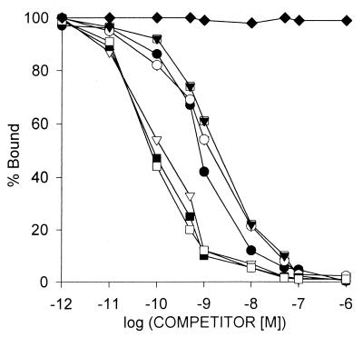
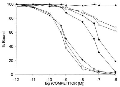
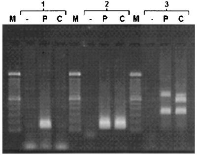
 ). VIP (♦) as well as glucagon, PACAP, JV-1–53, and PG 97–269 and other unrelated peptides, such as luteinizing hormone-releasing hormone, epidermal growth factor, [Tyr11]somatostatin-14, [Tyr4]bombesin, and IGF-I, did not displace the radioligand (data not shown). One hundred percent specific binding is defined as difference between binding in absence and in presence of 10−5 M JV-1–42. Each data point represents mean of at least two experiments, done in duplicate or triplicate.
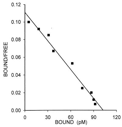
References
-
- Schally A V, Comaru-Schally A M. In: Cancer Medicine. 5th Ed. Holland J F, Frei E III, Bast R C Jr, Kufe D W, Pollock R E, Weichselbaum R R, editors. Ontario: Decker; 2000. pp. 715–729.
-
- Schally A V, Kovacs M, Toth K, Comaru-Schally A M. In: Growth Hormone Secretagogues in Clinical Practice. Bercu B B, Walker R F, editors. New York: Dekker; 1998. pp. 145–162.
-
- Schally A V, Varga J L. Trends Endocrinol Metab. 1999;10:383–391. - PubMed
-
- Zarandi M, Kovacs M, Horvath J E, Toth K, Halmos G, Groot K, Nagy A, Kele Z, Schally A V. Peptides. 1997;18:423–430. - PubMed
Publication types
MeSH terms
Substances
LinkOut - more resources
Full Text Sources
Other Literature Sources
Medical
