. 2000 Aug 29;97(18):10050-5.
doi: 10.1073/pnas.97.18.10050.

Neural crest-directed gene transfer demonstrates Wnt1 role in melanocyte expansion and differentiation during mouse development

Affiliations

Neural crest-directed gene transfer demonstrates Wnt1 role in melanocyte expansion and differentiation during mouse development

K J Dunn et al. Proc Natl Acad Sci U S A. .

Abstract

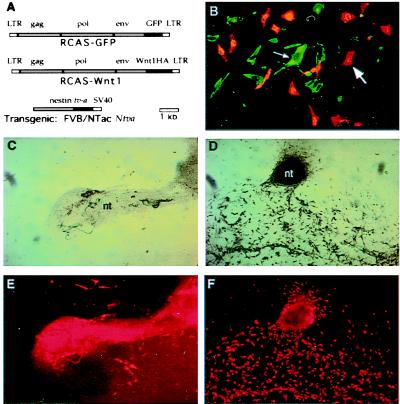
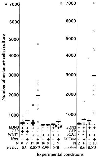
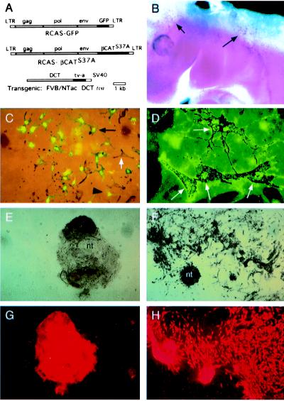
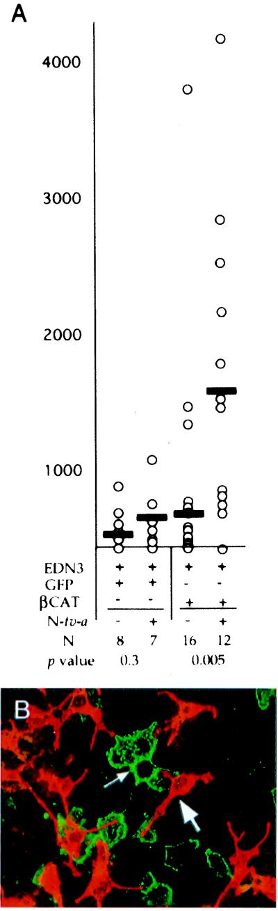
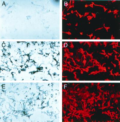
Wnt1 signaling has been implicated as one factor involved in neural crest-derived melanocyte (NC-M) development. Mice deficient for both Wnt1 and Wnt3a have a marked deficiency in trunk neural crest derivatives including NC-Ms. We have used cell lineage-directed gene targeting of Wnt signaling genes to examine the effects of Wnt signaling in mouse neural crest development. Gene expression was directed to cell lineages by infection with subgroup A avian leukosis virus vectors in lines of transgenic mice that express the retrovirus receptor tv-a. Transgenic mice with tva in either nestin-expressing neural precursor cells (line Ntva) or dopachrome tautomerase (DCT)-expressing melanoblasts (line DCTtva) were analyzed. We overstimulated Wnt signaling in two ways: directed gene transfer of Wnt1 to Ntva(+) cells and transfer of beta-catenin to DCTtva(+) NC-M precursor cells. In both methods, NC-M expansion and differentiation were effected. Significant increases were observed in the number of NC-Ms [melanin(+) and tyrosinase-related protein 1 (TYRP1)(+) cells], the differentiation of melanin(-) TYRP1(+) cells to melanin(+) TYRP1(+) NC-Ms, and the intensity of pigmentation per NC-M. These data are consistent with Wnt1 signaling being involved in both expansion and differentiation of migrating NC-Ms in the developing mouse embryo. The use of lineage-directed gene targeting will allow the dissection of signaling molecules involved in NC development and is adaptable to other mammalian developmental systems.


Figures

Figure 1
Figure 1
Infection of neural tube explants with RCAS-Wnt1 retrovirus. (A) Diagram of viral and transgene constructs. RCAS-GFP (Top) and RCAS-Wnt1(C-terminal HA epitope) (Middle) producing retrovirus constructs. (Bottom) The construct used to direct tv-a expression from the modified nestin promoter and enhancer sequences in transgenic mice (17). (B) Photomicrograph of Ntva transgenic neural tube explant infected with RCAS-Wnt1 and immunohistochemically stained for HA epitope representing Wnt1 expression from the retrovirus in green (small arrow) and a TYRP1 epitope representing NC-Ms in red (large arrow). Double staining revealed that Wnt1-expressing cells are a separate population from the pigmented or TYRP1+ cell population. The faint, yellow signals seen in B represent nonspecific binding of the secondary antibody to melanosomes, not coexpression of Wnt1 and TYRP1 in the same cells. (CF) Photomicrographs of nontransgenic (C and E) and Ntva transgenic (D and F) neural tube explants infected with RCAS-Wnt1. The remaining portion of the neural tube (nt) after 15 days of culture varies between experiments, resulting in the size differences denoted in C and D. Cultures show pigmented (C and D, bright field) and immunohistochemically stained TYRP1 NC-Ms (E and F, red). RCAS-Wnt1-infected cultures show an expansion of pigmented NC-Ms and TYRP1+ NC-Ms. The extensive red in the neural tube is a result of trapping of the secondary antibody in this multicellular structure, not TYRP1 antigenicity. All neural tube explants in this figure were cultured in the presence of EDN3.
Figure 2
Figure 2
Overexpression of Wnt signaling genes in neural tube explants results in increased numbers of pigmented NC-Ms. Results are shown from neural tube cultures derived from Ntva (A) and DCTtva (B) embryos. (A and B) Graphical representation of the number of pigmented NC-Ms (y axis) resulting from infection of nontransgenic and transgenic neural tube explants with RCAS viruses (x axis). Each open circle represents one neural tube explant culture. The NC-M numbers represent only the melanin+ cells that were not associated with the neural tube. The solid rectangles represent the average number of NC-Ms. For experimental conditions, the + or − represents the addition of EDN3 or the RCAS virus to the cultures, and the +# indicates the addition of EDN3 for only the first 4 days of the 15-day culture. The + or − for Ntva and DCTtva represents neural tubes isolated from transgenic and nontransgenic embryos, respectively. The number of neural tubes per condition is represented as N. The P values are comparisons of the numbers of NC-Ms observed between transgenic and nontransgenic cultures within each experimental condition (Mann–Whitney U analysis).
Figure 3
Figure 3
Lineage-directed infection of RCAS viruses into NC-M precursors using DCTtva transgenic mice. (A) Diagram of RCAS-GFP (Top) and RCASβCAT (Middle) viral constructs. The transgene construct (Bottom) used to direct expression of tv-a to melanoblasts in mouse embryos with the Dct promoter is shown. (B) Whole-mount in situ hybridization demonstrates expression of tv-a mRNA in migrating melanoblasts (arrows) of an embryonic day 10.5 DCTtva+ mouse embryo. (C) Photomicrograph demonstrating coexpression of GFP and melanin pigment in NC-Ms derived from neural tube explants of DCTtva embryos infected with RCAS-GFP. GFP expression is seen in both presumptive melanoblasts (large, black arrowhead; unpigmented cell) and pigmented NC-M cells (small, black arrow). A class of melanin+ GFP NC-Ms also was observed (white arrow), indicating that not all NC-M precursor cells were infected. (D) Colocalization of the HA epitope located on βCAT with pigmented melanocytes (white arrows) in cultures derived from DCTtva+ embryos infected with RCAS-βCAT. (EH) Photomicrographs of nontransgenic (E and G) and DCTtva transgenic (F and H) neural tube explants infected with βCAT-expressing RCAS virus. Pigmented cultures (E and F, bright field) and immunohistochemically stained TYRP1+ NC-Ms (G and H) are shown. Nontransgenic, uninfected cultures show some melanin+ and TYRP1+ NC-Ms. RCAS-βCAT-infected cultures show an expansion of melanin+ and TYRP1+ NC-Ms.
Figure 4
Figure 4
Infection of Ntva+ cells with βCAT results in NC-M expansion because of a paracrine mode of action. (A) Graphical representation of results from neural tube cultures derived from Ntva embryos. The number of pigmented NC-Ms (y axis) resulting from infection of nontransgenic and transgenic neural tube explants with RCAS viruses (x axis) is shown. Details of the graph are described in the legend for Fig. 2. (B) Photomicrograph of a Ntva+ culture infected with RCAS-βCAT-HA and stained for HA (green, small arrow) and TYRP1 (red, large arrow). Only rarely did colocalization of the two occur in the same cell, demonstrating that the expansion of NC-M is caused by a paracrine effect of βCAT in progenitors of Ntva+ cells.
Figure 5
Figure 5
Stimulation of Wnt signaling increases differentiation and pigmentation of NC-Ms. Bright-field photomicrographs of melanin+ NC-Ms (A, C, and E) and photomicrographs of the same field demonstrating immunohistochemically stained TYRP1+ NC-Ms (B, D, and F) are shown. All are from neural tube explant cultures after treatment for 15 days. Cultures were derived from nontransgenic embryos (A and B), Ntva embryos infected with RCAS-Wnt1 (C and D), or with RCAS-βCAT (E and F). The majority of TYRP1+ cells are pigmented in Wnt-stimulated cultures, whereas most TYRP1+ cells are not overtly pigmented in control cultures (A and B). Pigmented NC-Ms also have an increased amount of pigment within each cell when Wnt1 signaling was stimulated by either Wnt1 (C) or βCAT (E).

Similar articles

Cited by

References

    1. Cadigan K M, Nusse R. Genes Dev. 1997;11:3286–3305. - PubMed
    1. Ikeya M, Lee S M, Johnson J E, McMahon A P, Takada S. Nature (London) 1997;389:966–970. - PubMed
    1. Dorsky R I, Moon R T, Raible D W. Nature (London) 1998;396:370–373. - PubMed
    1. Henion P D, Weston J A. Development. 1997;124:4351–4359. - PubMed
    1. Stemple D L, Anderson D J. Cell. 1992;71:973–985. - PubMed

MeSH terms

LinkOut - more resources