. 2000 Aug 29;97(18):10132-7.
doi: 10.1073/pnas.97.18.10132.

Retinoid-related orphan receptor gamma (RORgamma) is essential for lymphoid organogenesis and controls apoptosis during thymopoiesis

Affiliations

Retinoid-related orphan receptor gamma (RORgamma) is essential for lymphoid organogenesis and controls apoptosis during thymopoiesis

S Kurebayashi et al. Proc Natl Acad Sci U S A. .

Abstract

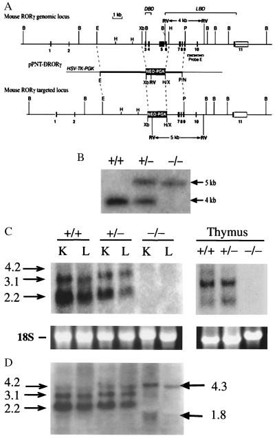
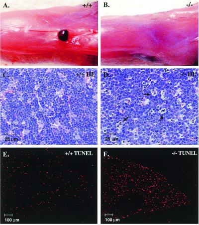
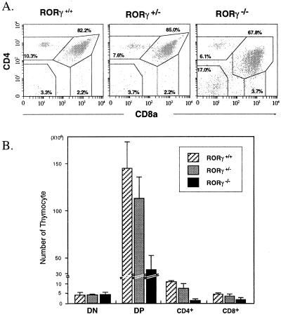
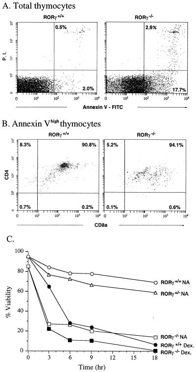
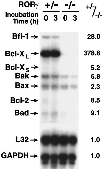
To identify the physiological functions of the retinoid-related orphan receptor gamma (RORgamma), a member of the nuclear receptor superfamily, mice deficient in RORgamma function were generated by targeted disruption. RORgamma(-/-) mice lack peripheral and mesenteric lymph nodes and Peyer's patches, indicating that RORgamma expression is indispensable for lymph node organogenesis. Although the spleen is enlarged, its architecture is normal. The number of peripheral blood CD3(+) and CD4(+) lymphocytes is reduced 6- and 10-fold, respectively, whereas the number of circulating B cells is normal. The thymus of RORgamma(-/-) mice contains 74.4% +/- 8.9% fewer thymocytes than that of wild-type mice. Flow cytometric analysis showed a decrease in the CD4(+)CD8(+) subpopulation. Terminal deoxynucleotidyltransferase-mediated dUTP nick end labeling (TUNEL) staining demonstrated a 4-fold increase in apoptotic cells in the cortex of the thymus of RORgamma(-/-) mice. The latter was supported by the observed increase in annexin V-positive cells. RORgamma(-/-) thymocytes placed in culture exhibit a dramatic increase in the rate of "spontaneous" apoptosis. This increase is largely associated with CD4(+)CD8(+) thymocytes and may, at least in part, be related to the greatly reduced level of expression of the anti-apoptotic gene Bcl-X(L). Flow cytometric analysis demonstrated a 6-fold rise in the percentage of cells in the S phase of the cell cycle among thymocytes from RORgamma(-/-) mice. Our observations indicate that RORgamma is essential for lymphoid organogenesis and plays an important regulatory role in thymopoiesis. Our findings support a model in which RORgamma negatively controls apoptosis in thymocytes.


Figures

Figure 1
Figure 1
Targeting the RORγ locus. (A) Schematic representation of the mouse RORγ gene locus, the pPNT-ΔRORγ targeting vector, and the recombination at the RORγ locus. In the targeted locus the region from exon 3 through exon 6 is deleted. DBD, DNA-binding domain; LBD, ligand-binding domain. (B) Diagnostic Southern blot analysis. Genomic DNA was cut by EcoRV, electrophoresed, and hybridized to the 3′-flanking probe E (indicated in A), which detected fragments of the expected size of 4.0 kb for wild-type and 5.0 kb for the mutant allele. Lanes indicate digested DNA from wild-type (wt; +/+), heterozygous (+/−), and homozygous mutant (−/−) mice. (C and D) Northern blot analysis was performed with RNA isolated from liver (L), kidney (K), and thymus of wt, RORγ+/−, and RORγ−/− mice by using either a probe encompassing the deleted region encoded by exons 3–6 (C) or a full-length RORγ probe (D).
Figure 2
Figure 2
RORγ−/− mice lack peripheral lymphoid organs and manifest increased apoptosis in the thymus. (A and B) India ink was injected into the hind footpads and the popliteal fossae were dissected. A lymph node is clearly visible in wt mice (A), whereas RORγ−/− mice did not contain detectable lymph node tissue (B). (C–F) Sections of thymus from RORγ+/+ (C and E) and RORγ−/− (D and F) mice stained by hematoxylin/eosin (C and D) or by TUNEL staining using TMR red-dUTP (E and F). Arrows indicate apoptotic cells.
Figure 3
Figure 3
Flow cytometric analysis of CD4 and CD8 expression. (A) Isolated thymocytes from RORγ+/+, RORγ+/−, and RORγ−/− mice stained with anti-CD4-PE and anti-CD8-FITC cell surface markers and then analyzed by flow cytometry. Numbers within each of the four outlined areas indicate the percentage of the respective subpopulation. The results shown are representative for four independent analyses. (B) Comparison of the total cell number in each CD4/CD8 subpopulation.
Figure 4
Figure 4
Apoptosis is largely associated with DP thymocytes. (A) Thymocytes were isolated and treated with annexin V-FITC, anti-CD4-PE and anti-CD8-Cy. Total thymocytes were first analyzed by flow cytometry for annexin V binding. The lower and upper right quadrants indicate early and late apoptotic cells, respectively. (B) Thymocytes highly labeled by annexin V (early and late apoptotic cells) were separated further by flow cytometry on the basis of anti-CD4 and anti-CD8 labeling. (C) The rate of apoptosis in cultured ROR−/− thymocytes is greatly increased. Thymocytes were cultured in RPMI medium 1640 plus 10% FBS in the presence (Dex.) or absence (NA) of 0.1 μM dexamethasone. At the indicated times, samples were removed and analyzed for annexin V binding by flow cytometry as in A. The percentage of annexin Vlow cells (% viability) was calculated and plotted as a function of time in culture.
Figure 5
Figure 5
Effect of RORγ disruption on the expression of anti- and pro-apoptotic genes. RNA was prepared from freshly isolated thymocytes from RORγ+/− or RORγ−/− mice, and thymocytes were cultured for 3 hr. RNA was then analyzed by RPA. The positions of mRNAs encoding Bcl-2, Bfl-1, Bcl-XL, Bak, Bax, Bad, L32, and glyceraldehyde-3-phosphate dehydrogenase (GAPDH) are indicated. The results are representative for two independent experiments. The level of each RNA relative to the level of GAPDH was determined, and the ratio between the level of each RNA in RORγ+/− versus RORγ−/− thymocytes was calculated. The ratios are shown on the right.

References

    1. Willy P J, Mangelsdorf D J. In: Hormones and Signaling. O'Malley B W, editor. Vol. 1. San Diego: Academic; 1998. pp. 308–358.
    1. Kumar R, Thompson E B. Steroids. 1999;64:310–319. - PubMed
    1. McKenna N J, Xu J, Nawaz Z, Tsai S Y, Tsai M J, O'Malley B W. J Steroid Biochem Mol Biol. 1999;69:3–12. - PubMed
    1. Xu L, Glass C K, Rosenfeld M G. Curr Opin Genet Dev. 1999;9:140–147. - PubMed
    1. Giguere V, Tini M, Flock G, Ong E, Evans R M, Otulakowski G. Genes Dev. 1994;8:538–553. - PubMed

Publication types

MeSH terms

Substances