. 2000 Aug 29;97(18):10231-5.
doi: 10.1073/pnas.97.18.10231.

Establishment of an animal-bacterial association: recruiting symbiotic vibrios from the environment

Affiliations

Establishment of an animal-bacterial association: recruiting symbiotic vibrios from the environment

S V Nyholm et al. Proc Natl Acad Sci U S A. .

Abstract

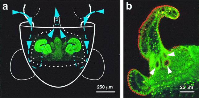
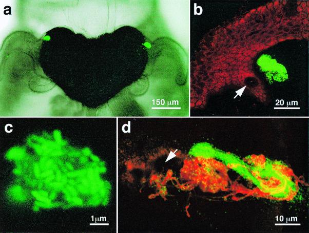
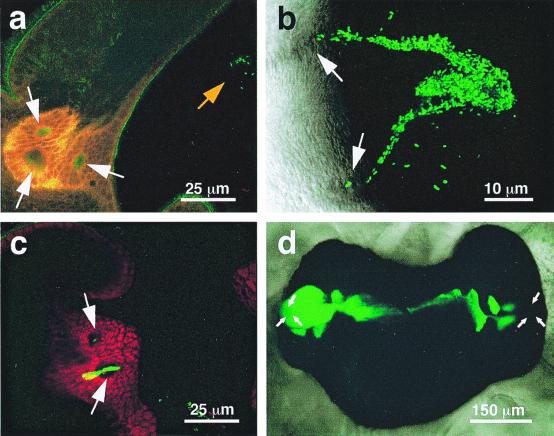
While most animal-bacterial symbioses are reestablished each successive generation, the mechanisms by which the host and its potential microbial partners ensure tissue colonization remain largely undescribed. We used the model association between the squid Euprymna scolopes and Vibrio fischeri to examine this process. This light organ symbiosis is initiated when V. fischeri cells present in the surrounding seawater enter pores on the surface of the nascent organ and colonize deep epithelia-lined crypts. We discovered that when newly hatched squid were experimentally exposed to natural seawater, the animals responded by secreting a viscous material from the pores of the organ. Animals maintained in filtered seawater produced no secretions unless Gram-negative bacteria, either living or dead, were reintroduced. The viscous material bound only lectins that are specific for either N-acetylneuraminic acid or N-acetylgalactosamine, suggesting that it was composed of a mucus-containing matrix. Complex ciliated fields on the surface of the organ produced water currents that focused the matrix into a mass that was tethered to, and suspended above, the light organ pores. When V. fischeri cells were introduced into the seawater surrounding the squid, the bacteria were drawn into its fluid-filled body cavity during ventilation and were captured in the matrix. After residing as an aggregate for several hours, the symbionts migrated into the pores and colonized the crypt epithelia. This mode of infection may be an example of a widespread strategy by which aquatic hosts increase the likelihood of successful colonization by rarely encountered symbionts.


Figures

Figure 1
Figure 1
The path of V. fischeri cells to the site of inoculation of the E. scolopes light organ. (a) Diagram illustrating an outline of the host's body (solid white lines), superimposed over a laser-scanning confocal micrograph (LSM) of the nascent light organ, indicating the relative size and position of the organ within the host's mantle cavity. The organ is circumscribed by the posterior portion of the excurrent funnel (dotted white lines). Ventilatory movements of the host draw ambient seawater (blue arrows and lines) containing V. fischeri cells into the mantle cavity. The water travels into the funnel where, before being vented back into the environment, it encounters complex ciliated fields (bright green) on the lateral surfaces of the organ. The fields entrain water into the vicinity of pores on the light organ surface. (b) Higher-magnification LSM of one side of a hatchling light organ, showing the location of the three pores (arrows) that lie at the base of the appendages of each ciliated field.
Figure 2
Figure 2
Bacterial aggregations that form during inoculation of the E. scolopes light organ. (a) Differential interference contrast image superimposed over a fluorescent image of the organ of a newly hatched host squid that had been exposed to GFP-labeled V. fischeri. Within 3 h after inoculation, the labeled bacteria have formed two dense aggregations on either side of the organ near the base of the anterior appendage of the ciliated field. (b) In a higher-magnification LSM, an aggregation of GFP-labeled bacteria could be seen suspended just above a light-organ pore (arrow). (c) A very highly magnified LSM of one of these aggregates confirmed that it was a dense assemblage of the GFP-labeled V. fischeri. (d) An LSM of an aggregate containing GFP-labeled V. fischeri cells 8 h after inoculation. The mucus-like matrix originating from the host's pore (arrow) was stained with fluorescently labeled wheat germ agglutinin (red).
Figure 3
Figure 3
The aggregation and segregation of polystyrene beads within the mucus-like secretions induced by the presence of V. fischeri cells. (a) Red-fluorescent polystyrene beads (Molecular Probes), 1 μm in diameter, were incubated with GFP-labeled V. fischeri and visualized by laser-scanning microscopy. After a 2- to 4-h incubation, bacteria and beads were found randomly distributed in aggregations. (b) Six hours after inoculation, the bacteria and beads had become segregated as the V. fischeri cells migrated in the direction of the pores (arrows).
Figure 4
Figure 4
Stages in the process of infection and colonization of the squid light organ. (a) After a 1-h exposure to GFP-labeled V. fischeri, an LSM revealed a small aggregate (orange arrow) forming above a pore of the light organ. (b) Between 2 and 4 h after inoculation, bacteria were seen as streams migrating from the aggregate to the pores. (c) Between 4 and 6 h after inoculation, a mass of GFP-labeled V. fischeri cells appeared to be migrating through a pore and into a duct of the light organ. Cells within the duct appear yellow. (d) Differential interference contrast image of the fully colonized light organ of E. scolopes, showing the population of GFP-labeled symbionts (green). The location of the pores is indicated by white arrows in all panels.

References

    1. VanRhijn P, Vanderleyden J. Microbiol Rev. 1995;59:124–142. - PMC - PubMed
    1. Fisher R F, Long S R. Nature (London) 1992;357:655–660. - PubMed
    1. Haygood M G. Crit Rev Microbiol. 1993;19:191–216. - PubMed
    1. Gros O, Darrasse A, Durand P, Frenkiel L, Moueza M. Appl Environ Microbiol. 1996;62:2324–2330. - PMC - PubMed
    1. Muller-Parker G, D'Elia C F. In: Life and Death of Coral Reefs. Birkeland C, editor. New York: Chapman and Hall; 1997. pp. 96–113.

Publication types

LinkOut - more resources