. 2000 Sep 1;20(17):6517-28.
doi: 10.1523/JNEUROSCI.20-17-06517.2000.

Laminin expression in adult and developing retinae: evidence of two novel CNS laminins

Affiliations

Laminin expression in adult and developing retinae: evidence of two novel CNS laminins

R T Libby et al. J Neurosci. .

Abstract

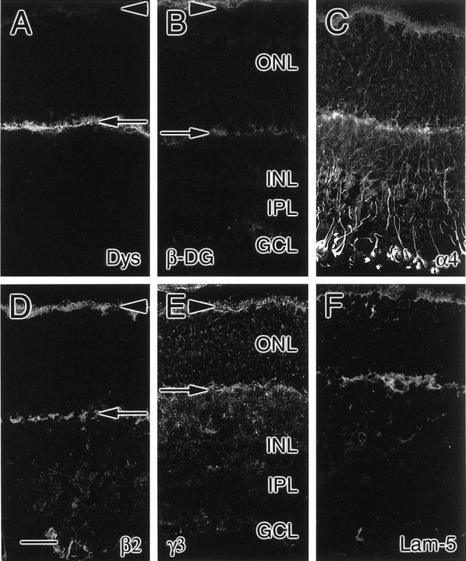
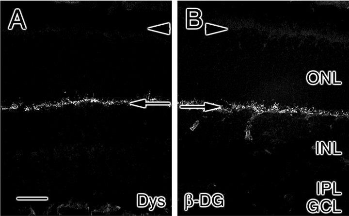
Components of the extracellular matrix exert myriad effects on tissues throughout the body. In particular, the laminins, a family of heterotrimeric extracellular glycoproteins, have been shown to affect tissue development and integrity in such diverse organs as the kidney, lung, skin, and nervous system. Of these, we have focused on the roles that laminins play in the differentiation and maintenance of the nervous system. Here, we examine the expression of all known laminin chains within one component of the CNS, the retina. We find seven laminin chains-alpha3, alpha4, alpha5, beta2, beta3, gamma2, and gamma3-outside the retinal basement membranes. Anatomically, these chains are coexpressed in one or both of two locations: the matrix surrounding photoreceptors and the first synaptic layer where photoreceptors synapse with retinal interneurons. Biochemically, four of these chains are coisolated from retinal extracts in two independent complexes, confirming that two novel heterotrimers-alpha4beta2gamma3 and alpha5beta2gamma3-are present in the retinal matrix. During development, all four of these chains, along with components of laminin 5 (the alpha3, beta3, and gamma2 chains) are also expressed at sites at which they could exert important effects on photoreceptor development. Together, these data suggest the existence of two novel laminin heterotrimers in the CNS, which we term here laminin 14 (composed of the alpha4, beta2, and gamma3 chains) and laminin 15 (composed of the alpha5, beta2, and gamma3 chains), and lead us to hypothesize that these laminins, along with laminin 5, may play roles in photoreceptor production, stability, and synaptic organization.


Figures

Fig. 1.
Fig. 1.
Expression patterns of laminin α chains in mature rat and human retinae. Unfixed frozen sections of rat (top row; A, C, E, G, I) and human (bottom row; B, D, F, H, J) retinae were probed for the presence of the laminins α1β1γ1 (A, B), α2 (C, D), α3 (E, F), α4 (G, H), and α5 (I, J) chains by the use of chain-specific (or trimer-specific for α1β1γ1) antibodies. Although several chains are present in the vasculature, only three (α3, α4, and α5) are expressed within the interphotoreceptor matrix (arrowheads) and outer plexiform layer (arrows). In addition, α4 is present in fibers extending through the outer nuclear layer (ONL) and inner nuclear layer (INL). GCL, Ganglion cell layer. Scale bar, 25 μm.
Fig. 2.
Fig. 2.
Expression patterns of laminin β chains in mature rat and human retinae. Unfixed frozen sections of rat (top row; A, C, E) and human (bottom row; B, D, F) retinae were probed for the presence of the laminin β1 (A), α1β1γ1 (B), β2 (C, D), and β3 (E, F) chains by the use of chain-specific (or trimer-specific for α1β1γ1) antibodies. Two β chains, β2 and β3, are expressed within the neural retina, specifically, in the interphotoreceptor matrix (arrowheads); in addition, the β2 chain is present in the outer plexiform layer (arrows). GCL, Ganglion cell layer;INL, inner nuclear layer; ONL, outer nuclear layer. Scale bar, 25 μm.
Fig. 3.
Fig. 3.
Expression patterns of laminin γ chains in mature rat and human retinae. Unfixed frozen sections of rat (top row; A, C, E) and human (bottom row; B, D, F) retinae were probed for the presence of the laminin γ1 (A, B), γ2 (C, D), and γ3 (E, F) chains by the use of chain-specific antibodies. Two γ chains, γ2 and γ3, are expressed within the neural retina, specifically in the interphotoreceptor matrix (arrowheads); in addition, the γ3 chain is present in the outer plexiform layer (arrows). GCL, Ganglion cell layer;INL, inner nuclear layer; ONL, outer nuclear layer. Scale bar, 25 μm.
Fig. 4.
Fig. 4.
Expression patterns of the RNA encoding laminin α chains in mature rat and human retinae. Fixed sections of rat (top row; A, C, E, G, I) and human (bottom row; B, D, F, H, J) retinae were probed for the presence of RNA encoding the laminin α1 (A, B), α2 (C, D), α3 (E, F), α4 (G, H), and α5 (I, J) chains by the use of chain-specific cRNA probes. Two RNAs (those encoding α3 and α4) are expressed within the neural retina of both species; in addition, the RNA encoding α5 is readily detected in human retina.GCL, Ganglion cell layer; INL, inner nuclear layer; ONL, outer nuclear layer. Scale bar, 25 μm.
Fig. 5.
Fig. 5.
Expression patterns of the RNA encoding laminin β chains in mature rat and human retinae. Fixed sections of rat (top row; A, C, E, G) and human (bottom row; B, D, F, H) retinae were probed for the presence of the RNA encoding laminin β1 (A, B), β2 (C, D), and β3 (G, H) chains by the use of chain-specific cRNA probes. RNA encoding the β2 and β3 chains is expressed within the neural retina. Expression of an RNA encoding a Müller cell antigen, cellular retinaldehyde-binding protein (CRALBP), is shown for comparison (E, F). GCL, Ganglion cell layer; INL, inner nuclear layer;ONL, outer nuclear layer. Scale bar, 25 μm.
Fig. 6.
Fig. 6.
Expression patterns of the RNA encoding laminin γ chains in mature rat and human retinae. Fixed sections of rat (top row; A, C, E) and human (bottom row; B, D, F) retinae were probed for the presence of the RNA encoding laminin γ1 (A, B), γ2 (C, D), and γ3 (E, F) chains by the use of chain-specific cRNA probes. Only the RNA encoding the γ3 chain is readily detected within the neural retina, although γ2 can be detected as well. GCL, Ganglion cell layer; INL, inner nuclear layer;ONL, outer nuclear layer. Scale bar, 25 μm.
Fig. 7.
Fig. 7.
Purification of laminins 14 and 15 from retinal extracts. Glycoproteins were isolated from retinal matrix by concanavalin A chromatography, separated on a nonreducing polyacrylamide gel, and stained with Coomassie blue (CB). Two components (A, B) were further separated on reducing polyacrylamide gels and then probed for the presence of the α4, β2, and γ3 chains by protein transfer blots. Both A and B contain the β2 and γ3 chains; B additionally contains the α4 chain. The γ3 chain antibody reacts with two bands: the full-length γ3 chain and a faster-migrating protein, a degradation product of the γ3 chain (or, potentially, cross-reactivity with the β2 chain) induced by the multiple purification steps used to prepare these proteins. Position of 200 kDa marker is shown.
Fig. 8.
Fig. 8.
Expression patterns of laminin chains in P0 rat retina. At PO, the outer plexiform layer has not yet formed, as demonstrated by the lack of dystrophin (Dys) expression in the retina (A). However, the laminin α4 (B), β2 (C), and γ3 (D) chains are prominently expressed in the subretinal space (SRS) and in fibers spanning the neuroepithelium (NE) and penetrating through the inner plexiform layer (IPL) and ganglion cell layer (GCL). In addition, the components of laminin 5—the α3 (E), β3 (F), and γ2 (G) chains—are expressed at P0, as is immunoreactivity for laminin 5 (Lam-5;H). Scale bar, 25 μm.
Fig. 9.
Fig. 9.
Expression patterns of laminin chains in central (top row; A, C, E, G, I) and peripheral (bottom row; B, D, F, H, J) P5 rat retina. At P5, the outer plexiform layer has begun to form in the central retina, as judged by dystrophin (Dys) expression (A), whereas it has not yet formed in the peripheral retina, as demonstrated by the lack of dystrophin expression (B). The laminin α4 (C, D), β2 (E, F), and γ3 (G, H) chains are expressed in the subretinal space (SRS) and in fibers spanning, centrally, the outer nuclear layer (ONL) and inner nuclear layer (INL) and, peripherally, the neuroepithelium (NE). These fibers penetrate through the inner plexiform layer (IPL) and ganglion cell layer (GCL). All three chains are also present in the developing outer plexiform layer (arrows). In addition, immunoreactivity for laminin 5 (Lam-5) is present at P5 (I, J). Scale bar, 25 μm.
Fig. 10.
Fig. 10.
Expression patterns of laminin chains in P10 rat retina. At P10, the outer plexiform layer has formed throughout the retina, as judged by dystrophin (Dys) expression (A). The laminin α4 (B), α5 (C), β2 (D), and γ3 (E) chains are expressed most prominently in the interphotoreceptor matrix (arrowheads) and outer plexiform layer (arrows) and are weakly present in fibers spanning the outer nuclear layer (ONL) and inner nuclear layer (INL); these fibers penetrate through the inner plexiform layer (IPL) and ganglion cell layer (GCL). In addition, laminin 5 (Lam-5) immunoreactivity is present at P10 (F). Scale bar, 25 μm.
Fig. 11.
Fig. 11.
Expression patterns of laminin chains in P15 rat retina. At P15, the outer plexiform layer not only contains dystrophin (Dys; A) but has also started to contain β-dystroglycan (β-DG; B). The laminin α4 (C), β2 (D), and γ3 (E) chains are expressed most prominently in the interphotoreceptor matrix (arrowheads) and outer plexiform layer (arrows); in addition, the α4 chain is prominent in fibers spanning the outer nuclear layer (ONL), inner nuclear layer (INL), inner plexiform layer (IPL), and ganglion cell layer (GCL). In addition, laminin 5 (Lam-5) immunoreactivity remains at P15 (F). Scale bar, 25 μm.
Fig. 12.
Fig. 12.
Expression patterns of components of the dystrophin complex in P26 rat retina. At P26, the outer plexiform layer (arrows) not only contains dystrophin (Dys; A) but also β-dystroglycan (β-DG; B). In this regard, unlike the P15 retina, the outer plexiform layer resembles that in the mature retina. GCL, Ganglion cell layer; INL, inner nuclear layer; IPL, inner plexiform layer;ONL, outer nuclear layer; arrowheads, interphotoreceptor matrix. Scale bar, 25 μm.
Fig. 13.
Fig. 13.
Summary of laminin expression in the retina. At least three distinct laminin compartments are present in the retina: the basement membranes, the interphotoreceptor matrix, and the matrices of the outer plexiform layer (OPL) and of the inner plexiform layer (IPL). Retinal basement membranes, that is, vitreal and Bruch's membranes (red lines), contain the laminin α1, α5, β1, β2, and γ1 chains; the exact composition of laminin isoforms in these membranes awaits the isolation of native laminins from this compartment. The interphotoreceptor matrix (and, during early development, the subretinal space) has a different complement of laminins (green shading/hatched); it contains the laminin α3, α4, α5, β2, β3, γ2, and γ3 chains. This suggests the presence of three laminins: laminin 5 (α3β3γ2), isolated previously from other tissues, and laminin 14 (α4β2γ3) and laminin 15 (α5β2γ3), newly isolated here (Fig.7). These same laminin chains are expressed in the outer plexiform layer as it develops; this expression becomes restricted to the α3, α4, α5, β2, and γ3 chains in the adult (green shading). The matrix of the inner plexiform layer (blue shading), on the other hand, contains only the α4 chain and no other known laminin chains, suggesting the presence of at least two as-yet-identified laminin chains that would partner with the α4 chain to form heterotrimeric laminins in the inner plexiform layer.BM, Bruch's membrane; RPE, retinal pigmented epithelium; OS, outer segments, ELM, external limiting membrane; GCL, ganglion cell layer;ILM, internal limiting membrane; INL, inner nuclear layer; ONL, outer nuclear layer.

References

    1. Anton ES, Sandrock AW, Jr, Matthew WD. Merosin promotes neurite growth and Schwann cell migration in vitro and nerve regeneration in vivo: evidence using an antibody to merosin, ARM-1. Dev Biol. 1994;164:133–146. - PubMed
    1. Arahata K, Ishii H, Hayashi YK. Congenital muscular dystrophies. Curr Opin Neurol. 1995;8:385–390. - PubMed
    1. Bunt-Milam AH, Saari JC. Immunocytochemical localization of two retinoid-binding proteins in vertebrate retina. J Cell Biol. 1983;97:703–712. - PMC - PubMed
    1. Burgeson RE, Chiquet M, Deutzmann R, Ekblom P, Engel J, Kleinman H, Martin GR, Meneguzzi G, Paulsson M, Sanes J, Timpl R, Tryggvason K, Yamada Y, Yurchenco PD. A new nomenclature for the laminins. Matrix Biol. 1994;14:209–211. - PubMed
    1. Chait BT, Kent SB. Weighing naked proteins: practical, high-accuracy mass measurement of peptides and proteins. Science. 1992;257:1885–1894. - PubMed

Publication types

MeSH terms

LinkOut - more resources