. 2000 Sep 1;20(17):6540-50.
doi: 10.1523/JNEUROSCI.20-17-06540.2000.

Netrin-G1: a novel glycosyl phosphatidylinositol-linked mammalian netrin that is functionally divergent from classical netrins

Affiliations

Netrin-G1: a novel glycosyl phosphatidylinositol-linked mammalian netrin that is functionally divergent from classical netrins

T Nakashiba et al. J Neurosci. .

Abstract

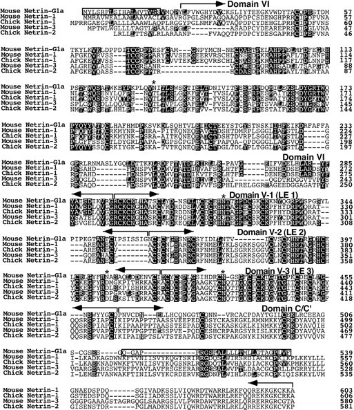
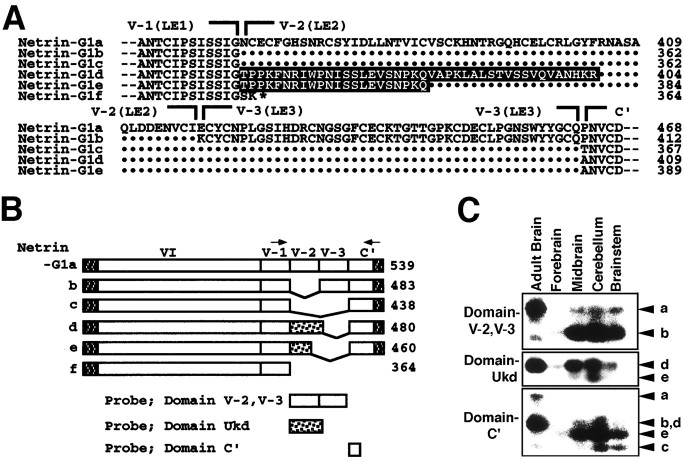
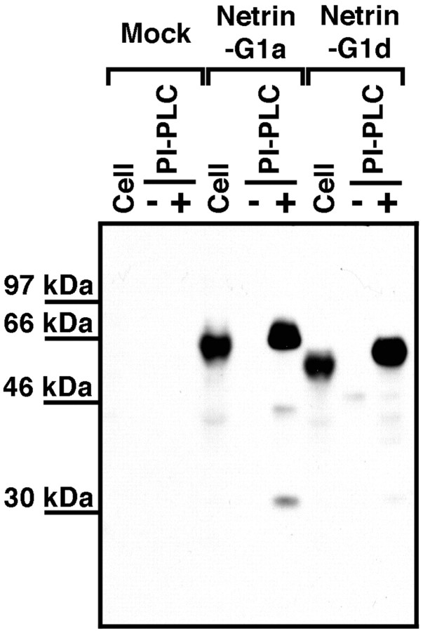
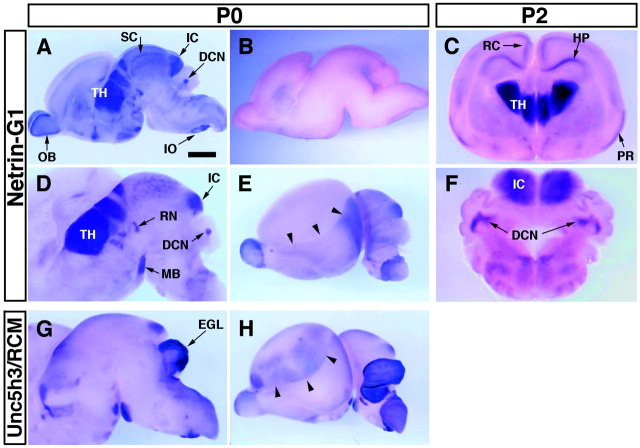
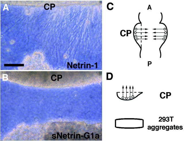
UNC-6/netrins compose a small phylogenetically conserved family of proteins that act as axon guidance cues. With a signal sequence trap method, we isolated a cDNA encoding a novel member of the UNC-6/netrin family, which we named netrin-G1. Unlike classical netrins, netrin-G1 consists of at least six isoforms of which five were predominantly anchored to the plasma membrane via glycosyl phosphatidyl-inositol linkages. Netrin-G1 transcripts were first detected in midbrain and hindbrain regions by embryonic day 12 and reached highest levels at perinatal stages in various brain regions, including olfactory bulb mitral cells, thalamus, and deep cerebellar nuclei. Its expression was primarily restricted to the CNS. Interestingly, netrin-G1 proteins did not show appreciable affinity to any netrin receptor examined. Unlike netrin-1, a secreted form of netrin-G1 consistently failed to attract circumferentially growing axons from the cerebellar plate. Our findings suggest that netrin-G1 and its putative receptors have coevolved independently from the classical netrins. The expression pattern of netrin-G1 and its predicted neuronal membrane localization suggest it may also have novel signaling functions in nervous system development.


Figures

Fig. 1.
Fig. 1.
Expression of 7D5 in the brain. A, Expression profile of 7D5 during cerebellar development. Poly(A+) RNAs from P0 and P21 mouse cerebellum were electrophoresed (2 μg/lane), transferred onto a nylon membrane, and hybridized with a 32P-labeled cDNA (7D5). The expression of 7D5 was characterized by downregulation in postnatal development. G3PDH was used as a control for loading of RNA. B, Tissue distribution of 7D5 in adult mice. An adult mouse multiple tissue Northern blot (Clontech) was hybridized with the probe mentioned above. 7D5 was specifically expressed in the brain. β-actin was used as a control for loading of RNA. Arrowheads inA and B indicate 7D5 signals.
Fig. 2.
Fig. 2.
Alignment of the deduced amino acid sequence of netrin-G1a (7D5) with vertebrate netrins. Amino acid residues identical between netrin-G1a and other netrins are in black. Hydrophobic stretches (boxed) of netrin-G1a appear to be signals for secretion and for GPI linkage predicted by the ω and ω + 2 rules (Gerber et al., 1992; von Heijne, 1996), respectively. Thebrackets above the sequences indicate domain structures (domains VI, V-1, V-2, V-3, and C′) of netrin-G1a defined by homology with other netrins. Asterisks indicate the putative N-glycosylation sites of netrin-G1a. The nucleotide sequences of netrin-G1a and other isoforms have been submitted to the GenBank (accession numbers AB038662, AB038663, AB038664, AB038665, AB038666, and AB038667).
Fig. 3.
Fig. 3.
Structural comparison of netrin-G1 and other UNC-6/netrins. A, Domain structures of members of the UNC-6/netrin family are schematically represented. The percent identity in amino acid sequence between homologous domains is shown. Netrin-G1a is anchored on cell membrane by a GPI linkage, whereas netrin-1 and netrin-3 are secreted. The C domains of classical netrins are highly charged (indicated by ++). The C-terminal sequence of netrin-G1a, named C′, is distinctly different from the C domain of classical netrins.B, Kyte-Doolittle hydrophobicity plot of deduced netrin-G1a and mouse netrin-1 amino acid sequences. Two hydrophobic stretches were observed at both N and C termini in netrin-G1a. The latter stretch is unique to netrin-G1 among members of the UNC-6/netrin family. C, A phylogenetic tree was constructed based on the amino acid sequences of C. elegans UNC-6 (P34710), chick netrin-1 (Q90922), chick netrin-2 (Q90923), mouse netrin-1 (AAC52971), mouse netrin-3 (AAD40063), human netrin-1 (NP004813), human NTN2L (NP006172), zebrafish netrin-1 (AAB70266), zebrafish netrin-1a (AAC60252), Drosophila netrin-A (Q24567),Drosophila netrin-B (Q24568), and netrin-G1a (AB038667) using CLUSTAL X program (ftp://ftp-igbmc.u-strasbg.fr/pub/ClustalX/). Netrin-G1 is evolutionarily distant from other members of the family, implying that the netrin-G1 may serve as a prototype of a novel subfamily.
Fig. 4.
Fig. 4.
Structure and expression of G1 isoforms.A, Six isoforms are named netrin-G1a to netrin-G1f. Sequences around and including variable regions of the isoforms are aligned. Netrin-G1a most resembles mouse netrin-1 and netrin-3 in terms of modular structure and predicted sequences of LE modules. Netrin-G1b and netrin-G1c lack domain V-2 (LE2) and domains V-2 and V-3 (LE3), respectively. Netrin-G1d has an insert of 42 aa, tentatively named Ukd for “Unknown domain.” Sequences are in black, between domains V-1 (LE1) and C′. Netrin-G1e has a shorter Ukd. All five of these isoforms (netrin-G1a to netrin-G1e) contain domains VI, V-1, and C′. Netrin-G1f lacks domains V-2, V-3, and C′. This isoform is exceptional in that it does not contain the C-terminal hydrophobic stretch. Dots indicate missing residues.Hyphens at both ends represent extending sequences.B, Schematic representation of netrin-G1 isoforms. N- and C-terminal hydrophobic stretches are indicated as shaded boxes, and domain Ukd of netrin-G1d and netrin-G1e is indicated as dotted boxes. C, RT-PCR analysis of five isoforms in adult whole brain and various regions of P0 brain. RT-PCR was performed using a set of primers, indicated byarrows in B, corresponding to nucleotides of LE1 and domain C′, respectively. This set of primers amplified the fragments of all netrin-G1s, ex-ceptnetrin-G1f, with the expected sizes of 520, 352, 217, 343, and 283 bp for netrin-G1a,netrin-G1b, netrin-G1c,netrin-G1d, and netrin-G1e, respectively. To discriminate between these bands, isoform-specific probes indicated to the left in C were used for Southern blot hybridization. Isoforms are indicated witharrowheads at the right.
Fig. 5.
Fig. 5.
PI-PLC sensitivity of recombinant netrin-G1a and netrin-G1d. Netrin-G1a and netrin-G1d were transiently expressed in HEK293T cells. At 60 hr after transfection, transfected HEK293T cells were incubated with or without 200 mU/ml PI-PLC in OptiMEM at 37°C for 2 hr. The supernatants were clarified by ultracentrifugation and TCA-precipitated. The cell lysates were obtained from cells not treated with PI-PLC. These samples were fractionated by SDS-PAGE and probed with affinity-purified anti-netrin-G1 polyclonal antibodies against domain VI. Note that these isoforms were released from transfected cells by PI-PLC treatment.
Fig. 6.
Fig. 6.
Regional distribution ofnetrin-G1 transcripts in mouse brain revealed byin situ hybridization. Parasagittal cuts of the P0 mouse brain (A, B, D,E, G, H) and P2 coronal vibratome slices of the P2 mouse brain (C,F) were hybridized with digoxigenin-labeled antisense cRNA probes specific for netrin-G1(A–F, except for B) orUnc5h3/rcm (G, H). Sense probe for netrin-G1 showed no signal under the conditions used (B). In the lateral views of cerebral hemisphere, distributions of netrin-G1(E) and Unc5h3/rcm(H) were complementary and delineated the boundary (arrowhead) between neocortex and allocortex. Scale bar: A, B, E,H, 1 mm ; C, 0.85 mm; F, 0.65 mm; D, G, 0.8 mm.DCN, Deep cerebellar nuclei; EGL, external germinal layer; HP, hippocampal formation;IC, inferior colliculus; IO, inferior olive; MB, mammillary body; OB, olfactory bulb; PR, piriform cortex; RN, red nucleus; RC, retrosplenial cortex; SC, superior colliculus; TH, thalamus.
Fig. 7.
Fig. 7.
Ontogenic expression of netrin-G1. Ontogenic expression of netrin-G1 was examined byin situ hybridization using frozen parasagittal sections, except for B. The digoxigenin-labeled antisense cRNA for netrin-G1 was used as a probe. At E14, expression of netrin-G1 was initially detected in accessory olfactory bulb (OB), restricted in mitral cell layers (ML) and tufted cells (TF) at P0, and persisted to P21. In thalamus, expression was segmental in dorsal thalamus (DT) and pretectum (PT) at E14, peaked at perinatal stages and downregulated in postnatal development. In cerebellum, expression of netrin-G1 was also detected at E14, restricted in deep nucleus (DCN) and downregulated postnatally by P21 in contrast to olfactory bulb. IC, Inferior colliculus;TH, thalamus. Scale bar: A, 0.42 mm; B, E, H, 1 mm;C, 0.5 mm; D, 0.38 mm; F, 0.6 mm; G, 0.27 mm; I, 1.2 mm.
Fig. 8.
Fig. 8.
Lack of affinity of netrin-G1 to netrin receptors. UNC5H3/RCM, with its cytoplasmic domain replaced with ECFP, was transiently expressed in COS7 cells. The receptor protein expression was detected with ECFP fluorescence (blue;A, C, E). DCC was expressed in HEK293EBNA cells and detected using mouse monoclonal anti-DCC antibody and Alexa 488-conjugated anti-mouse IgG (green; G,I). In the same field, binding of myc-tagged chick netrin-1 (Netrin-1/Myc) on the cells was detected by immunocytochemistry using a monoclonal anti-myc antibody (9E10) and Alexa 546-conjugated anti-mouse IgG (B) in the case of UNC5H3/RCM, and rabbit polyclonal anti-myc antibody and Alexa 546-conjugated anti-rabbit IgG (H) in the case of DCC (red). However, secreted forms of myc-tagged netrin-G1a (sNetrin-G1a/Myc) and Netrin-G1d (sNetrin-G1d/Myc) did not show binding to the cells expressing both receptors, even at the higher concentrations (D, F, J). Similar results were obtained with cells expressing UNC5H1 or UNC5H2 receptors. sNetrin-G1c and sNetrin-G1e also did not show binding to cells expressing any type of receptor examined (data not shown).K represents relative concentrations of test ligands used in binding experiments, revealed by anti-myc immunoblotting. Shown are representative results from one of three independent experiments. Scale bar, 10 μm.
Fig. 9.
Fig. 9.
Netrin-G1 cannot compensate for netrin-1 activity in collagen gel cerebellar plate explant culture. Cerebellar plates (CP) from E12 mice were cocultured with aggregates of HEK293T cells expressing either netrin-1 (A) or secreted forms of netrin-G1a (sNetrin-G1a;B) in collagen gels. Whereas netrin-1-expressing cells attracted axons from cerebellar plates (A), sNetrin-G1a did not elicit neurite outgrowth of cerebellar plate axons (B) (n = 8). Scale bar:A, B, 100 μm. C, Diagram showing a flat whole-mount preparation of embryonic mouse brain and the area used for explant coculture experiments. CP, Cerebellar plate. Arrows indicate the axon orientation in the cerebellar plate. D, The lateral surface of the cerebellar plate explant was faced with the HEK293T aggregates.

References

    1. Ackerman SL, Kozak LP, Przyborski SA, Rund LA, Boyer BB, Knowles BB. The mouse rostral cerebellar malformation gene encodes an UNC-5-like protein. Nature. 1997;386:838–842. - PubMed
    1. Altman J, Bayer SA. Afferent and efferent connections of the cerebellar deep nuclei. In: Altman J, Bayer SA, editors. Development of the cerebellar system. CRC; Boca Raton, FL: 1997. pp. 66–79.
    1. Araujo M, Piedra ME, Herrera MT, Ros MA, Nieto MA. The expression and regulation of chick EphA7 suggests roles in limb patterning and innervation. Development. 1998;125:4195–4204. - PubMed
    1. Beck K, Hunter I, Engel J. Structure and function of laminin: anatomy of a multidomain glycoprotein. FASEB J. 1990;4:148–160. - PubMed
    1. Brown D. The tyrosine kinase connection: how GPI-anchored proteins activate T cells. Curr Opin Immunol. 1993;5:349–354. - PubMed

Publication types

MeSH terms