. 2000 Aug 2;79(2):213-24.
doi: 10.1002/1097-4644(20001101)79:2<213::aid-jcb50>3.0.co;2-g.

The multi-PDZ domain protein MUPP1 is a cytoplasmic ligand for the membrane-spanning proteoglycan NG2

Affiliations

The multi-PDZ domain protein MUPP1 is a cytoplasmic ligand for the membrane-spanning proteoglycan NG2

D S Barritt et al. J Cell Biochem. .

Abstract

A yeast two-hybrid screen was employed to identify ligands for the cytoplasmic domain of the NG2 chondroitin sulfate proteoglycan. Two overlapping cDNA clones selected in the screen are identical in sequence to a DNA segment coding for the most amino-terminal of the 13 PDZ domains found in the multi-PDZ-protein MUPP1. Antibodies made against recombinant polypeptides representing these two clones (NIP-2 and NIP-7) are reactive with the same 250-kDa molecule recognized by anti-MUPP1 antibodies, confirming the presence of the NIP-2 and NIP-7 sequences in the MUPP1 protein. NIP-2 and NIP-7 GST fusion proteins effectively recognize NG2 in pull-down assays, demonstrating the ability of these polypeptide segments to interact with the intact proteoglycan. The fusion proteins fail to bind NG2 missing the C-terminal half of the cytoplasmic domain, emphasizing the role of the NG2 C-terminus in the interaction with MUPP1. The existence of an NG2/MUPP1 interaction in situ is demonstrated by the ability of NG2 antibodies to co-immunoprecipitate both NG2 and MUPP1 from detergent extracts of cells expressing the two molecules. MUPP1 may serve as a multivalent scaffold that provides a means of linking NG2 with key structural and/or signaling components in the cytoplasm.


Figures

Fig. 1
Fig. 1
Sequence homology of NIP-2 and NIP-7 with MUPP1. The nucleotide sequence of the NIP-2 and NIP-7 cDNA clones are compared with the nucleotide sequences of the appropriate segments of mouse (m) and rat (r) MUPP1. The amino acid sequence of the mouse molecule is also shown. Residues in bold type correspond to the first PDZ domain of MUPP1 (Ullmer et al, 1998). Nucleotide and amino acid numbers are listed in the righthand margin. Superscript numerals 2 and 7 mark the beginning and end of the NIP-2 and NIP-7 cDNA clones.
Fig. 2
Fig. 2
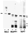
Identification of molecules containing NIP-2 and NIP-7 segments. NP40 extracts of U251 cells transfected with NG2 were treated with chondroitinase ABC (+) or left untreated (−) and then electrophoresed on 3–20% SDS-PAGE gels. After transfer to Immobilon P, samples were subjected to immunoblot analysis with rabbit antibodies against NG2, MUPP1, NIP-7, and NIP-2 (not shown). The 300 kDa NG2 core protein (arrow) becomes much more prominent following removal of chondroitin sulfate chains from the intact proteoglycan. In contrast, the 250 kDa MUPP1 band, recognized by both the MUPP1 and NIP-7 antisera (asterisk), is insensitive to chondroitinase treatment. Arrowheads at right indicate positions of 200, 116, and 92 kDa molecular weight standards.
Fig. 3
Fig. 3
Pull-down of intact NG2 by NIP-2 and NIP-7 fusion proteins. NP40 extracts of 125I-labelled U251 cells expressing either wild type NG2 (wt) or the NG2/t3 mutant (t3) were incubated with glutathione-agarose beads carrying GST fusion proteins of NIP-2, NIP-7, or lamin (as a negative control). A positive control is provided by incubation of the extract with a rabbit antibody against NG2 (αNG2) followed by Protein A-Sepharose beads. Samples were treated with chondroitinase ABC (+) or left untreated (−) and analyzed by electrophoresis on 3–20% SDS-PAGE gels. Arrow marks position of 300 kDa NG2 core protein. Arrowheads indicate positions of 200, 116, 92, and 67 kDa molecular weight standards. NIP-2 and NIP-7 are very effective binders of wild type NG2, but fail to recognize NG2/t3, which is missing the C-terminal half of the cytoplasmic domain. The antibody against NG2 recognizes both the wild type and mutant NG2 species.
Fig. 4
Fig. 4
Comparison of NG2 binding by different MUPP1 PDZ domains. A. NP40 extracts of 125I-labelled U251 cells expressing wild type NG2 were incubated with glutathione-agarose beads carrying GST alone, the NIP-7 GST fusion protein, or GST fusion proteins representing MUPP1 PDZ pairs 2/3, 10/11 and 12/13. A positive control is provided by use of rabbit anti-NG2 (αN). Washed samples were analyzed by electrophoresis on 3–20% SDS-PAGE gels. Samples marked as (+) were treated with chondroitinase ABC to facilitate visualization of the 300 kDa NG2 core protein (arrowheads). NIP-7 and PDZ 2/3 are effective in pulling down full length NG2, while PDZ 10/11 and 12/13 are comparable to the GST negative control. B. Equal quantities of glutathione-agarose beads carrying GST (b), PDZ 10/11 (c), PDZ 12/13 (d), PDZ 2/3 (e), and NIP-7 (f) were boiled in SDS-PAGE sample buffer and electrophoresed on 3–20% SDS-PAGE gels. Staining with Coomassie Blue shows that comparable amounts of protein are carried by the various types of beads. Lane (a) contains molecular weight standards of 116, 92, 67, 45, 31, and 21 kDa.
Fig. 5
Fig. 5
Co-immunoprecipitation of NG2 and MUPP1. Chondroitinase-treated NP40 extracts of NG2-transfected U251 cells were subjected to immunoprecipitation with affinity-purified rabbit antibody against NG2 (N) or with control nonimmune immunoglobulin (C). Immunoprecipitates and samples of the crude NP40 extract (X) were run on 3%–20% SDS-PAGE gels, transferred to Immobilon P, and immunoblotted with antibodies against NG2 (blot NG2) or MUPP1 (blot MUPP1). Anti-NG2 immunoprecipitates contain both NG2 (arrow at right) and MUPP1 (large arrowhead at left). Arrowheads at left indicate positions of 200, 116, and 92 kDa molecular weight standards.

Similar articles

Cited by

References

    1. Altschul S, Gish W, Miller W, Myers E, Lipman D. Basic local alignment search tool. J Mol Biol. 1990;215:403–410. - PubMed
    1. Ausubel F, Brent R, Kingston R, Moore D, Seidman J, Smith J, Struhl J, editors. Current protocols in molecular biology. New York: John Wiley and Sons, Inc; 1997.
    1. Bartel P, Chien C, Sternglanz R, Fields S. Using the two-hybrid system to detect protein-protein interactions. In: Hartley DA, editor. Cellular interaction in development: a practical approach. Oxford: IRL Press; 1993. pp. 153–179.
    1. Behm F, Smith F, Raimondi S, Pui C, Bernstein I. Human homologue of the rat chondroitin sulfate proteoglycan NG2, detected by monoclonal antibody 7.1, identifies childhood acute lymphoblastic leukemias with t(4;11)(q21;q23) or t(11;19)(q23;13) and MLL gene rearrangements. Blood. 1996;87:1134–1139. - PubMed
    1. Burg M, Grako K, Stallcup W. Expression of the NG2 proteoglycan enhances the growth and metastatic properties of melanoma cells. J Cell Physiol. 1998;177:299–312. - PubMed

Publication types

LinkOut - more resources