. 2000 Sep 1;527 Pt 2(Pt 2):203-12.
doi: 10.1111/j.1469-7793.2000.00203.x.

Differential modulation of N-type 1B and P/Q-type 1A calcium channels by different G protein subunit isoforms

Affiliations

Differential modulation of N-type 1B and P/Q-type 1A calcium channels by different G protein subunit isoforms

M I Arnot et al. J Physiol. .

Abstract

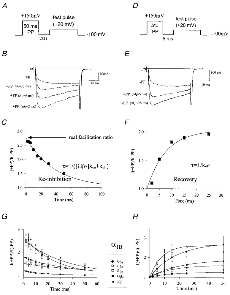
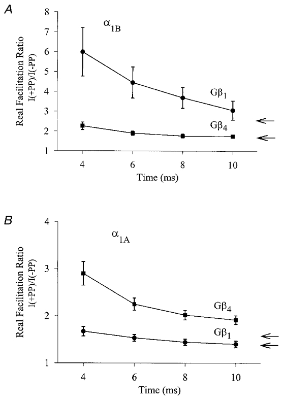
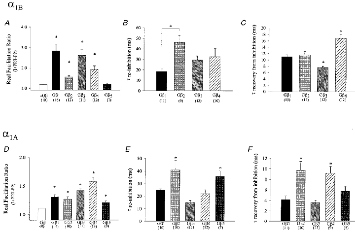
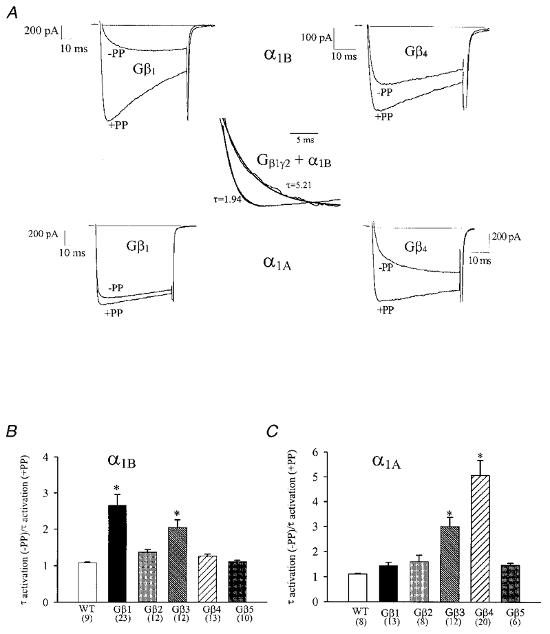
Using transient calcium phosphate transfection into the human embryonic kidney tsa-201 cell line and subsequent whole-cell patch-clamp protocols, we examined the tonic modulation of cloned N- and P/Q-type calcium channels by five different G protein beta subunits via strong depolarizing voltage prepulses. For N- and P/Q-type channels, the magnitude of inhibition was dependent on the Gbeta subtype co-expressed. Both the absolute and relative magnitudes of Gbeta subunit-induced inhibition of P/Q-type channels differed from those observed with the N-type channel. For each calcium channel subtype, kinetics of both the prepulse-mediated recovery from inhibition and the re-inhibition following the prepulse were examined for each of the Gbeta subunits by varying either the duration between the pre- and the test pulse or the length of the prepulse. For each channel subtype, we observed a differential Gbeta subunit rank order with regard to the rates of re-inhibition and recovery from inhibition. On average, P/Q-type channels exhibited more rapid rates of recovery from inhibition than those observed with N-type channels. Different Gbeta subtypes mediated different degrees of slowing of activation kinetics. The differential modulation of P/Q- and N-type channels by various Gbeta subtypes may provide a mechanism for fine tuning the amount of calcium entering the presynaptic nerve termini.


Figures

Figure 1
Figure 1. Determination of G protein inhibition kinetics with PP protocols
A, pulse protocol used to determine G protein re-inhibition kinetics following a strong depolarizing PP, with Δt1 being varied as outlined in Methods. B, representative current traces obtained from N-type channels co-expressed with Gβ3γ2 either without a PP, or in the presence of a 50 ms PP to +150 mV at variable intervals prior to the test pulse. C, dependence of PP relief on Δt1. The symbols reflect peak current levels normalized to those obtained at Δt1 = 1000 ms. The continuous line is a mono-exponential fit to the data. The time constant obtained from the fit was 38.7 ms, the y intercept (i.e. the real facilitation ratio without contamination from re-inhibition) was 2.76. D, pulse protocol used to determine the rates of recovery from G protein inhibition developing during a strong depolarizing PP. A +150 mV PP of variable duration (see Methods) preceded a test depolarization by a fixed interval of 5 ms. E, current record obtained from N-type channels co-expressed with Gβ3γ2 in the absence of a PP, or in the presence of a PP of either 5 ms or 25 ms duration. F, dependence of PP relief on the duration of the PP (Δt2). Peak currents were normalized to the levels obtained in the absence of the PP and plotted as a function of Δt2. The data were fitted mono-exponentially (continuous line), the time constant obtained from the fit was 7.6 ms. G, re-inhibition kinetics of four different types of Gβ subunits with N-type calcium channels, using the protocol described in Fig. 1A. For comparison, the data obtained in the absence of exogenous Gβγ are included. Error bars are standard errors, and data were fitted mono-exponentially. The time constants/y intercept values obtained from the fits were as follows: Gβ1, 24.9 ms/2.69; Gβ2, 66.4 ms/1.50; Gβ3, 34.8 ms/2.55; Gβ4, 42.0 ms/1.76. H, recovery from inhibition of N-type channels by the various Gβ isoforms using the pulse protocol described in Fig. 1D. The data were fitted exponentially with a single time constant. The time constants obtained from the fit were as follows: Gβ1, 17.9 ms; Gβ2, 15.0 ms; Gβ3, 8.3 ms; Gβ4, 20.61 ms. For comparison, data obtained in the absence of exogenous Gβγ are included.
Figure 4
Figure 4. Effect of kinetic slowing on the degree of G protein inhibition
Real facilitation ratios for N-type (A) and P/Q-type (B) channels in the presence of Gβ1γ2 or Gβ4γ2, obtained via isochronal measurements of current amplitude ratios I(+PP)/I(-PP) at various time points, t, into the test depolarization. In each individual experiment, the real facilitation ratio was obtained separately for each time point, t, using the protocols described in Fig. 1. Each point reflects means of 9 to 12 experiments and the error bars reflect standard errors. For comparison, the arrows represent the true facilitation ratios obtained from non-isochronal measurements of the absolute peak current levels (see Fig. 2A and D).
Figure 2
Figure 2. Calcium channel modulation by different Gβ subunit isoforms
Real facilitation ratios (A and D), mean re-inhibition time constants (B and E) and time constants for recovery from inhibition (C and F) for inhibition of N-type (top row) and P/Q-type (bottom row) calcium channels by various Gβ isoforms. The data were obtained from fits to individual experiments as described in Fig. 1. The numbers in parentheses reflect the number of experiments, error bars indicate standard errors, *Significance (P < 0.05) relative to control (A and D) or among the complete data set (B, C, E and F).
Figure 3
Figure 3. Kinetic slowing of channel kinetics by different Gβ subunit isoforms
A, current traces illustrating the kinetic slowing of N-type (top row) and P/Q-type (bottom row) channels by Gβ1γ2 and Gβ4γ2. Records shown were obtained either in the absence of a PP, or 2 ms subsequent to a 50 ms long PP to +150 mV. Inset: mono-exponential fits of the activation time course as a semi-quantitative measure of activation time constants. The records shown in the inset are from the same experiment as that in A1B/Gβ1γ2), but here the currents have been arbitrarily scaled to further illustrate the kinetic slowing. B and C, the Gβγ mediated slowing of the activation properties of (α1B) N-type (B) and (α1A) P/Q-type channel (C) is demonstrated for each Gβ subtype. The bar graphs reflect the mean change in activation time constant following the PP from 50 ms to +150 mV obtained from mono-exponential fits to the raw current data as illustrated in the inset. *Significance (P < 0.05) within the data set (ANOVA).

References

    1. Bean BP. Neurotransmitter inhibition of neuronal calcium currents by changes in channel voltage-dependence. Nature. 1989;340:153–155. - PubMed
    1. Bertram R, Behan M. Implications of G-protein-mediated Ca2+ channel inhibition for neurotransmitter release and facilitation. Journal of Computational Neuroscience. 1999;7:197–211. - PubMed
    1. Bourinet E, Soong T, Stea A, Snutch TP. Determinants of the G protein-dependent opioid modulation of neuronal calcium channels. Proceedings of the National Academy of Sciences of the USA. 1996;93:1486–1491. - PMC - PubMed
    1. Bourinet E, Soong TW, Sutton K, Slaymaker S, Mathews E, Monteil A, Zamponi GW, Nargeot J, Snutch TP. Splicing of alpha 1A subunit gene generates phenotypic variants of P- and Q-type calcium channels. Nature Neuroscience. 1999;2:407–15. - PubMed
    1. Campbell V, Berrow NS, Fitzgerald EM, Brickley K, Dolphin AC. Inhibition of the interaction of G-protein Go with calcium channels by the calcium channel β-subunit in rat neurons. The Journal of Physiology. 1995;485:365–372. - PMC - PubMed

Publication types

Substances

LinkOut - more resources