. 2000 Sep 1;19(17):4473-84.
doi: 10.1093/emboj/19.17.4473.

Bacterial SLH domain proteins are non-covalently anchored to the cell surface via a conserved mechanism involving wall polysaccharide pyruvylation

Affiliations

Bacterial SLH domain proteins are non-covalently anchored to the cell surface via a conserved mechanism involving wall polysaccharide pyruvylation

S Mesnage et al. EMBO J. .

Abstract

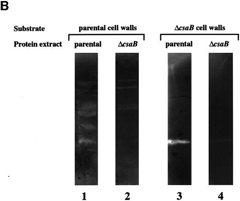
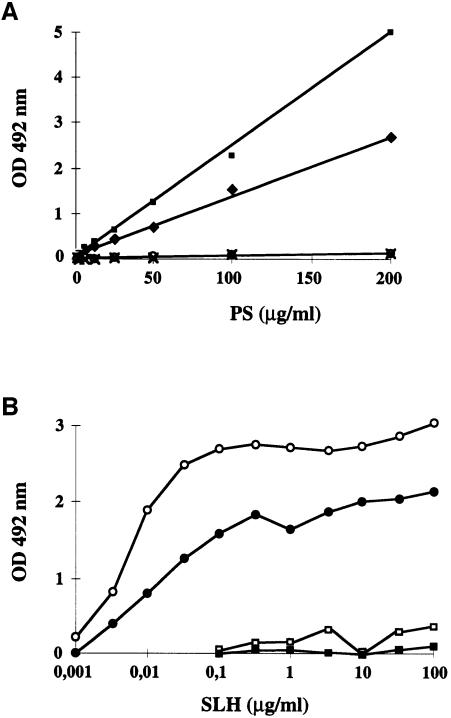
Several bacterial proteins are non-covalently anchored to the cell surface via an S-layer homology (SLH) domain. Previous studies have suggested that this cell surface display mechanism involves a non-covalent interaction between the SLH domain and peptidoglycan-associated polymers. Here we report the characterization of a two-gene operon, csaAB, for cell surface anchoring, in Bacillus anthracis. Its distal open reading frame (csaB) is required for the retention of SLH-containing proteins on the cell wall. Biochemical analysis of cell wall components showed that CsaB was involved in the addition of a pyruvyl group to a peptidoglycan-associated polysaccharide fraction, and that this modification was necessary for binding of the SLH domain. The csaAB operon is present in several bacterial species that synthesize SLH-containing proteins. This observation and the presence of pyruvate in the cell wall of the corresponding bacteria suggest that the mechanism described in this study is widespread among bacteria.


Figures

None
Fig. 1. Schematic diagram of the organization of the chromosomal region containing the B.anthracis S-layer genes sap and eag. Putative functions encoded by the ORFs flanking the S-layer genes, based on sequence comparison with protein databases, are shown.
None
Fig. 2. CsaB is necessary for the cell wall anchoring of B.anthracis S-layer proteins. (A) EA1 and (B) Sap were detected in crude extracts (CE, lanes 1, 3, 5 and 7) and TCA-precipitated supernatants (Sup, lanes 2, 4, 6 and 8) from parental (lanes 1 and 2), non-polar ΔcsaA (lanes 3 and 4), ΔcsaB (lanes 5 and 6) and ΔcsaAΔcsaB (lanes 7 and 8) strains by western blotting, using specific polyclonal sera.
None
Fig. 3. SLH-anchoring deficient mutants show aberrant morphology. (A) Sedimentation profile of liquid-grown parental (left panel) and ΔcsaB (right panel) strains. (B) Scanning electron microscopy of parental bacilli (left panel) and long twisted, septate (see arrows) filaments formed by the ΔcsaB mutant (right panel). (C) Thin sections showed that more of cell wall material accumulated in the ΔcsaB mutant (right panel) than in the parental strain (left panel), suggesting an autolysis defect. Bars represent 2.5 and 0.35 µm in (A) and (B), respectively.
None
Fig. 4. CsaB function is required for autolysis. (A) The autolytic kinetics of parental (+), non-polar ΔcsaA (diamonds), ΔcsaB (triangles) and ΔcsaAΔcsaB (circles) strains were compared. (B) The autolytic profiles of crude extracts from parental (lanes 1 and 3) and ΔcsaB (lanes 2 and 4) strains were analyzed by zymograms, with either parental (lanes 1 and 2) or ΔcsaB (lanes 3 and 4) cell walls as substrate.
None
Fig. 5. Contribution of CsaB to the biosynthesis of the peptidoglycan-associated PS fraction. The profiles from ion-exchange chromatography (DEAE–Sephadex A50 column) of parental (A) and ΔcsaB (B). Total PS extracts are presented. The arrow indicates the addition of 500 mM NaCl to elute bound material.
None
Fig. 6. A pyruvate signal present in the fraction II (the parental strain-specific PS fraction) 1H NMR spectrum (A) is absent from that of fraction I (fraction common to both PS) (B). Spectra were recorded at 500 MHz and 50°C. The pyruvate signals are indicated with an arrow.
None
Fig. 7. Expansion of the 2D 13C-1H gHBMC spectrum of the parental PS fraction II that was absent from the ΔcsaB mutant, recorded with a 60 ms delay. The 13C links with the pyruvate proton are indicated with a dashed line; C2 corresponds to the ketal carbon [C3H3–C2(O2)– C1OOH].
None
Fig. 8. In vitro binding of SLH domains to B.anthracis wall-associated PS. (A) The binding capacity of total PS from various B.anthracis strains [parental (diamonds), non-polar ΔcsaA (squares), ΔcsaB (circles) and ΔcsaAΔcsaB (crosses)] to EA1 SLH was assayed. (B) Analysis of binding dose–response of EA1 (open symbols) and Sap (filled symbols) SLH domain to purified PS fractions I (squares) and II (circles).

References

    1. Altschul S.F., Madden,T.L., Schäffer,A.A., Zhang,J., Zhang,Z., Miller,W. and Lipman,D.J. (1997) Gapped BLAST and PSI-BLAST: a new generation of protein database search programs. Nucleic Acids Res., 25, 3389–3402. - PMC - PubMed
    1. Baba T. and Schneewind,O. (1996) Target cell specificity of a bacteriocin molecule: a C-terminal signal directs lysostaphin to the cell wall of Staphylococcus aureus. EMBO J., 15, 4789–4797. - PMC - PubMed
    1. Bowditch R.D., Baumann,P. and Yousten,A.A. (1989) Cloning and sequencing of the gene encoding a 125-kilodalton surface layer protein from Bacillus sphaericus 2362 and of a related cryptic gene. J. Bacteriol., 171, 4178–4188. - PMC - PubMed
    1. Bray D.F., Bagu,J. and Koegler,P. (1993) Comparison of hexamethyldisilane (HMDS), peldri II, and critical-point drying methods for scanning electron microscopy of biological specimens. Micro. Res. Tech., 26, 489–495. - PubMed
    1. Bugert P. and Geider,K. (1995) Molecular analysis of the ams operon required for expopolysaccharide synthesis of Erwinia amylovora. Mol. Microbiol., 15, 917–933. - PubMed

Publication types