. 2000 Sep 1;19(17):4493-502.
doi: 10.1093/emboj/19.17.4493.

The CXXCXXC motif determines the folding, structure and stability of human Ero1-Lalpha

Affiliations

The CXXCXXC motif determines the folding, structure and stability of human Ero1-Lalpha

A M Benham et al. EMBO J. .

Abstract

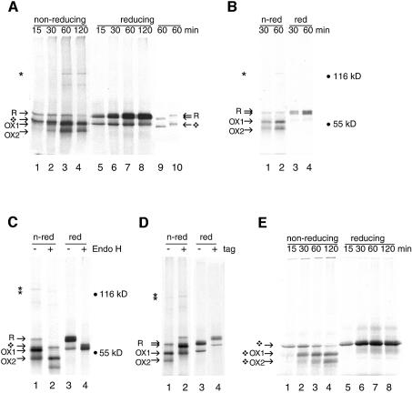
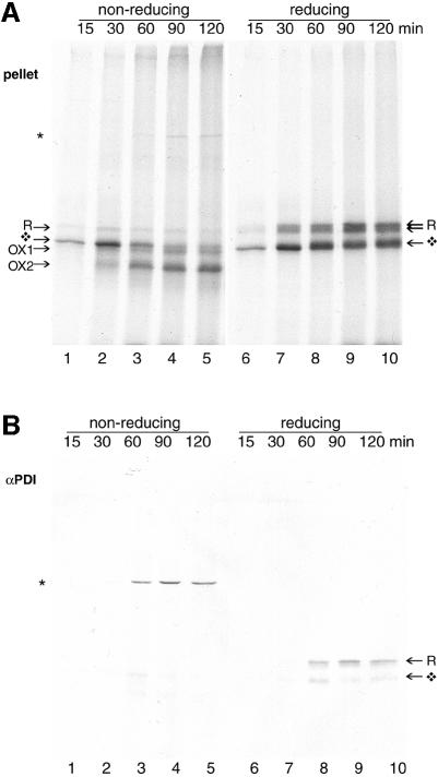
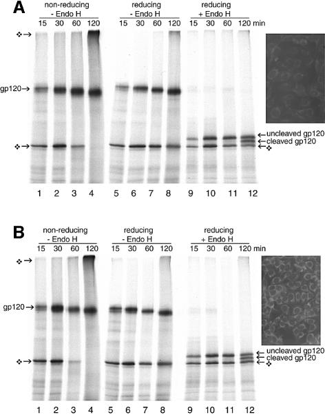
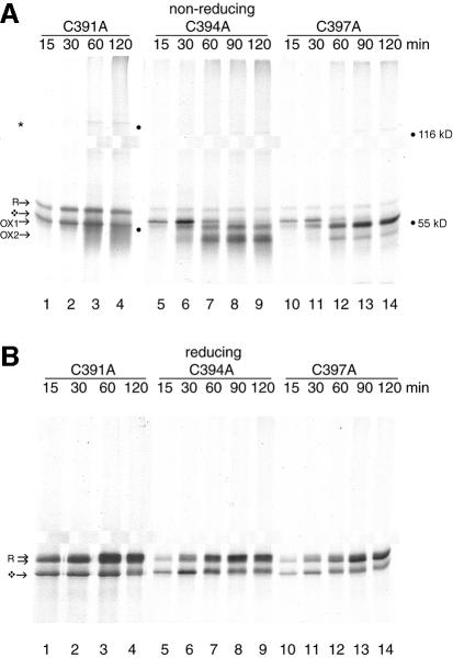
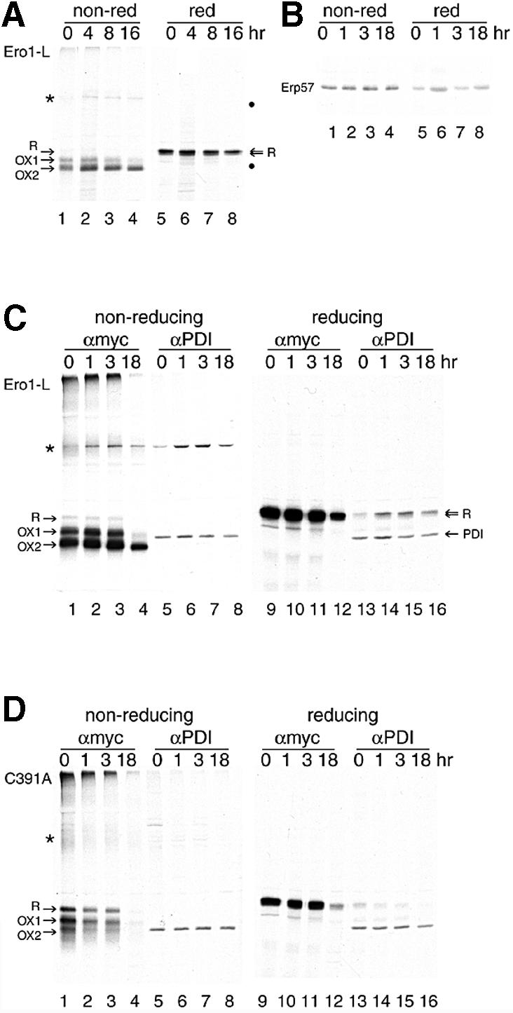
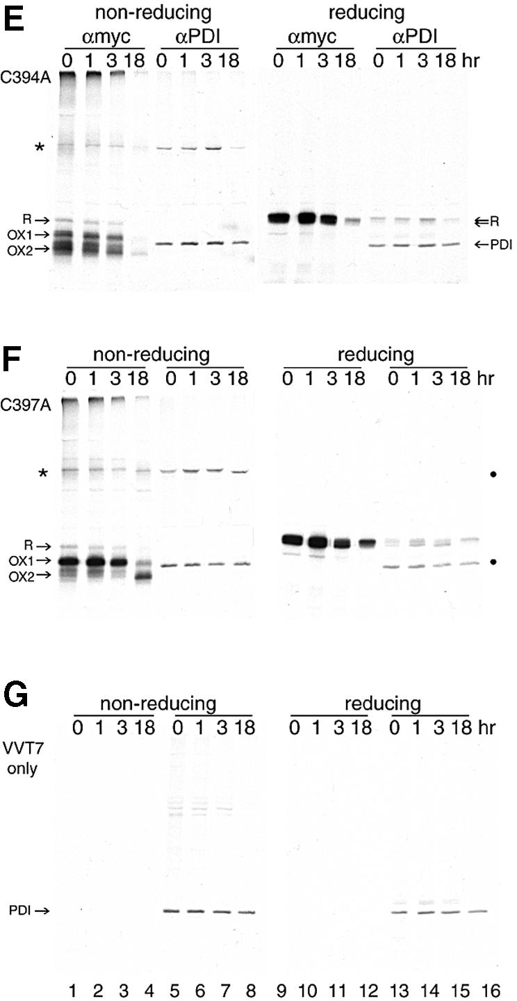
The presence of correctly formed disulfide bonds is crucial to the structure and function of proteins that are synthesized in the endoplasmic reticulum (ER). Disulfide bond formation occurs in the ER owing to the presence of several specialized catalysts and a suitable redox potential. Work in yeast has indicated that the ER resident glycoprotein Ero1p provides oxidizing equivalents to newly synthesized proteins via protein disulfide isomerase (PDI). Here we show that Ero1-Lalpha, the human homolog of Ero1p, exists as a collection of oxidized and reduced forms and covalently binds PDI. We analyzed Ero1-Lalpha cysteine mutants in the presumed active site C(391)VGCFKC(397). Our results demonstrate that this motif is important for protein folding, structural integrity, protein half-life and the stability of the Ero1-Lalpha-PDI complex.


Figures

None
Fig. 1. Folding of Ero1-Lα in semi-permeabilized cells. (A) Ero1-Lα mRNA was translated at 30°C in the presence of ∼2 × 10⁶ semi-permeabilized HT1080 cells for the times given, prior to detergent lysis of the cell pellets in sample buffer and analysis of samples by 7.5% SDS–PAGE. On a non-reducing gel (lanes 1–4), newly synthesized Ero1-Lα started as a reduced glycosylated protein R, then formed OX1 and OX2, as well as a 120 kDa complex (*). R formed a doublet on reducing gels (lanes 5–8). The diamond-shaped symbol designates non-glycosylated protein. Ero1-Lα protein was translated for 60 min and the reaction was stopped on ice in the absence of NEM. Reduced material in sample buffer was left unblocked (lane 9) or treated with excess NEM (lane 10). (B) Ero1-Lα mRNA was translated as in (A), but glycosylated proteins were isolated from the lysed ER pellets on concanavalin A–Sepharose beads, with subsequent analysis by non-reducing (lanes 1–2) and reducing (lanes 3–4) SDS–PAGE. (C) Ero1-Lα mRNA was translated for 60 min as in (A) and the pellets were either mock treated (lanes 1 and 3) or digested with Endo H (lanes 2 and 4) for 1 h at 37°C prior to non-reducing (lanes 1 and 2) or reducing (lanes 3 and 4) SDS–PAGE. The deglycosylated forms showed accelerated mobility. (D) Ero1-Lα mRNA (lanes 1 and 3) and Ero1-Lα-myc mRNA (lanes 2 and 4) were translated as in (A) for 60 min and analyzed on non-reducing (lanes 1 and 2) and reducing (lanes 3 and 4) SDS–PAGE. The tagged protein showed decreased mobility. (E) Translation of Ero1-Lα mRNA was performed in the absence of HT1080 cells. In the absence of ER membranes, folding of the non-glycosylated protein occurred (shown by diamond-shaped symbols) but complexes were not formed.
None
Fig. 2. PDI interacts specifically with Ero1-Lα. Approximately 2 × 10⁶ CHO DUKX cells were semi-permeabilized and incubated at 30°C in an in vitro translation reaction for the times indicated. Cell pellets were lysed in 1% CHAPS and the clarified lysate was split into two portions for direct analysis by SDS–PAGE (A) or immunoprecipitation with anti-PDI serum (B) prior to analysis by non-reducing (lanes 1–5) and reducing (lanes 6–10) 7.5% SDS–PAGE. Anti-PDI immunoprecipitation selectively recovered glycosylated Ero1-Lα in a complex (*).
None
Fig. 3. Ero1-Lα does not interact with HIV-1 gp120 or alter its maturation rate. Approximately 2 × 10⁶ HT1080 cells (A) and Ero1-Lα-overexpressing HT1080 cells (B) were used as a source of ER in an in vitro translation of gp120 mRNA. Aliquots at given time points were incubated without (lanes 1–8) or with (lanes 9–12) Endo H to remove N-linked glycans. By 120 min, folding and signal peptide cleavage had occurred at the same rate in both cell lines. No additional disulfide-linked complexes had formed. Reduced, non-glycosylated gp120 was also observed (diamond-shaped symbol). The inset shows the difference in ER-specific Ero1-Lα expression between the two cell lines by indirect immunofluorescence using antibody A29.
None
Fig. 4. Ero1-Lα CXXCXXC mutants have distinct folding patterns. Approximately 2 × 10⁶ HT1080 cells were used per translation to reconstitute the folding of mutants C391A (lanes 1–4), C394A (lanes 5–9) and C397A (lanes 10–14). On non-reducing gels (A), C391A ran as a smear, C394A behaved similar to wild-type protein and C397A was arrested in OX1. Each mutant formed the 120 kDa complex (*). On reducing gels (B), glycosylated (R) and non-glycosylated (diamond-shaped symbol) Ero1-Lα were recovered in each case (lanes 1–14).
None
Fig. 5. OX2 is the stable state of Ero1-Lα in the living cell. HeLa cells, grown in 6 cm dishes, were infected with VVT7 (A and C–G) and mock transfected (G) or transfected with pcDNA3.1 encoding (A and C) Ero1-Lα-myc, (D) C391A-myc, (E) C394A-myc or (F) C397A-myc. In (A), immunoprecipitation was with the anti-myc mAb 9E10 (lanes 1–4 non-reducing and lanes 5–8 reducing). In (C–G), immunoprecipitation was with either the anti-myc mAb 9E10 (lanes 1–4 non-reducing and lanes 9–12 reducing) or anti-PDI sera (lanes 5–8 non-reducing and lanes 13–16 reducing). 9E10 specifically isolated Ero1-Lα R, OX1 and OX2, along with the Ero1-Lα–PDI complex (*). Anti-PDI sera immunoprecipitated PDI in all cases, and the Ero1-Lα–PDI complex (*) from Ero1-Lα-myc, C391A-myc, C394A-myc and C397A-myc, but not the mock-transfected cells. In (B), Erp57 was immunoprecipitated from uninfected, untransfected HeLa cells after a pulse–chase and analyzed on non-reducing (lanes 1–4) and reducing (lanes 5–8) SDS–PAGE. ERp57 had no disulfide-bonded intermediates.

References

    1. Aslund F. and Beckwith,J. (1999) The thioredoxin superfamily: redundancy, specificity and gray-area genomics. J. Bacteriol., 181, 1375–1379. - PMC - PubMed
    1. Bader M., Muse,W., Ballou,D.P., Gassner,C. and Bardwell,J.C. (1999) Oxidative protein folding is driven by the electron transport system. Cell, 98, 217–227. - PubMed
    1. Bardwell J.C., McGovern,K. and Beckwith,J. (1991) Identification of a protein required for disulphide bond formation in vivo. Cell, 67, 581–589. - PubMed
    1. Beinert H. and Kiley,P.J. (1999) Fe–S proteins in sensing and regulatory functions. Curr. Opin. Chem. Biol., 3, 152–157. - PubMed
    1. Braakman I., Hoover-Litty,H., Wagner,K.R. and Helenius,A. (1991) Folding of influenza hemagglutinin in the endoplasmic reticulum. J. Cell Biol., 114, 401–411. - PMC - PubMed

Publication types