. 2000 Sep 1;19(17):4577-88.
doi: 10.1093/emboj/19.17.4577.

Localization of phosphatidylinositol 3-phosphate in yeast and mammalian cells

Affiliations

Localization of phosphatidylinositol 3-phosphate in yeast and mammalian cells

D J Gillooly et al. EMBO J. .

Abstract

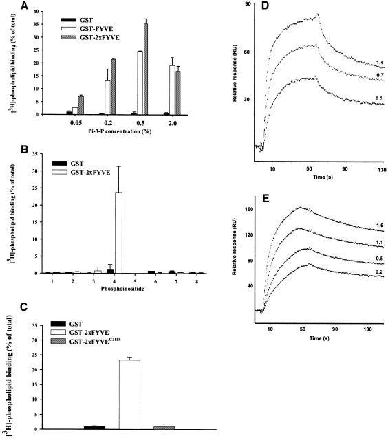
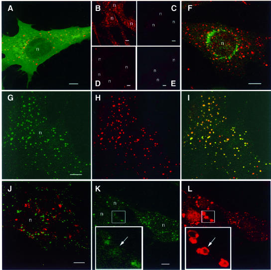
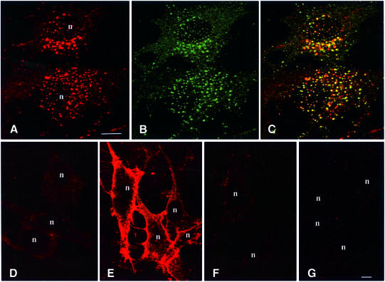
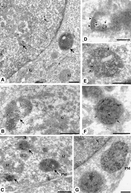
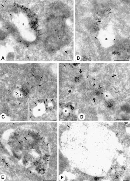
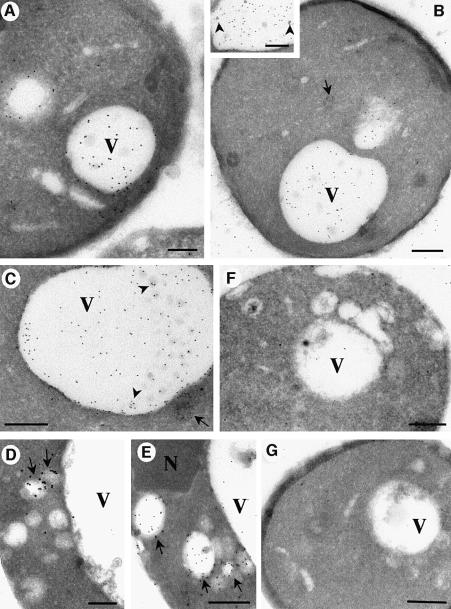
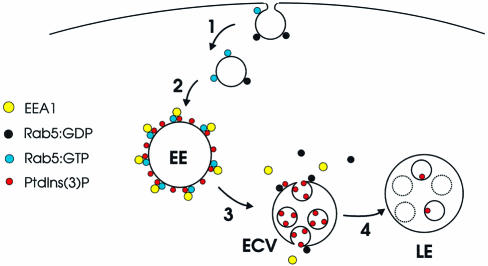
Phosphatidylinositol 3-kinase (PI3K) regulates several vital cellular processes, including signal transduction and membrane trafficking. In order to study the intracellular localization of the PI3K product, phosphatidylinositol 3-phosphate [PI(3)P], we constructed a probe consisting of two PI(3)P-binding FYVE domains. The probe was found to bind specifically, and with high affinity, to PI(3)P both in vitro and in vivo. When expressed in fibroblasts, a tagged probe localized to endosomes, as detected by fluorescence microscopy. Electron microscopy of untransfected fibroblasts showed that PI(3)P is highly enriched on early endosomes and in the internal vesicles of multivesicular endosomes. While yeast cells deficient in PI3K activity (vps15 and vps34 mutants) were not labelled, PI(3)P was found on intralumenal vesicles of endosomes and vacuoles of wild-type yeast. vps27Delta yeast cells, which have impaired endosome to vacuole trafficking, showed a decreased vacuolar labelling and increased endosome labelling. Thus PI(3)P follows a conserved intralumenal degradation pathway, and its generation, accessibility and turnover are likely to play a crucial role in defining the early endosome and the subsequent steps leading to multivesicular endosome formation.


Figures

None
Fig. 1. 2×FYVE binds specifically and with high affinity to PI(3)P. (A–C) Portions of GST alone or fused to FYVE, 2×FYVE or 2×FYVE with a double C215S mutation (Gaullier et al., 1998) were immobilized on glutathione–Sepharose beads and then incubated in the presence of 3H-labelled liposomes containing: (A) 0.05–2.0% of PI(3)P, (B) 0.2% of the inositol lipids indicated below or (C) 0.2% PI(3)P. All values are means ± SEM of multiple experiments performed in triplicate. 1, PI; 2, PI(4)P; 3, PI(5)P; 4, PI(3)P; 5, PI(3,4)P2; 6, PI(3,5)P2; 7, PI(4,5)P2; 8, PI(3,4,5)P3. (D) GST–FYVE or (E) GST–2×FYVE were injected into sensor chips that had been loaded with liposomes containing 2% PI(3)P. Sensorgrams at the indicated concentrations (in µM) of the GST fusion proteins are shown.
None
Fig. 2. Confocal fluorescence microscopy of cells expressing 2×FYVE. BHK cells were transfected with 2×FYVE constructs, then permeabilized with saponin and fixed for confocal microscopy. (A) myc-2×FYVE (red) and GFP-PLC δ1 (green). (B and C) myc-2×FYVE (red) in the absence (B) and presence (C) of 100 nm wortmannin for 30 min. (D and E) myc-2×FYVE (red) with a single (D) or double (E) C215S mutation. (F) myc-2×FYVE (red) and endogenous mannose 6-phosphate receptor (green). (G and H) myc-2×FYVE (red) and endogenous EEA1 (green). (J) GFP-2×FYVE (green) and endogenous LBPA (red). (K and L) myc-2×FYVE (red) and endogenous EEA1 (green). In the merged images (A, F, I and J), yellow colour indicates co-localization between 2×FYVE and the organelle marker molecule. The arrow in (K) and (L) points to an expanded vacuolar structure that contains a high level of 2×FYVE and a low level of EEA1. Bars, 5 µm. Nuclei are indicated with ‘n’.
None
Fig. 3. Confocal fluorescence microscopy of untransfected BHK cells with GST–2×FYVE as a probe. BHK cells on coverslips were either permeabilized by freeze–thawing (AC) or left unpermeabilized (DG). The unpermeabilized cells were incubated for 10 min at 37°C in the presence of buffer alone (D) or liposomes containing 10% PI(3)P (E), PI(3,4)P2 (F) or PI(3,4,5)P3 (G). The cells were then washed extensively prior to fixation. The coverslips were stained with biotinylated GST–2×FYVE (A and D–G) or anti-EEA1 (B) followed by Cy3–streptavidin or FITC–anti-IgG, respectively. Yellow colour in the merged image (C) indicates co-localization. Bar, 5 µm. Nuclei are indicated with ‘n’.
None
Fig. 4. Electron microscopic localization of PI(3)P using GST–2×FYVE as a probe. Ultrathin frozen sections of PFA/glutaraldehyde-fixed BHK cells were thawed and incubated with GST–2×FYVE. After washing and fixation, sections were incubated with antibodies to GST and then 10 nm protein A–gold. (A) A low-power overview demonstrating the specificity of the FYVE labelling. Specific labelling is associated with a multivesicular endosome and with a discrete region of the nucleus (arrows). Low or negligible labelling is associated with the Golgi complex (G), the nuclear envelope surrounding the nucleus (N) and mitochondria (M). (D–G) Higher magnification micrographs showing the specific labelling of early endosomes (D; recognizable by morphology and clear cytoplasmic coat, arrowheads), and multivesicular endosomes (B and C, arrows; and EG). Note that there is negligible labelling of the Golgi complex (G) and of the plasma membrane (PM). The multivesicular endosomes show labelling concentrated on the internal membranes (arrows in E and F) with low labelling of the limiting membrane. Bars (A), (C) and (G), 500 nm; (B), (D), (E) and (F), 200 nm.
None
Fig. 5. Early and late endosomal localization of PI(3)P. Sections were labelled for free PI(3)P using the 2×FYVE probe as described in the legend to Figure 4, or with antibodies to EEA1 (C, inset). BHK cells in (A–E) were incubated with 5 nm BSA–gold for 30 min at 37°C prior to fixation (Griffiths et al., 1989). (AD) Representative examples of early endosomes labelled with the 2×FYVE probe (large gold; indicated by small arrows in all panels) and internalized 5 nm BSA–gold. Typical tubular vesicular endosomes and ring-shaped early endosomes with an electron-lucent central region (asterisk) surrounded by a double membraned cisterna are evident. Labelling is associated with the multivesicular region (B and C), the cytoplasmic face of the cisternal region (C and D, inset) and with the inner cisternal membrane (see arrowheads indicating inner membrane in C). Labelling is also associated with BSA–gold-labelled tubules (see D; large arrows indicate peripheral BSA–gold-labelled tubules). Note the multivesicular endosome (putative endosome carrier vesicle) containing no internalized gold which is heavily labelled with the 2×FYVE probe (A, large arrow). EEA1 labelling (C, inset, arrows) is associated predominantly with the cytoplasmic face of the BSA–gold-labelled early endosome. (E and F) Late endosomes. (E) A BSA–gold-labelled late endosome showing labelling throughout the multivesicular structure. A heavily labelled small vesicle is also evident (large arrow). (F) Sections of control BHK cells were labelled with specific antibodies to LBPA (10 nm gold) and with the 2×FYVE probe (15 nm gold). Labelling for LBPA is concentrated in the electron-lucent region of the large late endosome. In contrast, the 2×FYVE labelling (arrows) is concentrated in regions showing lower LBPA labelling and more obvious membrane profiles. Bars, main panels, 200 nm; insets, 100 nm.
None
Fig. 6. Electron microscopic localization of PI(3)P in wild-type and mutant yeast cells. Saccharomyces cerevisae strains were fixed and processed for frozen sectioning. The Figure shows representative examples of wild-type (WT) cells (AC), vps27Δ (D and E), vps34 (F) and vps15 (G). Sections were thawed and incubated with GST–2×FYVE. After washing and fixation, sections were incubated with antibodies to GST and then 10 nm protein A–gold. (A and B) Overviews demonstrating the specificity of the 2×FYVE labelling. Specific labelling is associated with the vacuole (V). Low or negligible labelling is associated with the plasma membrane and other organelles. The inset and (C) show higher magnification views of the vacuole in which some labelling is seen associated with small internal vesicles (arrowheads). Labelling is also associated with cytoplasmic vesicles, some of which appear multivesicular (C, arrow). In contrast to the WT yeast, the vps34 and vps15 strains show very low labelling with the 2×FYVE probe (F and G), showing the specificity of the probe. As compared with wild-type strains, vps27Δ cells show a marked accumulation of labelling in cytoplasmic vesicles (D and E, arrows) and on average a decreased vacuolar labelling (see Table I for quantitation). Note that there is some labelling between the cell profiles of the wild-type cells (A and B) which was not observed in the vps34 and vps15 strains and therefore presumably arises from broken cells or diffusion of the lipid. N, nucleus. Bars (A) and (D), 200 nm; (B), (C) and (E–G), 500 nm.
None
Fig. 7. Model for the role of PI(3)P in endocytic traffic. (1) The endocytic vesicle, which is devoid of EEA1 (Christoforidis et al., 1999), contains an activity that converts Rab5:GDP into Rab5:GTP (Horiuchi et al., 1995). (2) The vesicle fuses with the early endosome (EE) in a process regulated by EEA1 and other proteins (Simonsen et al., 1998b; McBride et al., 1999). The limiting membrane of the EE is rich in PI(3)P, caused by Rab5:GTP-mediated PI 3-kinase recruitment (Christoforidis et al., 1999), and in EEA1, which binds to the EE via interaction with both PI(3)P and Rab5:GTP (Simonsen et al., 1998b). (3) On the ECV, GTP hydrolysis by Rab5 is accompanied by the dissociation of EEA1 and Rab5:GDP, and PI(3)P is internalized into intralumenal vesicles. (4) The ECV fuses with (or matures into) a late endosome (LE), in which PI(3)P is metabolized.

References

    1. Boronenkov I.V., Loijens,J.C., Umeda,M. and Anderson,R.A. (1998) Phosphoinositide signaling pathways in nuclei are associated with nuclear speckles containing pre-mRNA processing factors. Mol. Biol. Cell, 9, 3547–3560. - PMC - PubMed
    1. Burd C.G. and Emr,S.D. (1998) Phosphatidylinositol(3)-phosphate signaling mediated by specific binding to RING FYVE domains. Mol. Cell, 2, 157–162. - PubMed
    1. Christoforidis S., Miaczynska,M., Ashman,K., Wilm,M., Zhao,L., Yip,S.-C., Waterfield,M.D., Backer,J.M. and Zerial,M. (1999) Phosphatidylinositol-3-OH kinases are Rab5 effectors. Nature Cell Biol., 1, 249–252. - PubMed
    1. Corvera S., D’Arrigo,A. and Stenmark,H. (1999) Phosphoinositides in membrane traffic. Curr. Opin. Cell Biol., 11, 460–465. - PubMed
    1. Evan G.I., Lewis,G.K., Ramsay,G. and Bishop,J.M. (1985) Isolation of monoclonal antibodies specific for human c-myc proto-oncogene product. Mol. Cell. Biol., 5, 3610–3616. - PMC - PubMed

Publication types

Substances