. 2000 Sep 1;19(17):4759-73.
doi: 10.1093/emboj/19.17.4759.

Regulation of the 26S proteasome by adenovirus E1A

Affiliations

Regulation of the 26S proteasome by adenovirus E1A

A S Turnell et al. EMBO J. .

Abstract

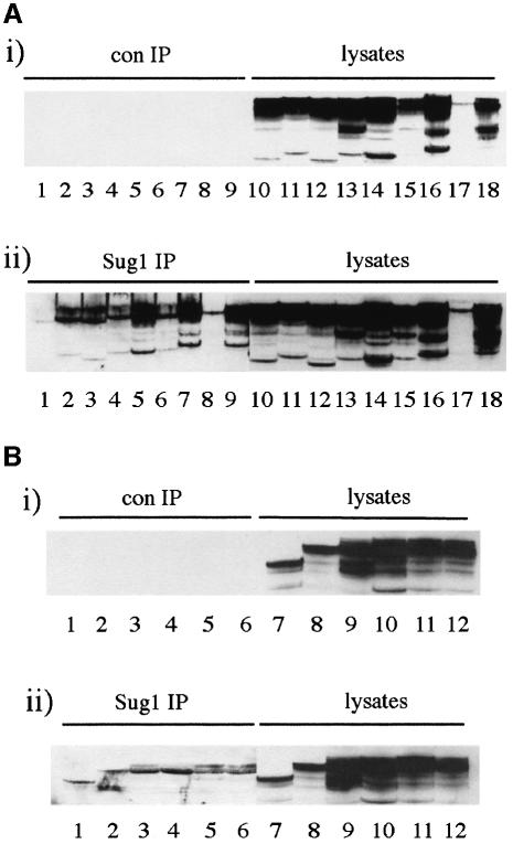
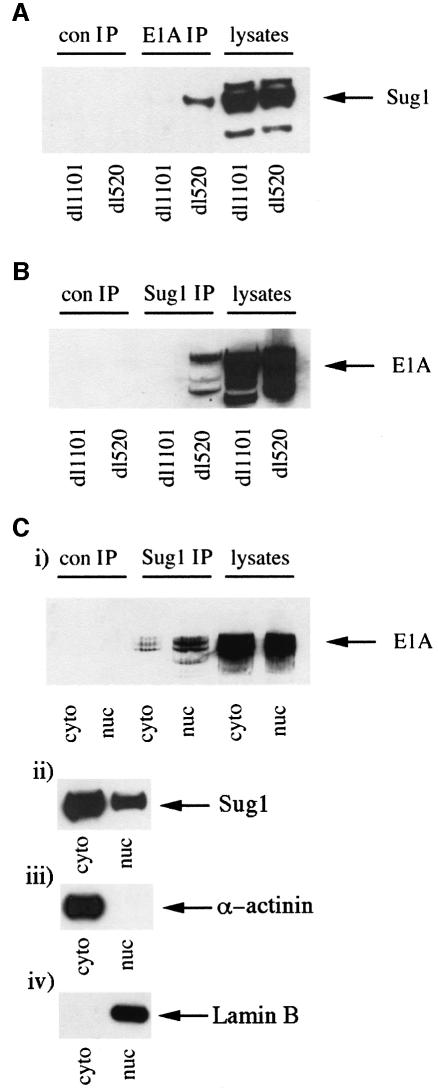
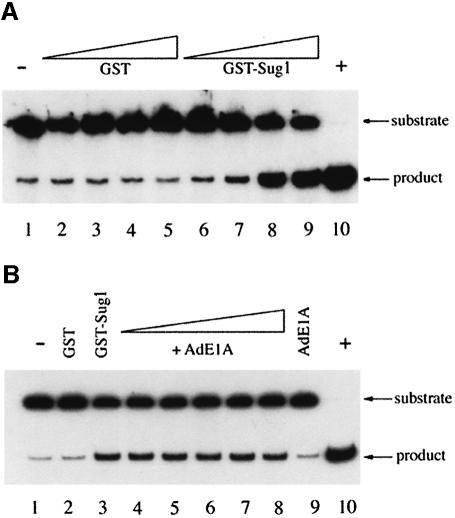
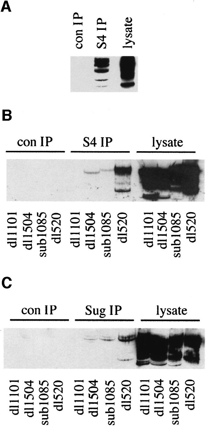
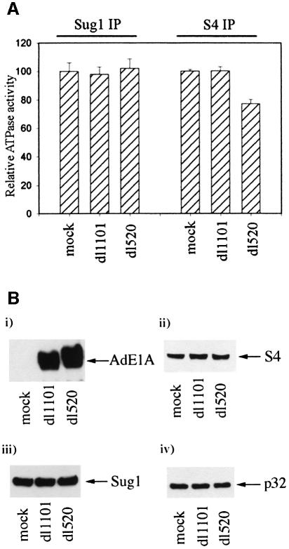
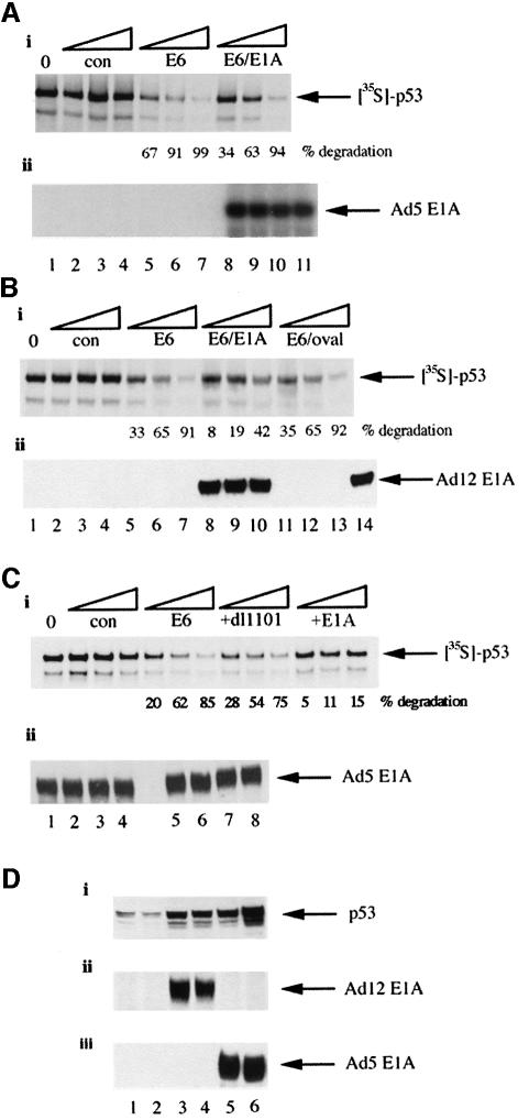
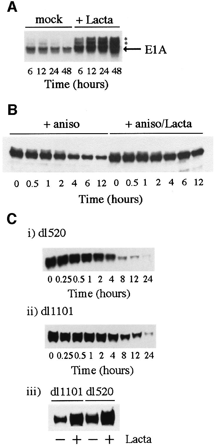
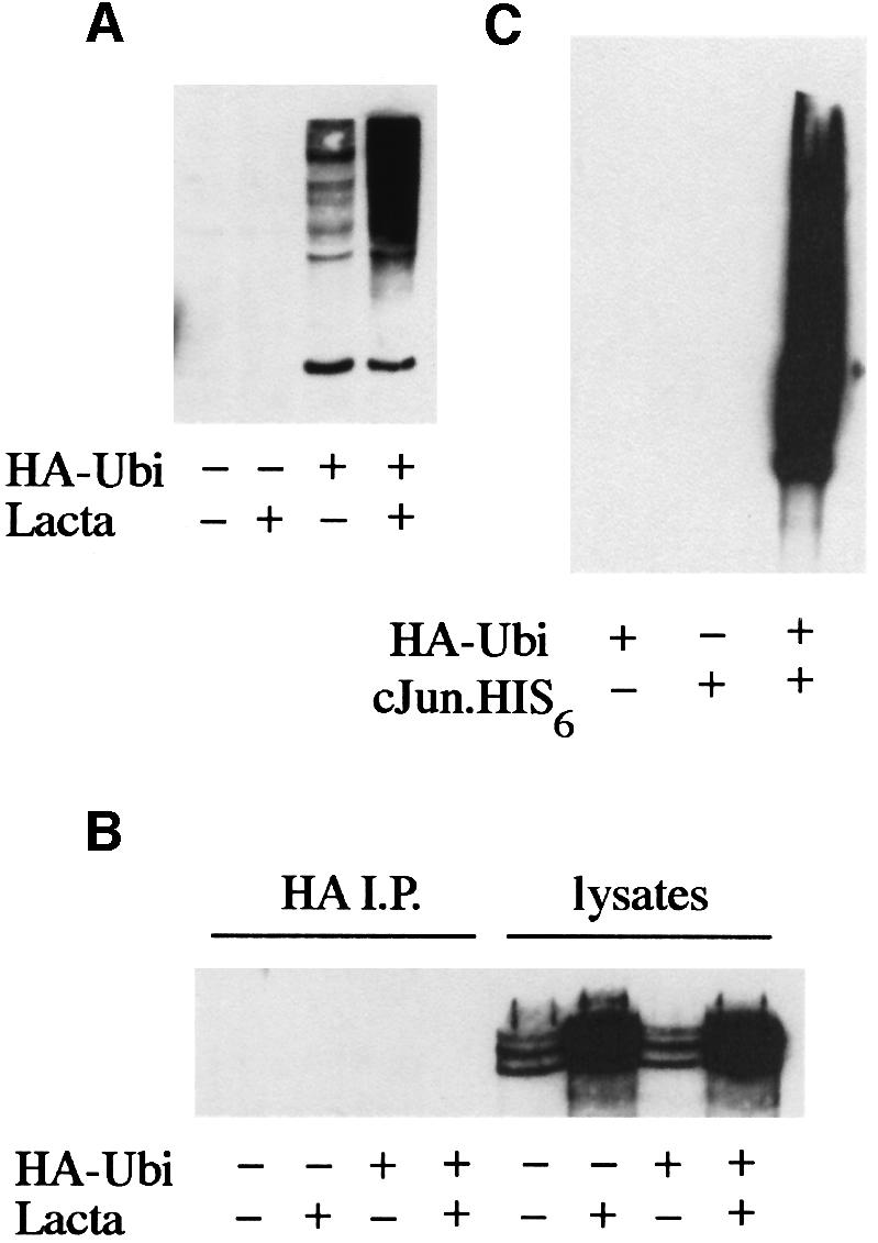
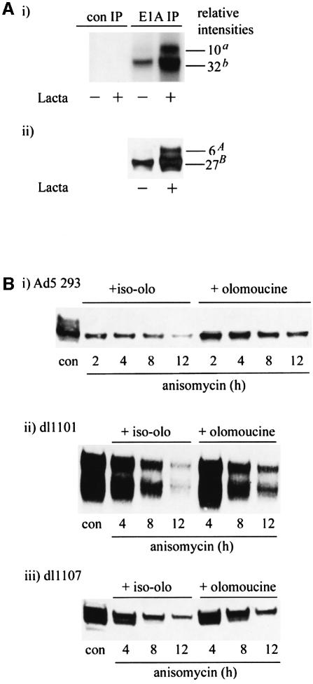
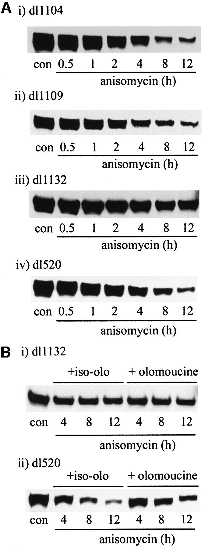
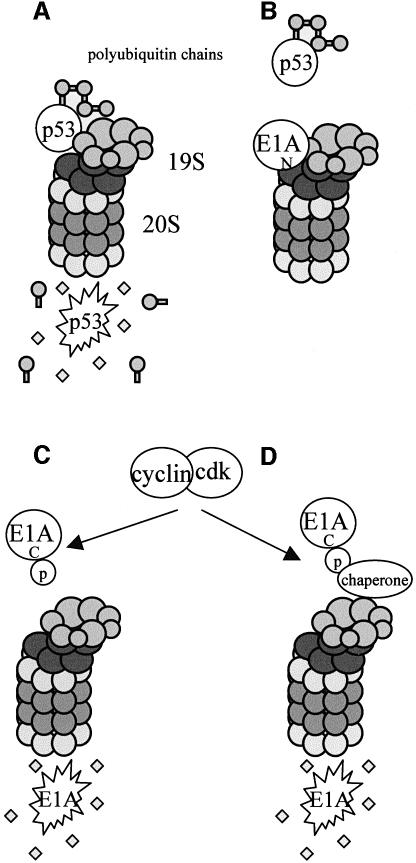
We have identified the N-terminus of adenovirus early region 1A (AdE1A) as a region that can regulate the 26S proteasome. Specifically, in vitro and in vivo co-precipitation studies have revealed that the 19S regulatory components of the proteasome, Sug1 (S8) and S4, bind through amino acids (aa) 4-25 of Ad5 E1A. In vivo expression of wild-type (wt) AdE1A, in contrast to the N-terminal AdE1A mutant that does not bind the proteasome, reduces ATPase activity associated with anti-S4 immunoprecipitates relative to mock-infected cells. This reduction in ATPase activity correlates positively with the ability of wt AdE1A, but not the N-terminal deletion mutant, to significantly reduce the ability of HPV16 E6 to target p53 for ubiquitin-mediated proteasomal degradation. AdE1A/proteasomal complexes are present in both the cytoplasm and the nucleus, suggesting that AdE1A interferes with both nuclear and cytoplasmic proteasomal degradation. We have also demonstrated that wt AdE1A and the N-terminal AdE1A deletion mutant are substrates for proteasomal-mediated degradation. AdE1A degradation is not, however, mediated through ubiquitylation, but is regulated through phosphorylation of residues within a C-terminal PEST region (aa 224-238).


Figures

None
Fig. 1. Mapping the binding site on AdE1A for Sug1. (A) Human A549 cells were infected with either dl1101, dl1102, dl1103, dl1141, dl1105, dl1142, dl1108, dl1109 or dl520 (lanes 1–9 and 10–18, respectively) at a multiplicity of 50 p.f.u. per cell. At 24 h post-infection, cell lysates were mock immunoprecipitated with pre-immune rabbit serum (i), or Sug1 was immunoprecipitated from infected cell lysates with an anti-Sug1 pAb (ii). Immunoprecipitates were separated on alkaline urea gels and AdE1A detected using M58 mAb. (B) Human A549 cells were infected with either dl177-9, dl1135, RG2, YH47, hr3 or wild-type Ad5 (lanes 1–6 and 7–12, respectively) at a multiplicity of 50 p.f.u. per cell. At 24 h post-infection, cell lysates were mock immunoprecipitated with pre-immune rabbit serum (i) or Sug1 was immunoprecipitated from infected cell lysates with an anti-Sug1 polyclonal antibody (ii). Immunoprecipitates were separated on alkaline urea gels and AdE1A detected using M58 mAb.
None
Fig. 2. The N-terminus of AdE1A binds to Sug1. Human A549 cells were infected with either dl1101 or dl520 at a multiplicity of 50 p.f.u. per cell. At 24 h post-infection, AdE1A was immunoprecipitated from infected cell lysates using M73 mAb. Immunoprecipitated protein complexes were separated by SDS–PAGE; Sug1 was identified by western blotting, using a rabbit anti-Sug1 pAb, (A). Sug1 was immunoprecipitated similarly from infected cell lysates 24 h post-infection, using a rabbit anti-Sug1 polyclonal antibody. Immunoprecipitates were separated on alkaline urea gels; AdE1A was identified by western blotting, using M73 mAb, (B). Human Ad5 293 cells were fractionated as described in Materials and methods. Sug1 was immunoprecipitated from cytoplasmic and nuclear cell equivalents; immunoprecipitates were separated on alkaline urea gels and AdE1A detected using M73 mAb, (C). The partitioning of AdE1A (i), Sug1 (ii), α-actinin (iii) and lamin B (iv) between the cytoplasm and nucleus by western blotting was also performed.
None
Fig. 3. AdE1A does not affect Sug1-associated DNA helicase activity in vitro. (A) DNA helicase activity associated with purified GST–Sug1 preparations. Lane 1, negative control (substrate alone); lanes 2–5, 1, 10, 100 and 200 ng of GST; lanes 6–9, 1, 10, 100 and 200 ng of GST–Sug1; lane 10, positive control (substrate heated to 95°C for 5 min). (B) GST–Sug1 DNA helicase activity assayed in the presence of purified Ad12 13S E1A. Lane 1, negative control; lane 2, 100 ng of GST; lane 3, 100 ng GST–Sug1; lanes 4–8, 100 ng GST–Sug1 with either 1, 10, 100, 200 or 1000 ng of Ad12 13S E1A; lane 9, 1000 ng of Ad12 13S E1A alone; lane 10, positive control.
None
Fig. 4. The N-terminus of AdE1A binds to the 19S proteasome. (A) S4 was immunoprecipitated from human Ad5 293 cells, and protein complexes separated by alkaline urea gels. AdE1A was detected using M73 mAb. (B and C) Human A549 cells were infected with either dl1101, dl1504, sub1085 or dl520 at a multiplicity of 50 p.f.u. per cell. At 24 h post-infection, S4 (B) or Sug1 (C) was immunoprecipitated from infected cell lysates, and protein complexes separated on alkaline urea gels; AdE1A was detected using M73 mAb. (DF) GST pull-downs separated by SDS–PAGE and subjected to fluorography, demonstrating in vitro binding capabilities between GST–Sug1 and 35S-labelled AdE1A mutants (D), GST–AdE1A mutants and 35S-labelled S4 (E), and 35S-labelled S5b and GST–AdE1A (F).
None
Fig. 5. AdE1A reduces ATPase activity associated with S4 immunoprecipitates, but does not affect activity associated with Sug1 immunoprecipitates. Human A549 cells were infected with either dl1101 or dl520 at a multiplicity of 50 p.f.u. per cell. At 24 h post-infection, S4 and Sug1 were immunoprecipitated from infected cells, and ATPase activity assayed as described in Materials and methods. (A) Relative ATPase activities associated with Sug1 immunoprecipitates: column 1, mock-infected cells; column 2, dl1101-infected cells; column 3, dl520-infected cells. Relative ATPase activities associated with S4 immunoprecipitates: column 4, mock-infected cells; column 5, dl1101-infected cells; column 6, dl520-infected cells; 100% activity corresponds to cellular ATPase activity associated with Sug1 and S4, respectively, from mock-treated cells. Error bars represent the mean ± SD from three independent experiments (= 9). (B) AdE1A expression does not alter expression of 26S proteasomal components. Aliquots taken from either mock-infected, dl1101-infected or dl520-infected cells were separated by SDS–PAGE and western blotted for (i) AdE1A, (ii) S4, (iii) Sug1 and (iv) p32.
None
Fig. 6. AdE1A reduces the ability of HPV16/18 E6 to target p53 for degradation. (A) (i) In vitro degradation of 35S-labelled p53 was performed as described in Materials and methods. Samples were taken at the appropriate times during the assay, separated by SDS–PAGE and subjected to fluorography to identify remaining p53: lane 1, control; lanes 2–4, no addition; lanes 5–7, addition of HPV16 E6; lanes 8–10, addition of HPV16 E6 + Ad5 E1A. Samples were taken 30, 60 and 120 min after the addition of unprogrammed lysate or HPV16 E6. (ii) As (i), but unlabelled p53 and [35S]Ad5E1A were used instead. SDS–PAGE and fluorography identify AdE1A. Lane 11 corresponds to input levels of Ad5E1A. (B) (i) As (A) (i) except that bacterially expressed Ad12 E1A was used (lanes 8–10) instead. Lanes 11–13, addition of HPV16 E6 + ovalbumin. Samples were taken 15, 30 and 60 min after the addition of unprogrammed lysate or HPV16 E6. (ii) Aliquots were taken and blotted for Ad12 E1A. Lane 14 corresponds to input levels of Ad12 E1A. (C) (i) As (A) (i) except that Ad5 dl1101-13S E1A (lanes 8–10) and Ad5 13S E1A (lanes 11–13) were used instead. (ii) Western blot indicating no degradation of Ad5 dl1101-13S E1A or Ad5 13S E1A during the time course of the experiment. Data shown in (A–C) are representative of several independent experiments. (D) AdE1A expression stabilizes p53 in HeLa cells. HeLa cells were either mock infected or infected with Ad12 dl620 or Ad5 dl1520 at a multiplicity of 50 p.f.u. per cell. At 24 and 48 h post-infection, cells were harvested and proteins were separated by SDS–PAGE and blotted for (i) p53, (ii) Ad12E1A or (iii) Ad5E1A. Lanes 1 and 2, mock-infected cells; lanes 3 and 4, Ad12 dl620-infected cells; lanes 5 and 6, Ad5 dl1520-infected cells.
None
Fig. 7. Factors governing AdE1A stability. (A) Stabilization of AdE1A. Human Ad5 293 cells were either mock treated or treated in the presence of 10 µM lactacystin for the times indicated. Proteins were separated by SDS–PAGE and western blotted for AdE1A using M73 mAb. The asterisk denotes slower migrating, stabilized forms of AdE1A. (B) Half-life of AdE1A in Ad5 293 cells in the absence or presence of lactacystin. Cells were treated with the protein synthesis inhibitor anisomycin (100 µM) in the absence or presence of lactacystin (10 µM). Proteins were separated by SDS–PAGE and western blotted for AdE1A using M73 mAb. (C) The N-terminus of AdE1A affects stability. Human A549 cells were infected with either dl1101 or dl520. At 12 h post-infection, cells were treated with anisomycin to gauge the half-life of different AdE1A species. Proteins were separated by SDS–PAGE and western blotted for AdE1A in Ad5 dl1101-infected cells (i), or Ad5 dl520-infected cells (ii), using M73 mAb. A549 cells were transfected with Ad5 dl1101 and dl520 AdE1A. At 12 h post-infection, cells were treated with 10 µM lactacystin for 12 h. A western blot is presented showing the levels of the respective AdE1A species in mock-treated and lactacystin-treated cells (iii), using M73 mAb.
None
Fig. 8. AdE1A is not a substrate for ubiquitylation. (A) Ad5 293 cells were transfected with 2 µg of an HA-tagged ubiquitin construct (HA-Ubi). At 24 h post-infection, cells were then either mock treated or treated in the presence of lactacystin for an additional 24 h. (A) Immunodetection of HA-Ubi conjugates using an anti-HA mAb (3F10). (B) HA-Ubi conjugates were immunoprecipitated from cell lysates, separated by alkaline urea gels and immunoblotted for AdE1A. No AdE1A was co-precipitated with HA-Ubi. (C) Ad5 293 cells were transfected with 2 µg of HA-Ubi alone (lane 1), 2 µg of cJun-His6 alone (lane 2) or co-transfected with 2 µg each of HA-Ubi and cJun-His6 (lane 3). At 48 h post-transfection, cJun-His6 conjugates were purified, separated by SDS–PAGE and immunoblotted for HA (using 3F10).
None
Fig. 9. (A) The minor higher molecular weight AdE1A species is hyperphosphorylated compared with the major lower molecular weight species. Ad5 293 cells were labelled with 100 µCi/ml [32P]orthophosphate for 18 h, in the absence or continued presence of lactacystin (10 µM). AdE1A species were immunoprecipitated from cell lysates, and separated by SDS–PAGE. (A) (i) An autoradiogram of the dried gel, indicating stabilized AdE1A species in the presence of lactacystin; (ii) a corresponding western blot for AdE1A using M73 mAB. (B) (i) Stabilization of AdE1A in Ad5 293 cells by the cdk inhibitor, olomoucine. Ad5 293 cells were treated for 1 h with 100 µM iso-olomoucine or 100 µM olomoucine, prior to treatment with 100 µM anisomycin. Western blot showing the respective levels of AdE1A after the indicated times of treatment, using mAb M73. (ii) Ad5 dl1101-13S and (iii) Ad5 dl1107 are stabilized by olomoucine treatment. A549 cells were transfected with the appropriate AdE1A construct. At 24 h post-infection, cells were pre-treated with 100 µM iso-olomoucine or olomoucine for 1 h prior to treatment with 100 µM anisomycin. Western blots are presented showing the effects of olomoucine on the half-life of different AdE1A species after the indicated times of treatment.
None
Fig. 10. (A) Role of PEST sequences in AdE1A degradation. A549 cells were infected with 50 p.f.u. per cell with different AdE1A deletion mutants. At 24 h post-infection, cells were treated with 100 µM anisomycin. Western blots are presented showing the effects of anisomycin on the levels of the respective AdE1A species, using mAb M73. AdE1A levels from dl1104 (i), dl1109 (ii), dl1132 (iii) and dl520 (iv) are shown. (B) Olomoucine treatment stabilizes the wild-type AdE1A species, dl520 (ii), but does not stabilize AdE1A from dl1132 (i). A549 cells were infected with 50 p.f.u. per cell with different AdE1A deletion mutants. At 24 h post-infection, cells were treated with 100 µM iso-olomoucine or olomoucine for 1 h, prior to treatment with 100 µM anisomycin. A western blot demonstrating the effects of olomoucine on the stability of the different AdE1A species is presented.
None
Fig. 11. Figure depicting the relationship between AdE1A and the proteasome. (A) Under normal circumstances, polyubiquitin modification of p53 targets p53 for degradation by the 26S proteasome. (B) The N-terminus of AdE1A inhibits 26S proteasomal-mediated degradation of p53, through binding to and inhibiting the 19S RC. (C and D) Ubiquitin-independent degradation of AdE1A through the 26S proteasome is dependent upon phosphorylation of a C-terminal PEST sequence by an olomoucine-sensitive kinase. Whether AdE1A utilizes a chaperone-like molecule (similar to ODC) as depicted in (D), is currently unknown.

References

    1. Ait Si Ali S. et al. (1998) Histone acetyltransferase activity of CBP is controlled by cycle-dependent kinases and oncoprotein E1A. Nature, 396, 184–186. - PubMed
    1. Asano K., Vornlocher, H.P., Richter-Cook,N.J., Merrick,W.C., Hinnebusch,A.G. and Hershey,J.W. (1997) Structure of cDNAs encoding human eukaryotic initiation factor 3 subunits. Possible roles in RNA binding and macromolecular assembly. J. Biol. Chem., 272, 27042–27052. - PubMed
    1. Bannister A.J. and Kouzarides,T. (1996) The CBP co-activator is a histone acetyltransferase. Nature, 384, 641–643. - PubMed
    1. Baumeister W., Walz,J., Zuhl,F. and Seemuller,E. (1998) The proteasome: paradigm of a self-compartmentalizing protease. Cell, 92, 367–380. - PubMed
    1. Bayley S.T. and Mymryk,J.S. (1994) Adenovirus E1A proteins and transformation. Int. J. Oncol., 5, 425–444. - PubMed

Publication types

MeSH terms

LinkOut - more resources