

Triamcinolone acetonide modulates permeability and intercellular adhesion molecule-1 (ICAM-1) expression of the ECV304 cell line: implications for macular degeneration
- PMID: 10971511
- PMCID: PMC1905725
- DOI: 10.1046/j.1365-2249.2000.01316.x
Triamcinolone acetonide modulates permeability and intercellular adhesion molecule-1 (ICAM-1) expression of the ECV304 cell line: implications for macular degeneration
Abstract
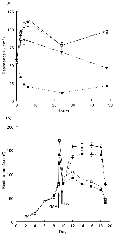
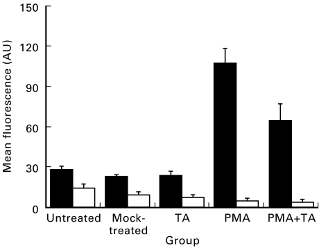
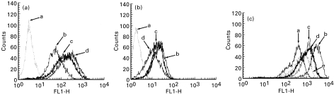
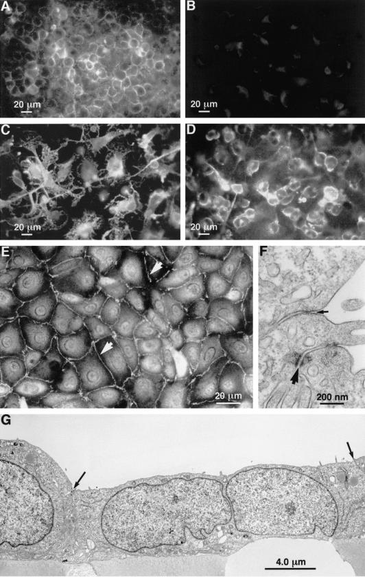
Whilst animal studies and a pilot clinical trial suggest that intravitreal triamcinolone acetonide (TA) may be useful in the treatment of age-related macular degeneration (AMD), its mode of action remains to be fully elucidated. The present study has investigated the capacity of TA to modulate the expression of adhesion molecules and permeability using a human epithelial cell line (ECV304) as a model of the outer blood-retinal barrier (BRB). The influence of TA on the expression of ICAM-1 and MHC-I was studied on resting and phorbol myristate acetate (PMA)- or interferon-gamma (IFN-gamma)- and/or tumour necrosis factor-alpha (TNF-alpha)-activated cells using flow cytometry and immunocytochemistry. Additionally, ECV304 cells were grown to confluence in uncoated Transwell chambers; transepithelial resistance (TER) across resting and PMA-activated cells was monitored. TA significantly decreased the paracellular permeability of ECV304 cells and down-regulated ICAM-1 expression, consistent with immunocytochemical observations. PMA-induced permeability changes were dose-dependent and TA decreased permeability of both resting and PMA-activated monolayers. MHC-I expression by ECV304 cells however, was not significantly affected by TA treatment. The modulation of TER and ICAM-1 expression in vitro correlate with clinical observations, suggesting re-establishment of the BRB and down-regulation of inflammatory markers are the principal effects of intravitreal TA in vivo. The results further indicate that TA has the potential to influence cellular permeability, including the barrier function of the retinal pigment epithelium (RPE) in AMD-affected retinae.
Figures
Similar articles
-
Modulation of permeability and adhesion molecule expression by human choroidal endothelial cells.Invest Ophthalmol Vis Sci. 2002 Sep;43(9):3125-30. Invest Ophthalmol Vis Sci. 2002. PMID: 12202538
-
Role of LFA-1, ICAM-1, VLA-4 and VCAM-1 in lymphocyte migration across retinal pigment epithelial monolayers in vitro.Immunology. 1996 Jul;88(3):456-62. doi: 10.1046/j.1365-2567.1996.d01-666.x. Immunology. 1996. PMID: 8774365 Free PMC article.
-
Triamcinolone Acetonide Suppresses Inflammation and Facilitates Vascular Barrier Function in Human Retinal Microvascular Endothelial Cells.Curr Neurovasc Res. 2017;14(3):232-241. doi: 10.2174/1567202614666170619081929. Curr Neurovasc Res. 2017. PMID: 28625129
-
Early effects of intravitreal triamcinolone acetonide on inflammation and proliferation in human choroidal neovascularization.Arch Ophthalmol. 2009 Mar;127(3):275-81. doi: 10.1001/archophthalmol.2008.602. Arch Ophthalmol. 2009. PMID: 19273790
-
[Intravitreal triamcinolone acetonide for the treatment of intraocular edematous and neovascular diseases].Ophthalmologe. 2004 Feb;101(2):113-20. doi: 10.1007/s00347-003-0982-0. Ophthalmologe. 2004. PMID: 14991306 Review. German.
Cited by
-
Intravitreal triamcinolone acetonide for exudative age related macular degeneration.Br J Ophthalmol. 2003 Apr;87(4):462-8. doi: 10.1136/bjo.87.4.462. Br J Ophthalmol. 2003. PMID: 12642311 Free PMC article.
-
Remodeling of the vascular channels in retinal angiomatous proliferations treated with intravitreal triamcinolone acetonide and photodynamic therapy.Graefes Arch Clin Exp Ophthalmol. 2006 Nov;244(11):1528-33. doi: 10.1007/s00417-006-0311-9. Epub 2006 Apr 12. Graefes Arch Clin Exp Ophthalmol. 2006. PMID: 16609904
-
Manufacturing of Dexamethasone-Poly(d,l-Lactide-co-Glycolide) Implants Using Hot-Melt Extrusion: Within- and Between-Batch Product Performance Comparisons.J Ocul Pharmacol Ther. 2020 Jun;36(5):290-297. doi: 10.1089/jop.2019.0074. Epub 2020 Apr 24. J Ocul Pharmacol Ther. 2020. PMID: 32330403 Free PMC article.
-
Extracellular Vesicles and MicroRNA: Putative Role in Diagnosis and Treatment of Diabetic Retinopathy.Antioxidants (Basel). 2020 Aug 4;9(8):705. doi: 10.3390/antiox9080705. Antioxidants (Basel). 2020. PMID: 32759750 Free PMC article. Review.
-
Retinal sensitivity improvement after intravitreal triamcinolone acetonide injection for macular edema secondary to branch retinal vein occlusion.Indian J Ophthalmol. 2013 Jan-Feb;61(1):3-7. doi: 10.4103/0301-4738.105048. Indian J Ophthalmol. 2013. PMID: 23275213 Free PMC article. Clinical Trial.
References
-
- Penfold PL, Provis JM, Billson FA. Age-related macular degeneration: ultrastructural studies of the relationship of leucocytes to angiogenesis. Graefes Arch Clin Exp Ophthalmol. 1987;225:70–76. - PubMed
-
- Lopez PF, Grossniklaus HE, Lambert HM, et al. Pathologic features of surgically excised subretinal neovascular membranes in age-related macular degeneration. Am J Ophthalmol. 1991;112:647–56. - PubMed
-
- Penfold PL, Liew SC, Madigan MC, Provis JM. Modulation of major histocompatibility complex class II expression in retinas with age-related macular degeneration. Invest Ophthalmol Vis Sci. 1997;38:2125–33. - PubMed
-
- Penfold PL, Provis JM, Furby JH, Gatenby PA, Billson FA. Autoantibodies to retinal astrocytes associated with age-related macular degeneration. Graefes Arch Clin Exp Ophthalmol. 1990;228:270–4. - PubMed
-
- Gurne DH, Tso MO, Edward DP, Ripps H. Antiretinal antibodies in serum of patients with age-related macular degeneration. Ophthalmology. 1991;98:602–7. - PubMed
Publication types
MeSH terms
Substances
LinkOut - more resources
Full Text Sources
Research Materials
Miscellaneous
