

The extracellular matrix can regulate vascular cell migration, proliferation, and survival: relationships to vascular disease
- PMID: 10971738
- PMCID: PMC2517724
- DOI: 10.1046/j.1365-2613.2000.00155.x
The extracellular matrix can regulate vascular cell migration, proliferation, and survival: relationships to vascular disease
Abstract
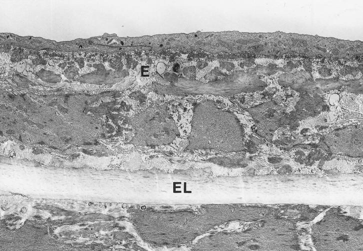
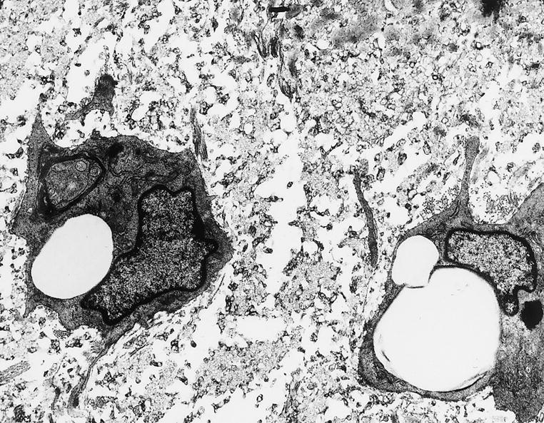
The extracellular matrix (ECM) of the normal artery wall is a collection of fibrous proteins and associated glycoproteins embedded in a hydrated ground substance of glycosaminoglycans and proteoglycans. These distinct molecules are organized into a highly ordered network that are closely associated with the vascular cells that produce them. In addition to providing the architectural framework for the artery wall that imparts mechanical support and viscoelasticity, the ECM can regulate the behaviour of vascular cells, including their ability to migrate, proliferate and survive injury. The composition of the ECM is different within intimal lesions of atherosclerosis, which are composed of monocytes and lymphocytes from the circulation and smooth muscle cells (SMC) that migrate from the media to the intima (Ross 1993, 1999), and these differences may contribute to the altered phenotype of vascular cells within lesions. This review will briefly outline the ECM changes observed in atherosclerosis and restenosis and the potential relationship of these changes to altered vascular cell functions.
Figures
References
-
- Aruffo A, Stamenkovic I, Melnick M. CD44 is the principal cell surface receptor for hyaluronate. Cell. 1990;61:1303–1313. - PubMed
-
- Beezhold DH, Personius C. Fibronectin fragments stimulate tumor necrosis factor secretion by human monocytes. J. Leukoc. Biol. 1992;51:59–64. - PubMed
-
- Bendeck MP, Irvin C, Reidy MA. Inhibition of matrix metalloproteinase activity inhibits smooth muscle cell migration but not neointimal thickening after arterial injury. Circ. Res. 1996;78:38–43. - PubMed
Publication types
MeSH terms
LinkOut - more resources
Full Text Sources
Other Literature Sources
