. 2000 Aug;81(4):271-82.
doi: 10.1046/j.1365-2613.2000.00161.x.

Demyelination and axonal dystrophy in alpha A-crystallin transgenic mice

Affiliations

Demyelination and axonal dystrophy in alpha A-crystallin transgenic mice

E P De Rijk et al. Int J Exp Pathol. 2000 Aug.

Abstract

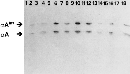
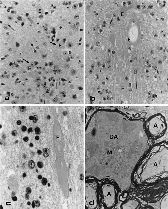
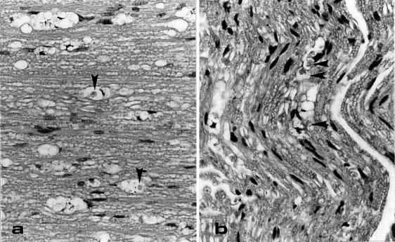
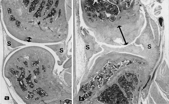
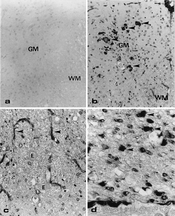
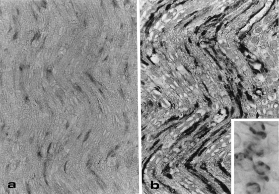
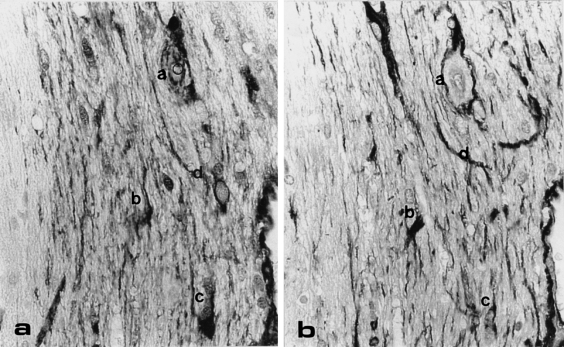
Homozygous mice transgenic for alphaA-crystallin, one of the structural eye lens proteins, developed hindlimb paralysis after 8 weeks of age. To unravel the pathogenesis of this unexpected finding and the possible role of alphaA-crystallin in this pathological process, mice were subjected to a histopathological and immunohistochemical investigation. Immunohistochemistry showed large deposits of alphaA-crystallin in the astrocytes of the spinal cord, and in the Schwann cells of dorsal roots and sciatic nerves. Additionally, microscopy showed dystrophic axons in the spinal cord and digestion chambers as a sign of ongoing demyelination in dorsal roots and sciatic nerves. Apart from a few areas with slight alphaA-crystallin-immunopositive structures, the brain was normal. Because the alphaA-crystallin protein expression appeared in specific cells of the nervous system (astrocytes and Schwann cells), the most plausible explanation for the paralysis is a disturbance of cell function caused by the excessive intracytoplasmic accumulation of the alphaA-crystallin protein. This is followed by a sequence of secondary changes (demyelination, axonal dystrophy) and finally arthrosis. In conclusion, alphaA-crystallin transgenic mice develop a peripheral and central neuropathy primarily affecting spinal cord areas at the dorsal side, dorsal root and sciatic nerve.


Figures

Figure 1
Figure 1
Immunoblot after SDS/PAGE of several tissues of wildtype (odd numbered lanes) and transgenic mice (even numbered lanes). Tissues in lanes are: 1,2: heart; 3,4: kidney; 5,6: lung; 7,8: liver; 9,10: spleen; 11,12: skeletal muscle; 13,14: brain; 15,16: uterus, and 17,18: spinal cord, with different expression levels of αA-crystallin and an elongated form αAins-crystallin of approximately 19.8 and 22.5 kDa, respectively. Detection was achieved with an affinity-purified anti-αA-crystallin antibody.
Figure 2
Figure 2
Light microscopical (LM) haematoxylin-eosine (HE)-stained section (a, b, c) and electron microscopical section (d) of abnormalities at the dorsal side of a transgenic mouse lumbar spinal cord. (a) Eosinophilic bodies/axonal swelling (E) scattered throughout the grey matter. Original Magnification (OM) = × 100. (b, c) Large eosinophilic bodies in the white matter, sometimes showing a torpedo-like shape (c) OM = 100x and × 250, respectively. (d) The degenerating axon (DA) in this electron micrograph is surrounded by normal axons (A). A clear difference in size and density of the cytoplasm and in number of mitochondria (M) in the degenerating and normal axon is shown. Note also the difference in thickness of the myelin surrounding the normal and degenerating axon (electron dense layered material in between the small arrowheads). OM = × 4.500.
Figure 3
Figure 3
Vacuolated structures in dorsal root (a) and sciatic nerve (b) of a transgenic mouse. In some of the vacuoles clear macrophages are present (arrowhead). LM-sections are HE-stained. OM = × 100.
Figure 4
Figure 4
A longitudinal HE-stained sections of the knee joint of a wildtype (a) and transgenic (b) mouse. In the transgenic mouse the amount of cartilage covering the joint (arrow) is increased significantly and more synovial tissue (S) is present which consists largely of cartilage. OM = × 6.6.
Figure 5
Figure 5
Cross-section of the lumbar dorsal spinal cord of a wildtype (a) and transgenic mouse (b, c, and d), immunostained with anti-αA-crystallin. (a) No immunostaining is observed in the wildtype mouse. OM = × 50. (b) Various sized anti-αA-positive structures are present in the grey matter of the transgenic mouse. OM = × 50. (c) In the white matter of the transgenic mouse, small dots and stellate cells (arrows) are positive with anti-αA-crystallin. The eosinophilic bodies/axonal swellings (E) are not immunopositive. OM = × 100. GM, grey matter; WM, white matter. (d) The dorsal root shows many large dot-like immunopositive structures (arrowheads). OM = × 250.
Figure 6
Figure 6
Longitudinal section of the sciatic nerve of a wildtype (a) and transgenic (b) mouse, immunostained with anti-αA-crystallin. (a) No immunostaining of nerve structures can be seen in the wildtype mouse. (b) The transgenic mouse shows a strong stripe-like staining which is running parallel to the nerve axons. OM = × 100. Inset: cross-section shows positive anti-αA-crystallin staining around the axon. OM = × 250.
Figure 7
Figure 7
Consecutive cross-section of the white matter in a transgenic mouse cerebellum, immunostained with anti-αA-crystallin (a) and anti-GFAP (b). The same stellate cells are anti-αA-crystallin and anti-GFAP positive (compare a, b, c, d in Figure 7a, b). OM = × 100.

Similar articles

Cited by

References

    1. Armand J. The origin, course and determination of corticospinal fibers in various mammals. In: KUypers HGJM, MArtin GF, editors. Progress in Brain Research: Anatomy of the Descending Pathways to the Spinal Cord. Amsterdam: Elsevier; 1982. pp. 329–360.
    1. Arrigo A-P, Previll X. Role of hsp27 and related proteins. In: LAtchman DS, editor. Stress Proteins. Vol. 136. Berlin: Springer Verlag; 1999. pp. 101–132.
    1. Bloemendal H. Lens proteins. Crit. Rev. Biochem. 1982;12:1–39. - PubMed
    1. Brady JP, Garland D, Douglas-Tabor Y, Jr Robinson WG, Groome A, Wawrousek EF. Targeted disruption of the mouse alpha A-crystallin gene induces cataract and cytoplasmic inclusion bodies containing the small heat shock protein alpha B-crystallin. Proc. Natl. Acad. Sci. USA. 1997;94:884–889. - PMC - PubMed
    1. Dahl D, Rueger DC, Bignami A, Weber A, Osborn M. Vimentin the 57000 daltons protein of fibroblast filaments is the major cytoskeletal component in immature glia. Eur. J. Cell Biol. 1981;24:191–196. - PubMed

Publication types

MeSH terms

LinkOut - more resources