Adenocarcinoma of the esophagogastric junction: results of surgical therapy based on anatomical/topographic classification in 1,002 consecutive patients
- PMID: 10973385
- PMCID: PMC1421149
- DOI: 10.1097/00000658-200009000-00007
Adenocarcinoma of the esophagogastric junction: results of surgical therapy based on anatomical/topographic classification in 1,002 consecutive patients
Abstract
Objective: To assess the outcome of surgical therapy based on a topographic/anatomical classification of adenocarcinoma of the esophagogastric junction.
Summary background data: Because of its borderline location between the stomach and esophagus, the choice of surgical strategy for patients with adenocarcinoma of the esophagogastric junction is controversial.
Methods: In a large single-center series of 1,002 consecutive patients with adenocarcinoma of the esophagogastric junction, the choice of surgical approach was based on the location of the tumor center or tumor mass. Treatment of choice was esophagectomy for type I tumors (adenocarcinoma of the distal esophagus) and extended gastrectomy for type II tumors (true carcinoma of the cardia) and type III tumors (subcardial gastric cancer infiltrating the distal esophagus). Demographic data, morphologic and histopathologic tumor characteristics, and long-term survival rates were compared among the three tumor types, focusing on the pattern of lymphatic spread, the outcome of surgery, and prognostic factors in patients with type II tumors.
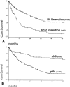
Results: There were marked differences in sex distribution, associated intestinal metaplasia in the esophagus, tumor grading, tumor growth pattern, and stage distribution between the three tumor types. The postoperative death rate was higher after esophagectomy than extended total gastrectomy. On multivariate analysis, a complete tumor resection (R0 resection) and the lymph node status (pN0) were the dominating independent prognostic factors for the entire patient population and in the three tumor types, irrespective of the surgical approach. In patients with type II tumors, the pattern of lymphatic spread was primarily directed toward the paracardial, lesser curvature, and left gastric artery nodes; esophagectomy offered no survival benefit over extended gastrectomy in these patients.
Conclusion: The classification of adenocarcinomas of the esophagogastric junction into type I, II, and III tumors shows marked differences between the tumor types and provides a useful tool for selecting the surgical approach. For patients with type II tumors, esophagectomy offers no advantage over extended gastrectomy if a complete tumor resection can be achieved.
Figures


References
-
- Devesa SS, Blot WJ, Fraumeni JF Jr. Changing patterns in the incidence of esophageal and gastric carcinoma in the United States. Cancer 1998; 83: 2049–2053. - PubMed
-
- Walsh TN, Noonan N, Hollywood D, et al. A comparison of multimodal therapy and surgery for esophageal adenocarcinoma. N Engl J Med 1996; 335: 462–467. - PubMed
-
- Parshad R, Singh RK, Kumar A, Gupta SD, Chattopadhyay TK. Adenocarcinoma of distal esophagus and gastroesophageal junction: long-term results of surgical treatment in a North Indian center. World J Surg 1999; 23: 277–283. - PubMed
-
- Wijnhoven BP, Siersema PD, Hop WC, van Dekken H, Tilanus HW. Adenocarcinomas of the distal oesophagus and gastric cardia are one clinical entity. Rotterdam Oesophageal Tumour Study Group. Br J Surg 1999; 86: 529–535. - PubMed
-
- Kajiyama Y, Tsurumaru M, Udagawa H, et al. Prognostic factors in adenocarcinoma of the gastric cardia: pathologic stage analysis and multivariate regression analysis. J Clin Oncol 1997; 15: 2015–2021. - PubMed
MeSH terms
LinkOut - more resources
Full Text Sources
Medical

