. 2000 Sep 12;97(19):10520-5.
doi: 10.1073/pnas.180130797.

Toll-related receptors and the control of antimicrobial peptide expression in Drosophila

Affiliations

Toll-related receptors and the control of antimicrobial peptide expression in Drosophila

S Tauszig et al. Proc Natl Acad Sci U S A. .

Abstract

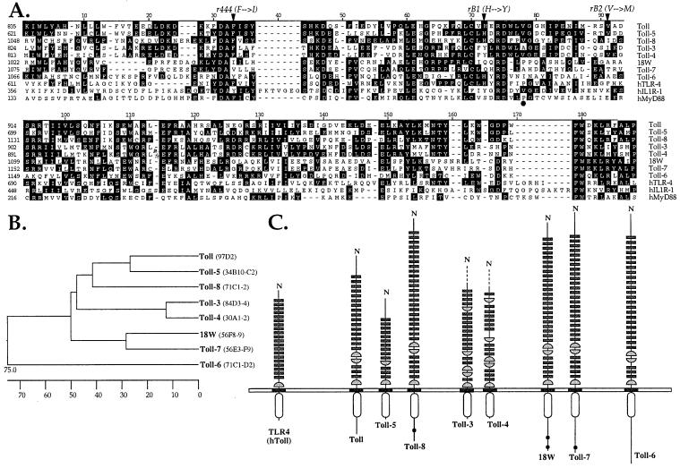
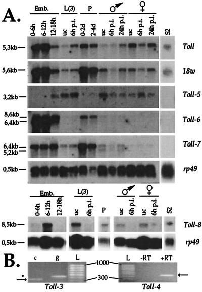
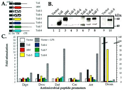
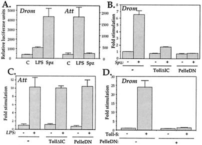
Insects defend themselves against infectious microorganisms by synthesizing potent antimicrobial peptides. Drosophila has appeared in recent years as a favorable model to study this innate host defense. A genetic analysis of the regulation of the antifungal peptide drosomycin has demonstrated a key role for the transmembrane receptor Toll, which prompted the search for mammalian homologs. Two of these, Toll-like receptor (TLR)2 and TLR4, recently were shown to play a critical role in innate immunity against bacteria. Here we describe six additional Toll-related genes (Toll-3 to Toll-8) in Drosophila in addition to 18-wheeler. Two of these genes, Toll-3 and Toll-4, are expressed at a low level. Toll-6, -7, and -8, on the other hand, are expressed at high levels during embryogenesis and molting, suggesting that, like Toll and 18w, they perform developmental functions. Finally, Toll-5 is expressed only in larvae and adults. By using chimeric constructs, we have tested the capacity of the signaling Toll/IL-1R homology domains of these receptors to activate antimicrobial peptide promoters and found that only Toll and Toll-5 can activate the drosomycin promoter in transfected cells, thus demonstrating specificity at the level of the Toll/IL-1R homology domain. In contrast, none of these constructs activated antibacterial peptide promoters, suggesting that Toll-related receptors are not involved in the regulation of antibacterial peptide expression. This result was independently confirmed by the demonstration that a dominant-negative version of the kinase Pelle can block induction of drosomycin by the cytokine Spaetzle, but does not affect induction of the antibacterial peptide attacin by lipopolysaccharide.


Figures

Figure 1
Figure 1
The Drosophila Toll family. (A) Alignment of the TIR domains from Drosophila Toll-related molecules and human TLR4, IL-1 receptor type I (IL-1RI), and MyD88. Sequences were aligned by using the clustal method. Conserved residues are shaded in black. Residues affected in the loss-of-function r444, rB1, and rB2 alleles of Toll are indicated by arrowheads (46). The black dot points to the position of the Pro residue critical for TLR4 signaling. (B) Phylogenetic relationship between Drosophila Toll molecules based on the alignment shown in A and generated by the neighbor-joining method. The scale beneath the tree measures the distance between the sequences. The chromosomal localization of the Toll-related genes is indicated (Right). (C) Schematic representation of domain organization of Drosophila Toll-related molecules and human TLR4. The receptors are grouped according to conservation of their TIR domains. The leucine-rich repeats are indicated by small rectangles, whereas cysteine-rich carboxyl-flanking motifs and cysteine-rich amino-flanking motifs are represented by half-circles. Black dots indicate polyglutamine stretches. Incomplete sequences of Toll-3 and Toll-4 are indicated by dashed lines.
Figure 2
Figure 2
Northern blot and RT-PCR analysis of Toll-related gene expression. (A) Poly(A)+ RNA extracted from embryos (Emb.), third-instar larvae [L(3)], pupae (P, 0–2 or 2–4 days), adult male or female Drosophila, and the cell line S2 were submitted to Northern blot analysis and hybridized to probes derived from Toll, 18w, and Toll-3 to Toll-8 gene sequences. No signal could be observed for the Toll-3 and Toll-4 probes. A probe recognizing RNA coding for ribosomal protein 49 (rp49) was used to ensure that comparable amounts of RNA were loaded in all lanes. uc, unchallenged; p.i., postinfection with a mixture of Gram-negative and Gram-positive bacteria; and d, days. (B) RT-PCR analysis of Toll-3 and Toll-4 expression. Primers specific for the Toll-3 gene were designed to flank a 104-bp intron sequence in the genomic DNA, resulting in the amplification of a 342-bp cDNA (c)-derived fragment (arrow) or a 446-bp genomic DNA (g)-derived fragment (*). Primers for the Toll-4 gene were designed to amplify a 480-bp intronless fragment. In this case, RT was omitted in a control reaction to ensure that the amplified band is derived from RNA. cDNA was prepared from mRNA derived from pupae (Toll-3) or third-instar larvae (Toll-4). L, 100-bp ladder.
Figure 3
Figure 3
Induction of antimicrobial peptide promoters by chimeric Toll receptors. (A) Schematic representation of the chimeric molecules. The constructs are based on a constitutively active version of Toll in which all LRR motifs have been deleted. The truncated ectodomain of Toll (black rectangle) is fused to the transmembrane and intracytoplasmic domains of the Toll-related molecules. EC, ectodomain; TM, transmembrane domain; CTE, C-terminal extension. (B) Expression of the chimeric proteins in transfected cells. Protein extracts prepared from S2 cells transfected by expression vectors either empty (Vector) or expressing TollΔLRR (Toll) or the various chimerae (18W, Toll-3 to -8) were submitted to Western blot analysis with an anti-FLAG mAb. Expected sizes for the various molecules are as follows: Toll, 50 kDa; 18W, 63 kDa; Toll-3, 40 kDa; Toll-4, 41 kDa; Toll-5, 41 kDa; Toll-6, 69 kDa; Toll-7, 63 kDa; and Toll-8, 57 kDa. (C) Induction of antimicrobial peptide promoters in transfected cells. S2 cells were cotransfected with 1 μg of expression vector either empty or expressing TollΔLRR (Toll) or the various chimerae (18W, Toll-5 to -8) and 0.1 μg of reporter plasmid encoding luciferase under the control of the diptericin (Dipt), drosocin (Droc), defensin (Def), cecropin (Cec), attacin (Att), or drosomycin (Drom) promoters. Transfections were repeated twice in S2 cells and l(2)mbn cells with identical results. A representative experiment is shown.
Figure 4
Figure 4
Activation of antimicrobial peptide promoters by Spaetzle and LPS in S2 cells. (A) S2 cells were cotransfected with 1 μg of either an empty expression vector or a vector expressing the carboxyl-terminal 106 aa of Spaetzle (Spz) (4, 30) and 0.1 μg of reporter constructs expressing luciferase under the control of the drosomycin (Drom) or attacin (Att) promoters. Cells transfected with the empty expression vector were either left untreated or exposed to LPS (10 μg/ml) for 16 h before harvesting and determination of luciferase activity in cell extracts. (B) S2 cells were cotransfected with 0.1 μg of drosomycin-luciferase reporter construct and 0.5 μg of expression vector encoding the C106 processed form of Spaetzle (Spz) together with 0.5 μg of expression vector empty or encoding a truncated Toll version deleted of its intracytoplasmic domain (TollΔIC) or a mutant Pelle version deleted of its kinase domain (PelleDN). (C) S2 cells were cotransfected with 0.1 μg of attacin-luciferase reporter construct and 1 μg of expression vector empty or encoding TollΔIC or PelleDN. Twenty-four hours after transfection, cells were either left untreated or exposed to LPS (10 μg/ml) for 16 h before harvesting and determination of luciferase activity in cell extracts. (D) S2 cells were cotransfected with 0.1 μg of drosomycin-luciferase reporter plasmid and 0.5 μg of expression vector encoding the TollΔLRR-Toll5 chimeric protein together with 0.5 μg of expression vector either empty or encoding PelleDN. All transfections were done in triplicate, and results represent means ± SD.

Comment in

  • Viral interference with IL-1 and toll signaling.
    Janeway C Jr, Medzhitov R. Janeway C Jr, et al. Proc Natl Acad Sci U S A. 2000 Sep 26;97(20):10682-3. doi: 10.1073/pnas.97.20.10682. Proc Natl Acad Sci U S A. 2000. PMID: 11005852 Free PMC article. No abstract available.

References

    1. Hoffmann J, Reichhart J. Trends Cell Biol. 1997;7:309–316. - PubMed
    1. Lemaitre B, Reichhart J, Hoffmann J. Proc Natl Acad Sci USA. 1997;94:14614–14619. - PMC - PubMed
    1. Lemaitre B, Nicolas E, Michaut L, Reichhart J, Hoffmann J. Cell. 1996;86:973–983. - PubMed
    1. DeLotto Y, DeLotto R. Mech Dev. 1998;72:141–148. - PubMed
    1. Levashina E A, Langley E, Green C, Gubb D, Ashburner M, Hoffmann J A, Reichhart J M. Science. 1999;285:1917–1919. - PubMed

Publication types

MeSH terms