. 2000 Sep 12;97(19):10643-8.
doi: 10.1073/pnas.180243697.

A calcium influx is triggered and propagates in the zygote as a wavefront during in vitro fertilization of flowering plants

Affiliations

A calcium influx is triggered and propagates in the zygote as a wavefront during in vitro fertilization of flowering plants

A F Antoine et al. Proc Natl Acad Sci U S A. .

Abstract

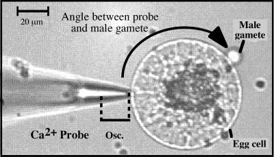
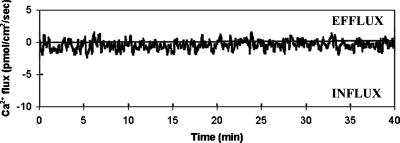
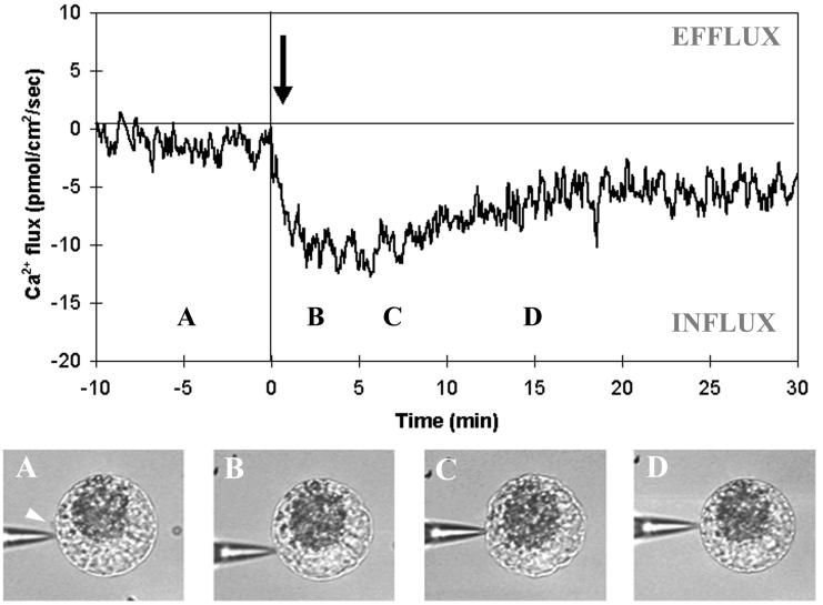
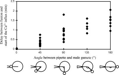
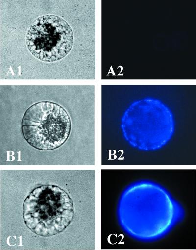
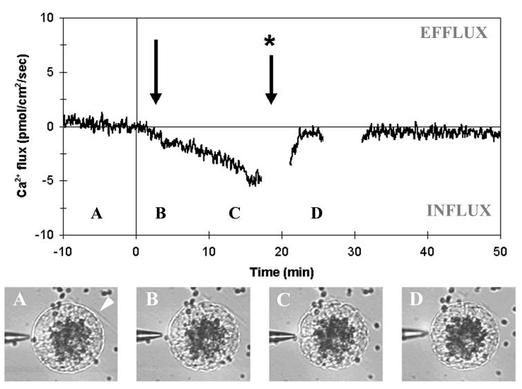
In this paper, we report direct measurement of an influx of extracellular Ca(2+) induced by gamete fusion in flowering plants. This result was obtained during maize in vitro fertilization with the use of an extracellular Ca(2+)-selective vibrating probe. Ca(2+) influx recorded at the surface of isolated egg cells, with or without adhesion of a male sperm cell, was close to zero and stable over time. Gamete fusion, however, triggered a Ca(2+) influx in the vicinity of the sperm entry site with a delay of 1.8 +/- 0.6 sec. The Ca(2+) influx spread subsequently through the whole egg cell plasma membrane as a wavefront, progressing at an estimated rate of 1.13 micrometer.(-1). Once established, Ca(2+) influx intensities were sustained, monotonic and homogeneous over the whole egg cell, with an average peak influx of 14.92 pmol .cm(-2).(-1) and an average duration of 24.4 min. The wavefront spread of channel activation correlates well with the cytological modifications induced by fertilization, such as egg cell contraction, and with the cytosolic Ca(2+) ((c)[Ca(2+)]) elevation previously reported. Calcium influx was inhibited effectively by gadolinium, possibly implicating mechanosensitive channels. Furthermore, artificial influxes created by incubation with Ca(2+) ionophores mimicked some aspects of egg activation. Taken together, these results suggest that, during fertilization in higher plants, gamete membrane fusion starts the first embryonic events by channel opening and Ca(2+) influx. In turn, (c)[Ca(2+)] may work as a trigger and possibly a space and time coordinator of many aspects of egg activation.


Figures

Figure 1
Figure 1
Micrograph from a typical Ca2+ flux measurement during maize IVF. The Ca2+-selective vibrating probe was positioned approximately 1 μm from the surface of the egg cell at its nearest vibration point. The probe oscillated with an excursion of 10 μm away from the cell (Osc.). The angle between the probe and the adhering male gamete is indicated by the black arrow.
Figure 2
Figure 2
Typical Ca2+ flux measurements on an egg cell adhered with a male gamete before fusion took place (n = 112). Only very small and stable influxes or effluxes were detected, which are close to the noise level independently assessed as 0.60 ± 0.08 pmol⋅cm−2⋅sec−1.
Figure 3
Figure 3
Ca2+ flux measurements during maize IVF. A typical recording is shown (n = 61) illustrating the onset of a Ca2+ influx after fusion. Time 0 is chosen arbitrarily as the time of gametic fusion, as asserted by direct microscopic observation. The arrow shows the detectable onset of a Ca2+ influx after fusion. A clear Ca2+ influx was always detected in the egg membrane, with a delay to fusion dependent on the relative position of the probe and fusion site (black arrow in Fig. 1). A, B, C, and D refer to the time when the pictures (Bottom) were taken. The following events are depicted: (A) egg cell before fusion (male gamete position is shown by a white arrow); (B) egg cell after fusion, just when contraction has started; (C) strong egg cell contraction; and (D) egg cell reshaping.
Figure 4
Figure 4
Influence of the position of the probe, respective to that of the adhering male gamete, on the measurement of the Ca2+ influx induced by fusion. Lag time between fusion and onset of Ca2+ influx is plotted against the angle between the male gamete and the Ca2+-selective electrode. The drawing (Lower) illustrates the different positions estimated visually during the adhesion procedure. Only differences of at least 45° have been taken into account.
Figure 5
Figure 5
Cell wall establishment around egg cells after fertilization or Ca2+ ionophore treatment. Transmission micrograph (A1) and fluorescent micrograph (A2) of an isolated egg cell. Transmission micrograph (B1) and fluorescent micrograph (B2) of a 2-h zygote. Transmission micrograph (C1) and fluorescent micrograph (C2) of egg cells 2 h after incubation for 40 min with A23187. (Bar = 20 μm.)
Figure 6
Figure 6
Effect of GdCl3 on the Ca2+ influx set after gametic fusion. Ca2+ fluxes were first recorded during IVF before addition of GdCl3 to the fusion medium. As in Fig. 3, time 0 is chosen arbitrarily as the time of gametic fusion. The single arrow indicates the onset of a Ca2+ influx. GdCl3 (10 μM final concentration) was then added (arrow with asterisk). A typical recording is shown (n = 17). GdCl3 inhibits 93.07% of the Ca2+ influx induced by fusion. A, B, C, and D refer to the time when the pictures (Lower) were taken. The following events are depicted: (A) egg cell before fusion (male gamete position is shown by white arrow); (B) egg cell after fusion, just when contraction has started; and (C) strong egg cell contraction, which was abolished immediately (D) by GdCl3 addition.

References

    1. Kranz E, Bautor J, Lörz H. Sex Plant Reprod. 1991;4:12–16.
    1. Kranz E, Lörz H. Zygote. 1994;2:125–128. - PubMed
    1. Faure J-E, Digonnet C, Dumas C. Science. 1994;263:1598–1600. - PubMed
    1. Kranz E, Von Wiegen P, Quader P, Lörz H. Plant Cell. 1998;10:511–524. - PMC - PubMed
    1. Kranz E, Dresselhaus T. Trends Plant Sci. 1996;1:82–87.

Publication types

LinkOut - more resources