. 2000 Sep 12;97(19):10454-9.
doi: 10.1073/pnas.180316397.

Essential role for p38alpha mitogen-activated protein kinase in placental angiogenesis

Affiliations

Essential role for p38alpha mitogen-activated protein kinase in placental angiogenesis

J S Mudgett et al. Proc Natl Acad Sci U S A. .

Abstract

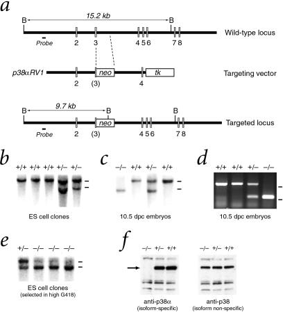
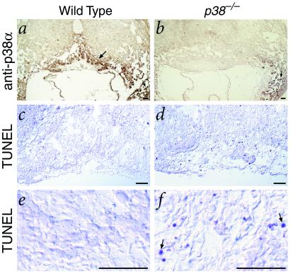
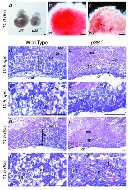
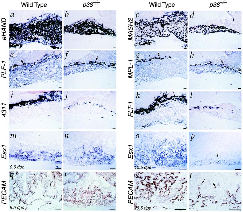
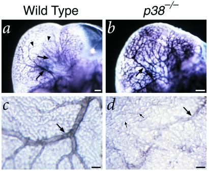
The p38 family of mitogen-activated protein kinases (MAPKs) mediates signaling in response to environmental stresses and inflammatory cytokines, but the requirements for the p38 MAPK pathway in normal mammalian development have not been elucidated. Here, we show that targeted disruption of the p38alpha MAPK gene results in homozygous embryonic lethality because of severe defects in placental development. Although chorioallantoic placentation is initiated appropriately in p38alpha null homozygotes, placental defects are manifest at 10.5 days postcoitum as nearly complete loss of the labyrinth layer and significant reduction of the spongiotrophoblast. In particular, p38alpha mutant placentas display lack of vascularization of the labyrinth layer as well as increased rates of apoptosis, consistent with a defect in placental angiogenesis. Furthermore, p38alpha mutants display abnormal angiogenesis in the embryo proper as well as in the visceral yolk sac. Thus, our results indicate a requirement for p38alpha MAPK in diploid trophoblast development and placental vascularization and suggest a more general role for p38 MAPK signaling in embryonic angiogenesis.


Figures

Figure 1
Figure 1
Targeted disruption of the p38α gene. (a) The gene-targeting vector p38αRV1 inserts an antisense PGKneo cassette into an EcoRI site in exon 3 and contains a 5′ flanking 4.5-kilobase (kb) EcoRI fragment and a 3′ flanking 3-kb SmaI–KpnI fragment, resulting in deletion of the 3′ half of exon 3 and part of intron 3. (b) Southern blot analysis of ES cell clones with a unique 5′ genomic probe distinguishes a wild-type 15.2-kb BamHI fragment from a 9.7-kb BamHI fragment generated by the targeted allele. Bars at right indicate positions of 15-kb wild-type and 10-kb targeted alleles. (c) Southern analysis of genomic DNA digested with BamHI from 10.5 dpc embryos obtained from a p38α heterozygous intercross. (d) PCR analysis of genomic DNA from visceral yolk sacs of 10.5 dpc embryos from a p38α heterozygous intercross. Bars at right indicate positions of markers at 500 and 250 bp. (e) Southern analysis of ES cell clones selected after growth in high concentrations (1 mg/ml) of G418 to obtain homozygosity for the p38α targeted allele. (f) Western analysis of total protein lysates from wild-type and targeted ES cells with antibodies that are specific for p38α or that crossreact with multiple p38 MAPK isoforms.
Figure 2
Figure 2
Expression of p38α MAPK and detection of apoptosis in wild-type and p38α mutant placentas. (a and b) Immunohistochemical detection of p38α in placenta and maternal decidua. Note the strong staining in the wild-type diploid trophoblast (arrows) in the wild type (a), but not the mutant (b); in contrast, staining in the maternal decidual cells is unaffected (arrowheads). (c–f) TUNEL analysis of wild-type and p38α mutant placentas shows nearly complete lack of apoptotic cells in the diploid trophoblast of the wild-type placenta (c and e), but abundant labeled cells in the mutant trophoblast (d and f). (Scale bars represent 100 μm.)
Figure 3
Figure 3
Morphology and histology of wild-type and p38α mutant embryos and placentas. (a) Gross morphology of a wild-type and p38α mutant littermate at 11.0 dpc, showing developmental retardation of the homozygote. (b and c) Ventral view of the placentas of a wild-type and p38α mutant, showing apparent decreased vascularization of the mutant placenta, as shown by its paler red color (arrow in c). (d–k) Hematoxylin and eosin-stained sections of placentas from wild-type (d, f, h, and j) and p38α homozygous mutant littermates (e, g, i, and k). (d and e) At 10.5 dpc, the developing labyrinth and spongiotrophoblast layers found in the wild type (d) are significantly decreased in thickness in the p38α mutant (e, arrow). (f and g) High-power views show a nearly complete lack of circulating fetal blood cells in the mutant. (h and i) By 11.5 dpc, the labyrinth layer is essentially missing and the spongiotrophoblast layer is greatly diminished in the p38α mutant compared with wild-type. (j and k) High-power views show that the placental vasculature with differentiated endothelial cells and circulating fetal blood cells (j) is nearly completely abolished in the p38α mutant, which instead contains apparent necrotic regions (k, arrows). (Scale bars in a–c represent 0.5 mm; bars in d–k represent 100 μm.) Abbreviations: al, allantois; ch, chorionic plate; en, endothelial cells; fb, fetal blood cells; gi, trophoblast giant cells; la, labyrinthine trophoblast; ma, maternal decidual tissue; sp, spongiotrophoblast; WT, wild type.
Figure 4
Figure 4
Marker analysis of wild-type and p38α mutant placentas at 10.5 dpc (a–l, o, p, s, and t) and 9.5 dpc (m, n, q, and r). (a–d) Expression of eHAND (a and b) and MASH2 (c and d) in diploid trophoblast is reduced in p38α mutant placentas. (e–h) Expression of PLF-1 (e and f) and MPL-1 (g and h) in trophoblast giant cells (arrows) is unaffected in p38α mutants. (i–l) Expression of 4311 and FLT-1 in spongiotrophoblast is greatly reduced in p38α mutant placentas. (mp) At 9.5 dpc, expression of Esx1 in the chorionic plate and labyrinth layer is relatively normal in p38α mutants (n) relative to wild type (m), whereas by 10.5 dpc, the expression of Esx1 is greatly diminished (p, arrow). (q–t) Immunohistochemical staining for PECAM marks endothelial cells in the allantoic plate and developing labyrinth at 9.5 dpc in both wild-type (q) and p38α mutant placentas (r). At 10.5 dpc, infiltration of endothelial cells into the labyrinth layer is well-advanced in the wild-type placenta (s), but only a few PECAM-positive regions can be detected in the labyrinth layer of the p38α mutant (t, arrows). (Scale bars represent 100 μm.)
Figure 5
Figure 5
PECAM whole-mount immunohistochemistry of embryos and visceral yolk sacs at 10.5 dpc. (a and b) Vasculature of the developing brain in a wild-type littermate (a) and p38α mutant embryo (b). Note that the tree-like architecture of the major vessels (arrows) connecting to minor branches (arrowheads) in the wild-type differs significantly from the relatively uniform size and disorganized pattern of the vasculature in the mutant embryo. (c and d) Vasculature of the visceral yolk sac in a wild-type littermate (c) and p38α mutant embryo (d). Although the wild-type yolk sac displays major vessels (arrows) and branches, the mutant yolk sac has relatively few major vessels with abnormal morphology (arrow), and retains a primitive capillary plexus (small arrows).

References

    1. Cross J C, Werb Z, Fisher S J. Science. 1994;266:1508–1518. - PubMed
    1. Risau W. Nature (London) 1997;386:671–674. - PubMed
    1. Han J, Lee J D, Bibbs L, Ulevitch R J. Science. 1994;265:808–811. - PubMed
    1. Rouse J, Cohen P, Trigon S, Morange M, Alonso-Llamazares A, Zamanillo D, Hunt T, Nebreda A R. Cell. 1994;78:1027–1037. - PubMed
    1. Lee J C, Laydon J T, McDonnell P C, Gallagher T F, Kumar S, Green D, McNulty D, Blumenthal M J, Heys J R, Landvatter S W, et al. Nature (London) 1994;372:739–746. - PubMed

Publication types

Substances