

Portosystemic shunting and persistent fetal vascular structures in aryl hydrocarbon receptor-deficient mice
- PMID: 10973493
- PMCID: PMC27043
- DOI: 10.1073/pnas.190256997
Portosystemic shunting and persistent fetal vascular structures in aryl hydrocarbon receptor-deficient mice
Abstract
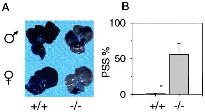
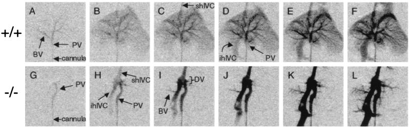
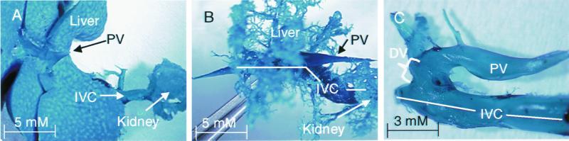
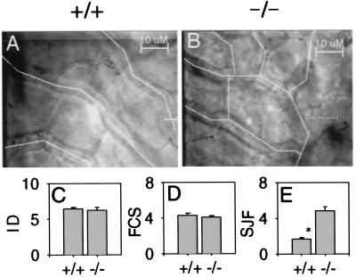
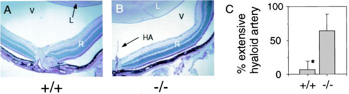
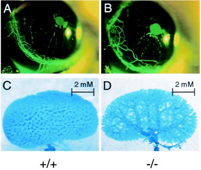
A physiological examination of mice harboring a null allele at the aryl hydrocarbon (Ah) locus revealed that the encoded aryl hydrocarbon receptor plays a role in the resolution of fetal vascular structures during development. Although the aryl hydrocarbon receptor is more commonly studied for its role in regulating xenobiotic metabolism and dioxin toxicity, a developmental role of this protein is supported by the observation that Ah null mice display smaller livers, reduced fecundity, and decreased body weights. Upon investigating the liver phenotype, we found that the decrease in liver size is directly related to a reduction in hepatocyte size. We also found that smaller hepatocyte size is the result of massive portosystemic shunting in null animals. Colloidal carbon uptake and microsphere perfusion studies indicated that 56% of portal blood flow bypasses the liver sinusoids. Latex corrosion casts and angiography demonstrated that shunting is consistent with the existence of a patent ductus venosus in adult animals. Importantly, fetal vascular structures were also observed at other sites. Intravital microscopy demonstrated an immature sinusoidal architecture in the liver and persistent hyaloid arteries in the eyes of adult Ah null mice, whereas corrosion casting experiments described aberrations in kidney vascular patterns.
Figures

Similar articles
-
Knockout of the aryl hydrocarbon receptor results in distinct hepatic and renal phenotypes in rats and mice.Toxicol Appl Pharmacol. 2013 Oct 15;272(2):503-18. doi: 10.1016/j.taap.2013.06.024. Epub 2013 Jul 13. Toxicol Appl Pharmacol. 2013. PMID: 23859880
-
Liver deformation in Ahr-null mice: evidence for aberrant hepatic perfusion in early development.Mol Pharmacol. 2006 May;69(5):1534-41. doi: 10.1124/mol.105.020107. Epub 2006 Jan 27. Mol Pharmacol. 2006. PMID: 16443691
-
Patent ductus venosus and dioxin resistance in mice harboring a hypomorphic Arnt allele.J Biol Chem. 2004 Apr 16;279(16):16326-31. doi: 10.1074/jbc.M400784200. Epub 2004 Feb 5. J Biol Chem. 2004. PMID: 14764592
-
Inherited liver shunts in dogs elucidate pathways regulating embryonic development and clinical disorders of the portal vein.Mamm Genome. 2012 Feb;23(1-2):76-84. doi: 10.1007/s00335-011-9364-0. Epub 2011 Nov 4. Mamm Genome. 2012. PMID: 22052005 Free PMC article. Review.
-
Evidence for the role of the Ah receptor in response to dioxin.Prog Clin Biol Res. 1994;387:139-54. Prog Clin Biol Res. 1994. PMID: 7972244 Review. No abstract available.
Cited by
-
Dioxin-dependent and dioxin-independent gene batteries: comparison of liver and kidney in AHR-null mice.Toxicol Sci. 2009 Nov;112(1):245-56. doi: 10.1093/toxsci/kfp191. Epub 2009 Sep 16. Toxicol Sci. 2009. PMID: 19759094 Free PMC article.
-
The aryl hydrocarbon receptor-interacting protein (AIP) is required for dioxin-induced hepatotoxicity but not for the induction of the Cyp1a1 and Cyp1a2 genes.J Biol Chem. 2010 Nov 12;285(46):35599-605. doi: 10.1074/jbc.M110.132043. Epub 2010 Sep 9. J Biol Chem. 2010. PMID: 20829355 Free PMC article.
-
Ah receptor binding to its cognate response element is required for dioxin-mediated toxicity.Toxicol Sci. 2008 Dec;106(2):301-3. doi: 10.1093/toxsci/kfn220. Epub 2008 Oct 16. Toxicol Sci. 2008. PMID: 18930947 Free PMC article. No abstract available.
-
The search for endogenous activators of the aryl hydrocarbon receptor.Chem Res Toxicol. 2008 Jan;21(1):102-16. doi: 10.1021/tx7001965. Epub 2007 Dec 13. Chem Res Toxicol. 2008. PMID: 18076143 Free PMC article. Review.
-
A novel role for the dioxin receptor in fatty acid metabolism and hepatic steatosis.Gastroenterology. 2010 Aug;139(2):653-63. doi: 10.1053/j.gastro.2010.03.033. Epub 2010 Mar 17. Gastroenterology. 2010. PMID: 20303349 Free PMC article.
References
-
- Hoffman E C, Reyes H, Chu F F, Sander F, Conley L H, Brooks B A, Hankinson O. Science. 1991;252:954–958. - PubMed
-
- Ema M, Sogawa K, Watanabe N, Chujoh Y, Matsushita N, Gotoh O, Funae Y, Fujii-Kuriyama Y. Biochem Biophys Res Commun. 1992;184:246–253. - PubMed
-
- Reyes H, Reisz-Porszasz S, Hankinson O. Science. 1992;256:1193–1195. - PubMed
Publication types
MeSH terms
Substances
Grants and funding
LinkOut - more resources
Full Text Sources
Molecular Biology Databases
