

doi: 10.1073/pnas.190268597.
Identification and characterization of a mammalian protein interacting with 20S proteasome precursors
Affiliations
- PMID: 10973495
- PMCID: PMC27027
- DOI: 10.1073/pnas.190268597
Item in Clipboard
Identification and characterization of a mammalian protein interacting with 20S proteasome precursors
Proc Natl Acad Sci U S A.
.
Abstract
The assembly of individual mammalian proteasome subunits into catalytically active 20S proteasome is not well understood. Herein, we report the identification and characterization of human and mouse homologues of the yeast proteasome maturating factor Ump1p. We delineate the region of hUMP1 implicated in the specific interaction with proteasome precursors and show that hUMP1 protein is absent from the mature form of the 20S proteasome. We also show that the transcript level of mammalian UMP1 is increased after IFN-gamma treatment and that mammalian UMP1 is functionally related to but not interchangeable with its yeast homologue.
Figures
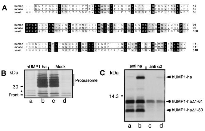
Identification of human and mouse homologue of the yeast Ump1p protein and its interaction with mammalian proteasome. (A) CLUSTALW sequences of human and mouse protein are aligned with yeast Ump1p. Identical amino acids are labeled in black, and similar amino acids are labeled in gray. Identical amino acids between human and mouse are boxed. (B) 293T cells were transiently transfected with recombinant plasmid directing the synthesis of hUMP1-ha (lanes a and b) or mock transfected (lanes c and d) and labeled after 24 h with [35S]methionine/cysteine. Cells were then solubilized, and cleared lysate was immunoprecipitated either with a monoclonal antibody against the ha epitope (lanes a and d) or with a polyclonal antibody against the proteasome (lanes b and c). The position of hUMP1-ha is indicated by an asterisk. (C) Fragments generated from internal initiation sites were used to identify the region of hUMP1-ha important for interaction with the proteasome. The cDNA encoding the wild-type hUMP1-ha (lanes b and d) or a mutant lacking the first initiation codon (lanes a and c) was transcribed and translated in vitro. Samples were immunoprecipitated either with the anti-ha antibody (lanes a and b) or with MCP21 (lanes c and d). The positions of full-length and truncated hUMP1-ha are indicated.
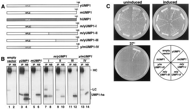
Mammalian and yeast UMP1 proteins are not functionally interchangeable. (A) Schematic representation of constructs. (B) Coimmunoprecipitation assay to detect association of various Ump1-ha variants with proteasomal complexes. Proteins were precipitated from extracts of the same strains shown in C (except for those expressing hUMP1) with antibodies against yeast 20S proteasomes. The precipitates (IP) as well as 25% of the nonbinding material (NB) were analyzed by Western blotting with anti-ha antibody. The positions of IgG heavy chains (HC), light chains (LC), and various Ump1-ha proteins are indicated. The position of a product of yUMP1, missing the domain necessary for interaction with the proteasome, is marked with an asterisk. (C) Growth assays of yeast strain JD59 (ump1-Δ) transformed with high-copy plasmids expressing, from the copper-inducible PCUP1 promoter, human (h), mouse (m), or yeast (y) UMP1 genes or various chimeric genes of the latter two (m/yUMP1-I to m/yUMP1-III or y/mUMP1 depicted in A; see Materials and Methods and main text for details). Transformants were streaked onto selective minimal medium, with (induced) or without (uninduced) 100 μM CuSO4, and incubated at 30°C for 3 days. To assay for complementation of ump1-Δ temperature-sensitive growth phenotype, transformants were streaked onto complete medium and incubated at 37°C for 2 days.
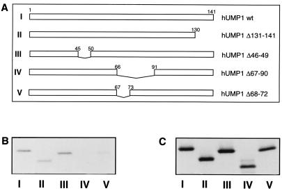
Delineation of the amino acids important for the interaction between hUMP1 and the proteasome. (A) Schematic description of the constructs used. Construct I represents the full-length hUMP1. Construct II depicts hUMP1 lacking the C-terminal 11 amino acids. Constructs III–V represent versions of hUMP1 lacking amino acids 46–49 (construct III), amino acids 67–90 (construct IV), or amino acids 68–72 (construct V). Constructs I–V were transcribed and translated in vitro and subjected to immunoprecipitation either by the monoclonal antibody against proteasome subunit α2 (B) or by the anti-ha antibody (C). Immunoprecipitated material was separated on SDS/15% PAGE. Constructs used are indicated at the bottom of each lane.
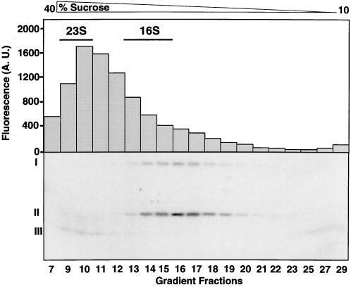
hUMP1-ha is a component of proteasome precursor complexes and is absent from active 20S proteasome. In vitro translated 35S-labeled hUMP1-ha was layered onto a 10–40% (vol/vol) sucrose gradient and centrifuged at 30,000 × g for 20 h. Fractions (350 μl) were collected and assayed for proteasomal activity by incubating them with Bz-LLE-AMC and monitoring the increased fluorescence in arbitrary units (A.U.) emitted by the released fluorogenic group AMC. Each fraction was simultaneously immunoprecipitated with the polyclonal anti-proteasome antibody, followed by SDS/15% PAGE analysis and fluorography. The fractions corresponding to enriched 23S and 16S ribosomal RNA are indicated. Radioactive bands corresponding to the full-length hUMP1-ha (I) and hUMP1-ha Δ1–61 (II) identified in an earlier experiment (Fig. 1C) are indicated. The position of a degradation product of hUMP1-ha is labeled (III). See text for details.
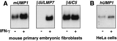
Transcript levels of mouse and human UMP1 increase after IFN-γ stimulation. Northern blot analysis of UMP1 transcripts in total RNA from mouse embryonic stem cells (A) and human HeLa cells (B) treated with 1,000 units/ml IFN-γ for 24 h where indicated. Membranes were hybridized with radiolabeled probes, stripped, and rehybridized as described in Materials and Methods. Quantification of signals revealed a 2-fold induction of mUMP1 transcript and a 2.1-fold induction of hUMP1. Transcript levels of β5i/LMP7, an immunoproteasome-specific subunit, increased by a factor of 85, whereas those of β6, encoding a subunit present in both standard and immunoproteasome, remained unchanged after IFN-γ treatment.
References
-
- Baumeister W, Walz J, Zühl F, Seemüller E. Cell. 1998;92:367–380. - PubMed
-
- Coux O, Tanaka K, Goldberg A L. Annu Rev Biochem. 1996;65:801–847. - PubMed
-
- Hilt W, Wolf D. Trends Biochem Sci. 1996;21:96–102. - PubMed
-
- Hershko A, Ciechanover A. Annu Rev Biochem. 1998;67:425–479. - PubMed
-
- Baumeister W, Cejka Z, Kania M, Seemüller E. Biol Chem. 1997;378:121–130. - PubMed
Publication types
MeSH terms
Substances
LinkOut - more resources
Full Text Sources
Other Literature Sources
Molecular Biology Databases
