. 2000 Sep 12;97(19):10418-23.
doi: 10.1073/pnas.190318397.

Nucleocytoplasmic translocation of Stat1 is regulated by a leucine-rich export signal in the coiled-coil domain

Affiliations

Nucleocytoplasmic translocation of Stat1 is regulated by a leucine-rich export signal in the coiled-coil domain

A Begitt et al. Proc Natl Acad Sci U S A. .

Abstract

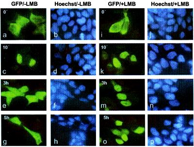
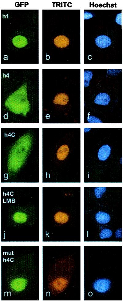
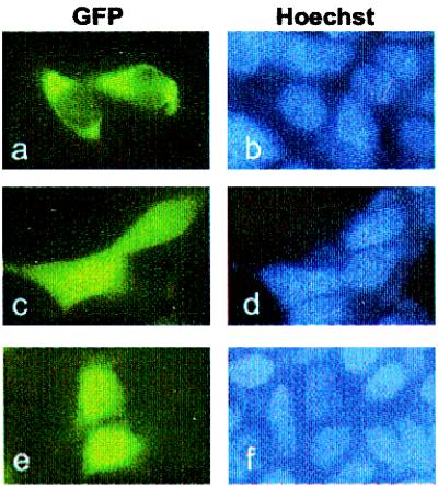
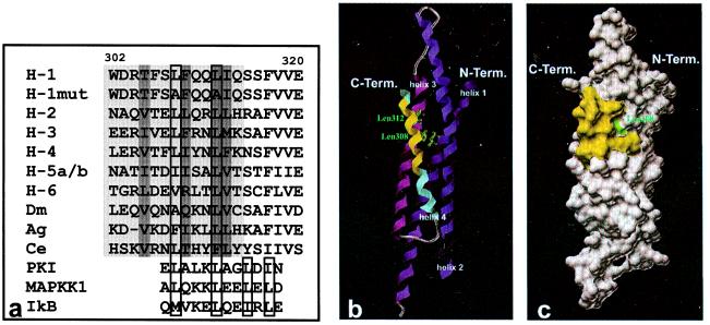
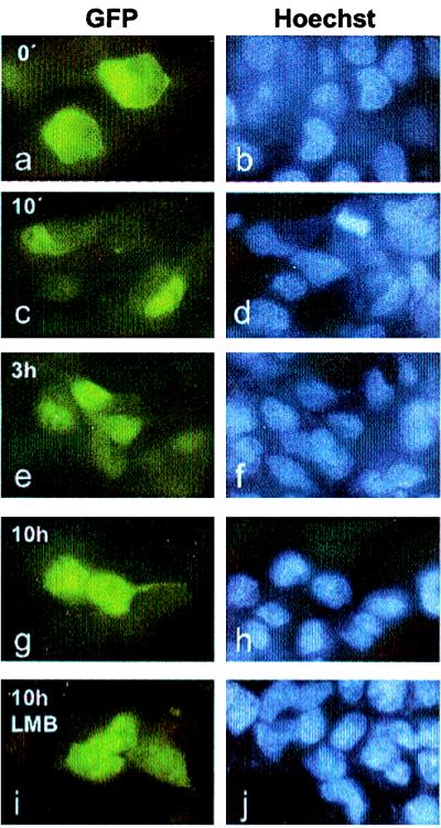
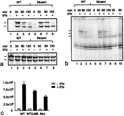
Signal transducer and activator of transcription (Stat) proteins are latent transcription factors that reside in the cytoplasm before activation. On cytokine-induced tyrosine phosphorylation, these molecules dimerize and accumulate transiently in the nucleus. No specific signals mediating these processes have been identified to date. In this report, we examine the nuclear export of Stat1. We find that treatment of cells with the export inhibitor leptomycin B does not affect steady-state localization of Stat1 but impedes nuclear export after IFNgamma-induced nuclear accumulation. We identify a conserved leucine-rich helical segment in the coiled-coil domain of Stat1, which is responsible for the efficient nuclear export of this protein. Mutation of two hallmark leucines within this segment greatly attenuate the back transport of Stat1 in the cytoplasm. When fused to a carrier protein, the Stat1 export sequence can mediate nuclear export after intranuclear microinjection. We show that prolonging the nuclear presence of Stat1 by inhibiting nuclear export reduces the transcriptional response to stimulation with IFNgamma. These data suggest that Stats are actively exported from the nucleus via several separate pathways and link this activity to transcriptional activation.


Figures

Figure 1
Figure 1
LMB inhibits nuclear export of Stat1. 293T cells transiently expressing Stat1α-GFP fusion proteins are shown before (a, b, i, j) and after the addition of IFNγ. Cells were stimulated with IFNγ for 30 min and fixed at the indicated time points (10 min, 3 h, and 5 h) after withdrawal of the cytokine. The subcellular localization of Stat1 in the absence (a–h) and presence (i–p) of 10 ng/ml LMB was determined by fluorescence microscopy. LMB does not influence nuclear accumulation of the fusion protein (c, k) but reduces the rate of cytoplasmic translocation (compare e and g to m and o).
Figure 2
Figure 2
Stat1 contains a NES at position 302–314. Affinity purified fusions of GFP and GST linked to Stat1 helix 1 (a–c), helix 4 (d–f), the C terminus of helix 4 comprising amino acids 302–314 (g–l), and a Leu308Ala mutant of the NES (m–o) were comicroinjected with TRITC-labeled goat immunoglobulins into the nuclei of HeLa S3 cells. Cells treated with LMB starting 1 h before microinjection are shown in j–l. After 3 h incubation at 37°C, the cells were fixed and the nuclei stained with Hoechst 33258 dye. The intracellular distribution of the GFP fusion proteins harboring Stat1 fragments, the TRITC-conjugated injection control, and positions of nuclei are indicated in the fluorescence micrographs for each Stat1 derivative. Only fusion proteins containing the intact NES from helix 4 of the coiled-coil domain of Stat1 are transported into the cytoplasm. The presence of LMB (j) and the mutation of leucine308 (m) inhibit nucleocytoplasmic translocation.
Figure 3
Figure 3
The NES in helix 4 of the coiled-coil domain of Stat1 leads to nuclear exclusion of a fusion protein. The fluorescence micrographs of transiently transfected 293T cells expressing GFP linked to helix 4 (a, b), helix 1 (c, d), or GFP alone (e, f) reveal the different subcellular distribution of chimeric constructs of low molecular weight. Only the fusion protein linked to helix 4 is excluded from the nucleus (a). The localization of GFP and the GFP fusions are depicted in a, c, and e, and the corresponding positions of nuclei stained with Hoechst 33258 are documented in b, d, and f.
Figure 4
Figure 4
(a) Aligned sequences of wild-type and NES mutant Stat1 (amino acids 302–320), of homologous regions of Stat family members, and of known leucine-rich export signals of other proteins. The NES sequence of Stat1 between amino acids 302 and 314 and the putative NESs of other Stat proteins are highlighted in light gray. Residues in the interior of the coiled-coil domain are marked with dark gray. Highly conserved hydrophobic residues in position 308 and 312 of the Stat1 NES and important hydrophobic residues in known NES sequences including hallmark residues indicative of the core tetramer are boxed. The following sequences from the National Center for Biotechnology Information Data Library are depicted: H-1, human Stat1, accession no. P42224; H-1mut, human Stat1 mutated in positions 308 and 312; H-2, human Stat2, no. P52630; H-3, human Stat3, no. NP_003141; H-4, human Stat4, no. Q14765; H-5a/b, human Stat5a/b, no. NP_003143/P51692; H-6, human Stat6, no. NP_003144; Dm, Drosophila melanogaster Stat, no. Q24151; Ag, Anopheles gambiae Stat, no. AJ010299; Ce, Caenorhabditis elegans Stat, no. AAD45535; PKI, human cAMP-dependent protein kinase inhibitor α, no. NP_006814; MAPKK1, human mitogen-activated protein kinase kinase 1, no. NP_002746; IkB, human IκBα, no. CAB65556. b and c depict the localization of the NES in the coiled-coil domain (amino acids 136–317) of Stat1. The ribbons drawing (b) shows the four-helix bundle, with amino acids 302–314 of the NES sequence in helix 4 (blue) marked in yellow. The side chains of the leucine residues in position 308 and 312 are included as atomic stick figures. A surface rendering of b is presented in c. The contributions to the surface of the coiled-coil domain by side chains of the NES sequence are highlighted in yellow, with the water accessible area of leucine 308 shown in green.
Figure 5
Figure 5
Mutation of two hallmark leucines in the NES impedes the nucleocytoplasmic transport of Stat1. 293T cells were transiently transfected with DNA constructs coding for a NES-minus Stat1 Leu308,312Ala double mutant linked to GFP. The cells were either left untreated (a, b) or were stimulated with IFNγ for 30 min (c–j) and fixed at the indicated time points after cytokine removal (10 min, 3 h, and 10 h). To investigate the influence of LMB on nuclear export of the Stat1 mutant, cells were incubated in the continuous presence of LMB starting 1 h before interferon treatment and were fixed 10 h after cytokine stimulation (i, j). The micrographs show the subcellular localization of the Stat1 fusion proteins (a, c, e, g, i) and the corresponding positions of nuclei stained with Hoechst 33258 (b, d, f, h, j).
Figure 6
Figure 6
Kinetics of tyrosine phosphorylation (a), DNA binding activity (b), and transcriptional activation of an IFNγ-responsive luciferase reporter gene (c) displayed by wild-type and export mutant Stat1. (a) 293T cells were transiently transfected with wild-type or Leu308,312Ala double mutant GFP-Stat1 DNA and pooled. Subsequently, equal numbers of cells were plated and 15 h later treated with or without IFNγ for 30 min. At this time point (30 min) as well as 1 hour (90 min) and 2 hours (150 min) later, the cells were lysed, and nuclear extracts were collected. Nuclear proteins were resolved on SDS/PAGE gels and blotted. Shown is a Western blot with a phosphospecific Stat1-Tyr701 antibody (Upper) and a reprobing with Stat1 antibody (Lower). The positions of the transfected proteins (upper mark) and the endogenous Stat1 (lower mark) are indicated. (b) Nuclear extracts prepared from the same cells described in a were subjected to DNA-binding analysis. Extracts were incubated with radiolabeled M67 probe and separated on 4% nondenaturing polyacrylamide gels. The positions of homodimers of endogenous Stat1 (bottom mark), of homodimers of transfected GFP-constructs (top mark), and of heterodimers of native and recombinant Stat1 proteins (middle mark) are indicated at the left edge of the gel. In lane 9, the DNA-binding pattern of interferon-stimulated (30 min) untransfected 293T cells is shown. Supershifting with a polyclonal GFP antibody (CLONTECH) of the same extract used in lane 7 specifies GFP-containing complexes (lane 10). (c) Effects of Leu308,312Ala mutations and LMB on transcription activation. Wild-type (WT) and mutant proteins (Mutant) were coexpressed with an IFNγ-inducible reporter plasmid in U3A cells. Luciferase activity was determined 36 h posttransfection in cells that had been stimulated with or without IFNγ for 6 h in the presence or absence of LMB. Error bars represent standard deviations for four independent experiments.

Similar articles

Cited by

References

    1. Darnell J E., Jr Science. 1997;277:1630–1635. - PubMed
    1. Darnell J E, Jr, Kerr I M, Stark G R. Science. 1994;264:1415–1421. - PubMed
    1. Leonard W J, O'Shea J J. Annu Rev Immunol. 1998;16:293–322. - PubMed
    1. Hoey T, Schindler U. Curr Opin Genet Dev. 1998;8:582–587. - PubMed
    1. Frank D A. Mol Med. 1999;5:432–456. - PMC - PubMed

MeSH terms

LinkOut - more resources