. 2000 Sep 4;150(5):939-48.
doi: 10.1083/jcb.150.5.939.

Atrophin-1, the dentato-rubral and pallido-luysian atrophy gene product, interacts with ETO/MTG8 in the nuclear matrix and represses transcription

Affiliations

Atrophin-1, the dentato-rubral and pallido-luysian atrophy gene product, interacts with ETO/MTG8 in the nuclear matrix and represses transcription

J D Wood et al. J Cell Biol. .

Abstract

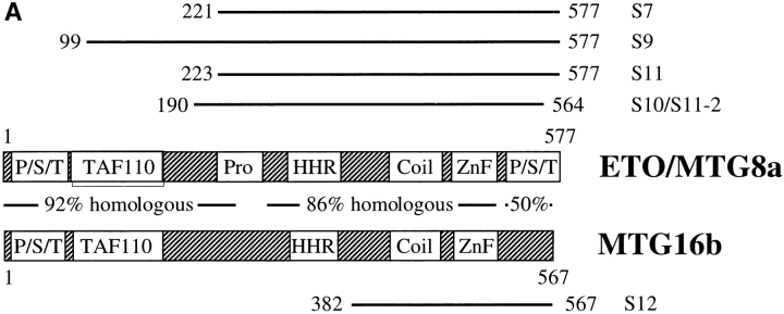
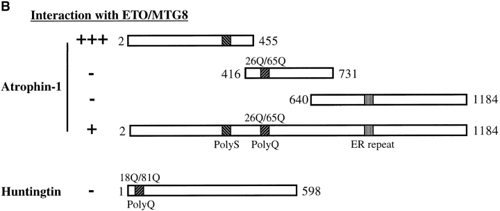
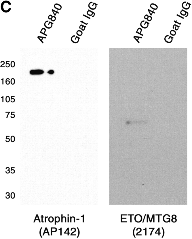
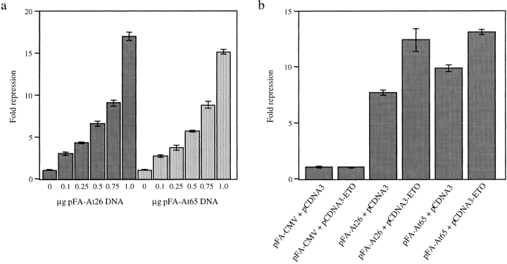
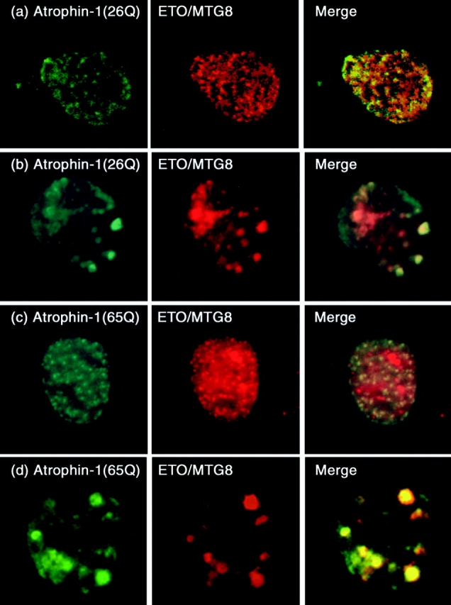
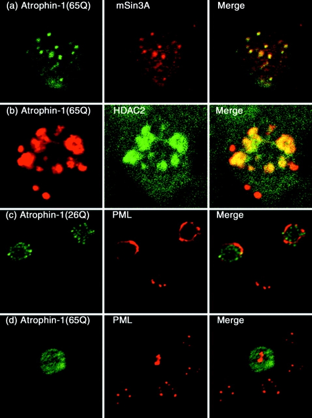
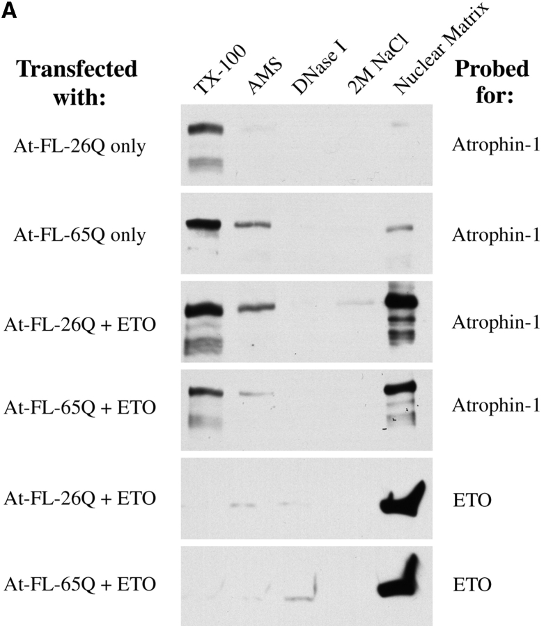
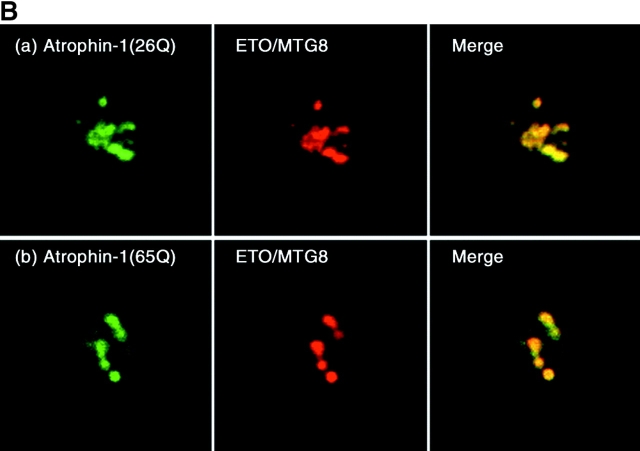
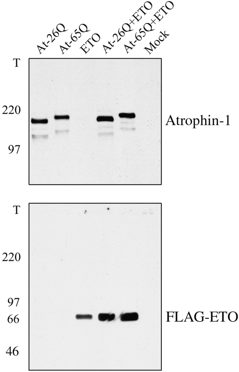
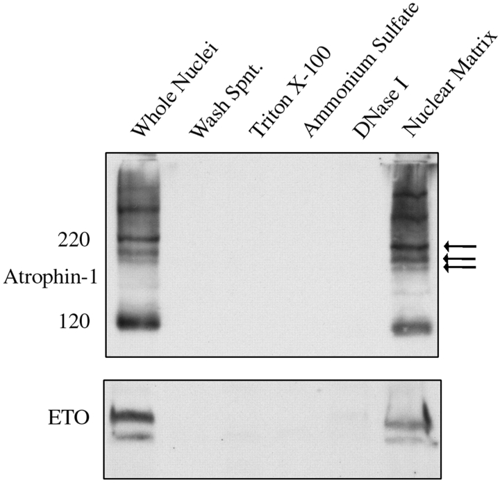
Dentato-rubral and pallido-luysian atrophy (DRPLA) is one of the family of neurodegenerative diseases caused by expansion of a polyglutamine tract. The drpla gene product, atrophin-1, is widely expressed, has no known function or activity, and is found in both the nuclear and cytoplasmic compartments of neurons. Truncated fragments of atrophin-1 accumulate in neuronal nuclei in a transgenic mouse model of DRPLA, and may underlie the disease phenotype. Using the yeast two-hybrid system, we identified ETO/MTG8, a component of nuclear receptor corepressor complexes, as an atrophin-1-interacting protein. When cotransfected into Neuro-2a cells, atrophin-1 and ETO/MTG8 colocalize in discrete nuclear structures that contain endogenous mSin3A and histone deacetylases. These structures are sodium dodecyl sulfate-soluble and associated with the nuclear matrix. Cotransfection of ETO/MTG8 with atrophin-1 recruits atrophin-1 to the nuclear matrix, while atrophin-1 and ETO/MTG8 cofractionate in nuclear matrix preparations from brains of DRPLA transgenic mice. Furthermore, in a cell transfection-based assay, atrophin-1 represses transcription. Together, these results suggest that atrophin-1 associates with nuclear receptor corepressor complexes and is involved in transcriptional regulation. Emerging links between disease-associated polyglutamine proteins, nuclear receptors, translocation-leukemia proteins, and the nuclear matrix may have important repercussions for the pathobiology of this family of neurodegenerative disorders.


Figures

Figure 1
Figure 1
Atrophin-1 interacts with ETO/MTG8. (A) Summary of data obtained from yeast two-hybrid screening. The degree of homology between the proteins is illustrated between the schematics. Lines represent clones isolated by yeast two-hybrid screening of a human cerebellum library with pPC97-At-2-455. P/S/T, proline/serine/threonine–rich domain; TAF110, region with homology to yeast transcription activating factor 110; Pro, proline-rich region; HHR, hydrophobic heptad repeat; Coil, coiled-coil domain; ZnF, zinc finger-like domains. (B) ETO/MTG8 interacts specifically with the NH2-terminal third of atrophin-1. No interaction was detected with the central portion of atrophin-1 containing the polyglutamine tract (with either 26 or 65 glutamines), the COOH-terminal half of atrophin-1, or the NH2-terminal 598 amino acids of huntingtin (with 18 or 81 glutamines). (C) Coimmunoaffinity purification of atrophin-1 and ETO/MTG8. Atrophin-1 protein complexes were purified with CarboLink-APG840 beads and immunoblotted for atrophin-1 (left) and ETO/MTG8 (right). Control immunoaffinity purifications were performed with CarboLink goat IgG.
Figure 1
Figure 1
Atrophin-1 interacts with ETO/MTG8. (A) Summary of data obtained from yeast two-hybrid screening. The degree of homology between the proteins is illustrated between the schematics. Lines represent clones isolated by yeast two-hybrid screening of a human cerebellum library with pPC97-At-2-455. P/S/T, proline/serine/threonine–rich domain; TAF110, region with homology to yeast transcription activating factor 110; Pro, proline-rich region; HHR, hydrophobic heptad repeat; Coil, coiled-coil domain; ZnF, zinc finger-like domains. (B) ETO/MTG8 interacts specifically with the NH2-terminal third of atrophin-1. No interaction was detected with the central portion of atrophin-1 containing the polyglutamine tract (with either 26 or 65 glutamines), the COOH-terminal half of atrophin-1, or the NH2-terminal 598 amino acids of huntingtin (with 18 or 81 glutamines). (C) Coimmunoaffinity purification of atrophin-1 and ETO/MTG8. Atrophin-1 protein complexes were purified with CarboLink-APG840 beads and immunoblotted for atrophin-1 (left) and ETO/MTG8 (right). Control immunoaffinity purifications were performed with CarboLink goat IgG.
Figure 4
Figure 4
Transcriptional repression by atrophin-1. (a) Dose-dependent repression of a luciferase reporter gene by GAL4 DBD-At26Q and GAL4 DBD-At65Q. The luciferase reporter construct contained four GAL4-binding sites upstream of a thymidine kinase promoter. Luciferase activity was normalized to β-galactosidase activity. n = 3 in both cases. (b) Cotransfection of ETO/MTG8 with GAL4 DBD-At26/65Q enhanced repression by atrophin-1. n = 3. Error bars represent SD.
Figure 2
Figure 2
Atrophin-1 and ETO/MTG8 colocalize in large discrete subnuclear structures in cotransfected Neuro-2a cells. At-26Q plus FLAG-ETO/MTG8 (a and b) and At-65Q plus FLAG-ETO/MTG8 (c and d) cotransfected cells were double-labeled for atrophin-1 (green, left) and FLAG (red, center). The merged images are illustrated on the right.
Figure 3
Figure 3
Atrophin-1– and ETO/MTG8-containing nuclear structures stain for mSin3A and histone deacetylases, but not PML. (a and b) At-65Q and FLAG-ETO/MTG8 cotransfected Neuro-2a cells were stained for atrophin-1 (green, left) and mSin3A (red, center) and for atrophin-1 (red, left) and HDAC2 (green, center). The merged images (right) show that endogenous mSin3A and HDAC2 colocalize with transfected atrophin-1. (c and d) At-26/65Q, FLAG-ETO/MTG8 and PML cotransfected Neuro-2a cells were stained for atrophin-1 (green, left) and PML (red, center). The merged images (right) show that PML does not colocalize with atrophin-1. Cells expressing PML alone contained PODs, whereas cells coexpressing atrophin-1 did not appear to contain PODs; PML was localized to larger nuclear structures or the nuclear periphery in the presence of atrophin-1. Similar results were obtained by double labeling for FLAG and PML.
Figure 6
Figure 6
Expression of ETO/MTG8 recruits atrophin-1 to the nuclear matrix fraction. (A) Nuclear matrix fractions were prepared from transfected Neuro-2a cells by sequential extraction or digestion with 0.5% Triton X-100, 250 mM (NH4)2SO4, 300 U/ml DNase I, and 2 M NaCl. The supernatants from these treatments and the final matrix preparations were immunoblotted for atrophin-1 and FLAG-ETO/MTG8. When transfected alone, most atrophin-1 was extracted with Triton X-100, although some atrophin-1 was associated with the insoluble matrix preparation (top two rows). When cotransfected with FLAG-ETO/MTG8, a substantial proportion of atrophin-1 was recruited to the matrix fraction (center two rows), where FLAG-ETO/MTG8 was mainly localized (bottom two rows). (B) Immunocytochemical staining of nuclear matrix preparations from transfected Neuro-2a cells in situ. The large structures containing atrophin-1 and FLAG-ETO/MTG8 remained attached to the nuclear matrix whereas the diffuse label observed after routine immunocytochemical staining was washed away by the extraction procedures.
Figure 6
Figure 6
Expression of ETO/MTG8 recruits atrophin-1 to the nuclear matrix fraction. (A) Nuclear matrix fractions were prepared from transfected Neuro-2a cells by sequential extraction or digestion with 0.5% Triton X-100, 250 mM (NH4)2SO4, 300 U/ml DNase I, and 2 M NaCl. The supernatants from these treatments and the final matrix preparations were immunoblotted for atrophin-1 and FLAG-ETO/MTG8. When transfected alone, most atrophin-1 was extracted with Triton X-100, although some atrophin-1 was associated with the insoluble matrix preparation (top two rows). When cotransfected with FLAG-ETO/MTG8, a substantial proportion of atrophin-1 was recruited to the matrix fraction (center two rows), where FLAG-ETO/MTG8 was mainly localized (bottom two rows). (B) Immunocytochemical staining of nuclear matrix preparations from transfected Neuro-2a cells in situ. The large structures containing atrophin-1 and FLAG-ETO/MTG8 remained attached to the nuclear matrix whereas the diffuse label observed after routine immunocytochemical staining was washed away by the extraction procedures.
Figure 5
Figure 5
Atrophin-1 and FLAG-ETO/MTG8 structures are SDS-soluble. Neuro-2a cells were transfected with 5 μg pCDNA3-At-26Q + 5 μg pCDNA3 (At-26Q), 5 μg pCDNA3-At-65Q + 5 μg pCDNA3 (At-65Q), 5 μg pCDNA3-FLAG-ETO/MTG8 + 5 μg pCDNA3 (ETO), 5 μg pCDNA3-At-26Q + 5 μg pCDNA3-FLAG-ETO/MTG8 (At-26Q+ETO), 5 μg pCDNA3-At-65Q + 5 μg pCDNA3-FLAG-ETO/MTG8 (At-65Q+ETO), or 10 μg pCDNA3 (mock). Total cell extracts were immunoblotted for atrophin-1 (top) and FLAG-ETO/MTG8 (bottom). No immunoreactivity was detected in the top of the wells in any of the sample lanes with either antibody, demonstrating that the atrophin-1/ETO structures are completely SDS-soluble.
Figure 7
Figure 7
Atrophin-1 and ETO/MTG8 cofractionate with the nuclear matrix fraction of DRPLA transgenic mouse brains. Nuclei were isolated from 6-mo-old AT-FL-65Q-150 DRPLA transgenic mice then extracted/digested sequentially with 0.5% Triton X-100, 250 mM (NH4)2SO4, and 300 U/ml DNase I. The supernatants from these treatments and the insoluble matrix fraction were immunoblotted for atrophin-1 (antibody AP142, top) and ETO/MTG8 (antibody 2174, bottom). The majority of the immunoreactivity for both proteins was associated with the matrix preparation. The atrophin-1 blot shows a triplet of bands at around 190–220 kD. The faint lower band (bottom arrow) corresponds to full-length endogenous atrophin-1, the middle band (center arrow) corresponds to full-length transgene-derived mutant atrophin-1, and the upper band (top arrow) is an expanded polyglutamine-specific modified form of atrophin-1. The identity of these bands was ascertained by comparing the banding patterns in extracts from nontransgenic mice, DRPLA transgenic mice, human DRPLA cerebella, and human control cerebella. Other high molecular weight species and truncated forms of atrophin-1 (the predominate band at ∼120 kD) were also associated with the matrix preparation.

Similar articles

Cited by

References

    1. Boutell J.M., Thomas P., Neal J.W., Weston V.J., Duce J., Harper P.S., Jones A.L. Aberrant interactions of transcriptional repressor proteins with the Huntington's disease gene product, huntingtin. Hum. Mol. Genet. 1999;8:1647–1655. - PubMed
    1. Cha J.H., Kosinski C.M., Kerner J.A., Alsdorf S.A., Mangiarini L., Davies S.W., Penney J.B., Bates G.P., Young A.B. Altered brain neurotransmitter receptors in transgenic mice expressing a portion of an abnormal human huntington disease gene. Proc. Natl. Acad. Sci. USA. 1998;95:6480–6485. - PMC - PubMed
    1. Chai Y., Koppenhafer S.L., Shoesmith S.J., Perez M.K., Paulson H.L. Evidence for proteasome involvement in polyglutamine diseaselocalization to nuclear inclusions in SCA3/MJD and suppression of polyglutamine aggregation in vitro. Hum. Mol. Genet. 1999;8:673–682. - PubMed
    1. Chen L.F., Ito K., Murakami Y., Ito Y. The capacity of polyomavirus enhancer binding protein 2alphaB (AML1/Cbfa2) to stimulate polyomavirus DNA replication is related to its affinity for the nuclear matrix. Mol. Cell. Biol. 1998;18:4165–4176. - PMC - PubMed
    1. Chevray P.M., Nathans D. Protein interaction cloning in yeastidentification of mammalian proteins that react with the leucine zipper of Jun. Proc. Natl. Acad. Sci. USA. 1992;89:5789–5793. - PMC - PubMed

Publication types

MeSH terms