. 2000 Sep 4;150(5):949-62.
doi: 10.1083/jcb.150.5.949.

The ER repeat protein YT521-B localizes to a novel subnuclear compartment

Affiliations

The ER repeat protein YT521-B localizes to a novel subnuclear compartment

O Nayler et al. J Cell Biol. .

Abstract

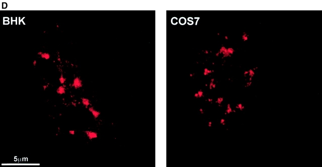
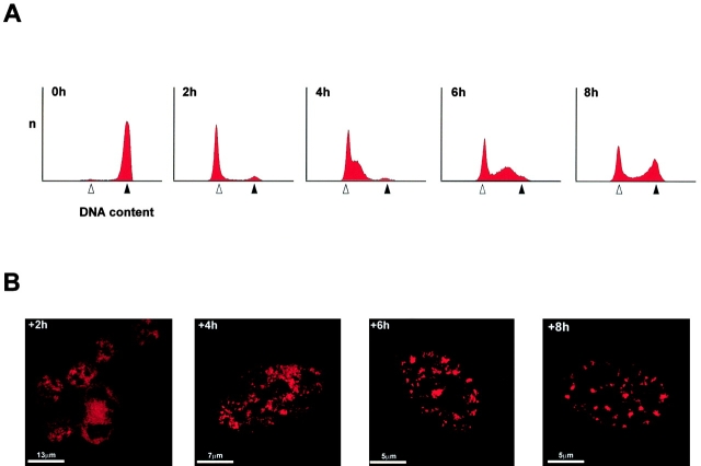
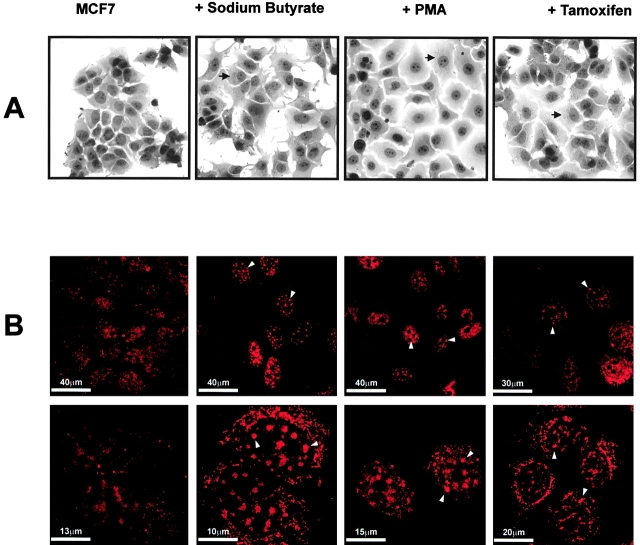
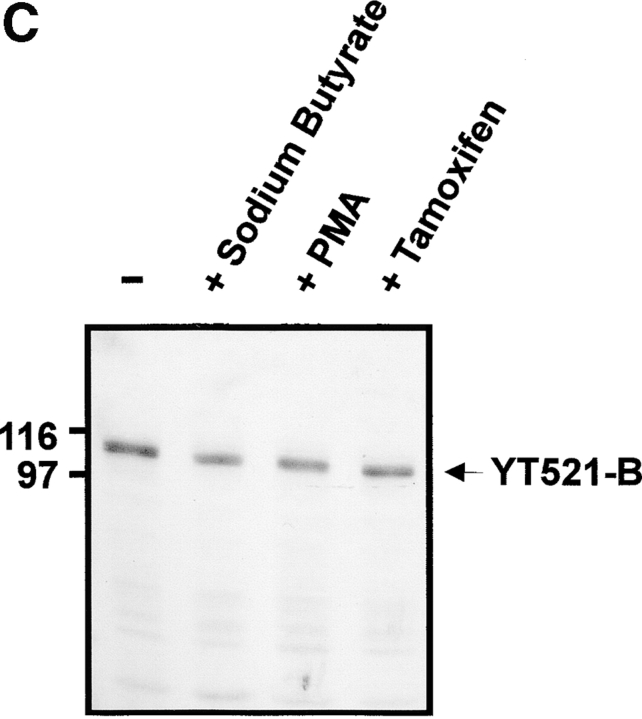
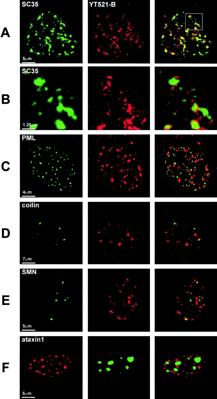
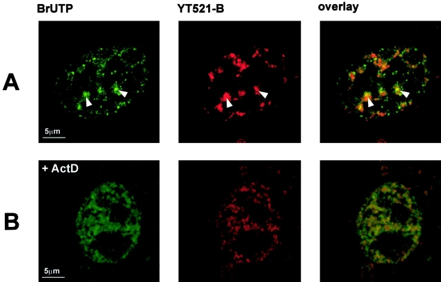
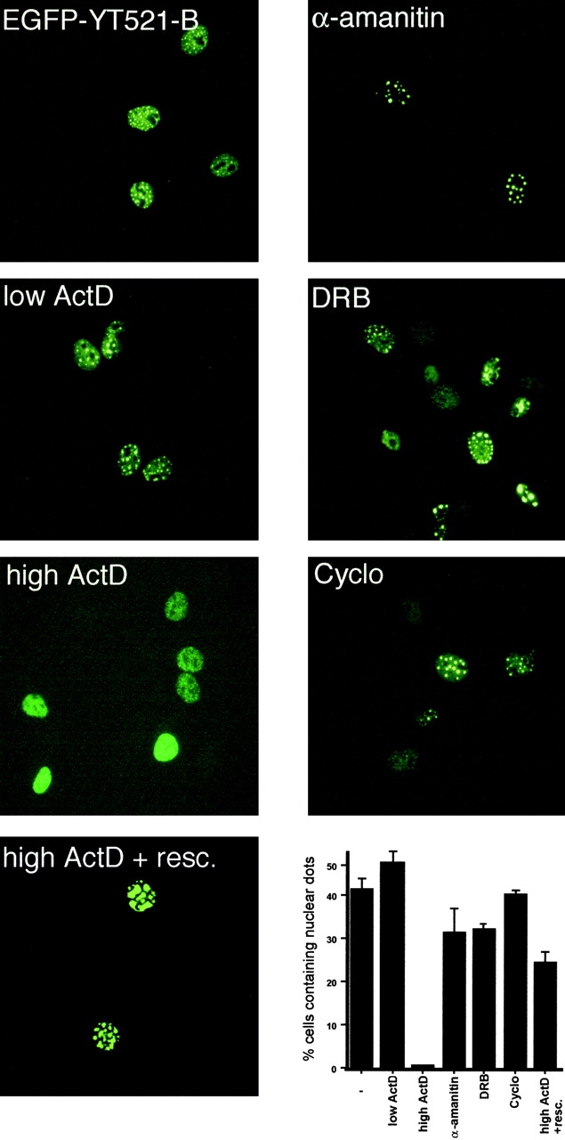
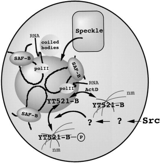
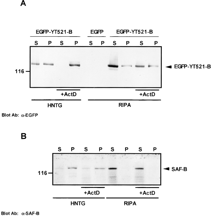
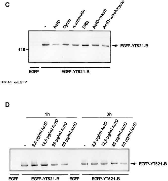
The characterization of distinct subnuclear domains suggests a dynamic nuclear framework supporting gene expression and DNA replication. Here, we show that the glutamic acid/arginine-rich domain protein YT521-B localizes to a novel subnuclear structure, the YT bodies. YT bodies are dynamic compartments, which first appear at the beginning of S-phase in the cell cycle and disperse during mitosis. Furthermore, in untreated cells of the human cell line MCF7 they were undetectable and appeared only after drug- induced differentiation. YT bodies contain transcriptionally active sites and are in close contact to other subnuclear structures such as speckles and coiled bodies. YT bodies disperse upon actinomycin D treatment, whereas other transcriptional inhibitors such as alpha-amanitin or DRB have little effect. On the basis of our experiments, we propose that YT521-B may participate in the assembly of genes into transcription centers, thereby allowing efficient regulation of gene expression.


Figures

Figure 1
Figure 1
Specificity of the anti–YT521-B antiserum. (A) Total RIPA/benzonase cell lysates of HEK293 cells expressing EGFP or EGFP-YT521-B were loaded onto 10% SDS-PAGE gels and analyzed by Western blot using the anti–YT521-B antiserum PK2 in the absence (−peptide) or presence (+peptide) of the antigenic YT521-B peptide. (B) Reblot of (A) using an anti-EGFP antibody. (C) Total COS7 RIPA and benzonase cell lysate (CL) was analyzed by SDS-PAGE and Western blot using anti–YT521-B antiserum PK2. The YT521-B proteins detected are indicated by arrows. Molecular mass standards are given in kD. (D) BHK (left) and COS7 cells (right) were fixed in 3.7% formaldehyde and immunostained with the anti–YT521-B antiserum PK2 using a secondary CY3-coupled antibody for detection.
Figure 1
Figure 1
Specificity of the anti–YT521-B antiserum. (A) Total RIPA/benzonase cell lysates of HEK293 cells expressing EGFP or EGFP-YT521-B were loaded onto 10% SDS-PAGE gels and analyzed by Western blot using the anti–YT521-B antiserum PK2 in the absence (−peptide) or presence (+peptide) of the antigenic YT521-B peptide. (B) Reblot of (A) using an anti-EGFP antibody. (C) Total COS7 RIPA and benzonase cell lysate (CL) was analyzed by SDS-PAGE and Western blot using anti–YT521-B antiserum PK2. The YT521-B proteins detected are indicated by arrows. Molecular mass standards are given in kD. (D) BHK (left) and COS7 cells (right) were fixed in 3.7% formaldehyde and immunostained with the anti–YT521-B antiserum PK2 using a secondary CY3-coupled antibody for detection.
Figure 2
Figure 2
YT521-B structures are regulated during the cell cycle. BHK cells were arrested at the G2/M phase using nocodazole and then incubated in absence of the drug for 2, 4, 6, and 8 h (as indicated). Cells were then fixed and used for FACS analysis or stained with anti–YT521 antiserum (PK2). A. FACS analysis of the cells. An open arrow indicates the G1, a closed arrow the G2/M population of cells. (B) Images of representative cells at the various time points.
Figure 3
Figure 3
YT521-B structures are markers for the drug-induced differentiation of the cell line MCF7. MCF7 cells were incubated in the presence of sodium butyrate, PMA, tamoxifen, or without drugs for 48 h. (A) The degree of differentiation was assessed by phase contrast microscopy after fixation in 2% formaldehyde with oil red O staining. The lipid droplet inclusions are shown with an arrow. (B) After drug treatment, MCF7 cells were fixed in 3.7% formaldehyde, and immunostaining was performed using the anti–YT521-B antiserum, followed by CY3-coupled secondary antibodies. The cells are shown at two different magnifications as indicated. Arrows indicate the formation of distinct YT bodies. The cytoplasmatic staining observed in differentiated cells is independent of the YT521-B antiserum and represents most likely an unspecific binding of the secondary antibody to lipid containing components. (C) A portion of the MCF7 cells were lysed in RIPA and benzonase after drug treatment and equal amounts (20 μg) of the soluble fraction were loaded on 10% SDS-PAGE, followed by Western blot analysis using the anti–YT521-B antiserum PK2. The detected endogenous YT521-B protein is indicated by an arrow; the molecular mass markers are given in kD.
Figure 3
Figure 3
YT521-B structures are markers for the drug-induced differentiation of the cell line MCF7. MCF7 cells were incubated in the presence of sodium butyrate, PMA, tamoxifen, or without drugs for 48 h. (A) The degree of differentiation was assessed by phase contrast microscopy after fixation in 2% formaldehyde with oil red O staining. The lipid droplet inclusions are shown with an arrow. (B) After drug treatment, MCF7 cells were fixed in 3.7% formaldehyde, and immunostaining was performed using the anti–YT521-B antiserum, followed by CY3-coupled secondary antibodies. The cells are shown at two different magnifications as indicated. Arrows indicate the formation of distinct YT bodies. The cytoplasmatic staining observed in differentiated cells is independent of the YT521-B antiserum and represents most likely an unspecific binding of the secondary antibody to lipid containing components. (C) A portion of the MCF7 cells were lysed in RIPA and benzonase after drug treatment and equal amounts (20 μg) of the soluble fraction were loaded on 10% SDS-PAGE, followed by Western blot analysis using the anti–YT521-B antiserum PK2. The detected endogenous YT521-B protein is indicated by an arrow; the molecular mass markers are given in kD.
Figure 4
Figure 4
YT521-B protein localizes to a novel subnuclear compartment. BHK cells were fixed with 3.7% formaldehyde and immunostained with antibodies against nuclear markers (left) and YT521-B (middle). The following proteins were used: (A and B) SC35, B is an enlargement of the area indicated with a box in A; (C) PML; (D) p80-coilin; (E) SMN. In (F), cells were cotransfected with pFlagSCA1[82] (red) and pEGFP-YT521-B (green). In A–E, YT521-B was detected using a CY3-coupled secondary antibody (red) and the other nuclear component was visualized with a FITC labeled secondary antibody (green).
Figure 5
Figure 5
YT bodies contain focal sites of transcription. (A and B) BHK cells were permeabilized with digitonin. After a 20-min incubation period at 30°C in transcription buffer in the absence of transcription inhibitors (A), or 50 μg/ml actinomycin D (B), cells were fixed. BrUTP incorporation was detected using anti-BrdU antibodies and CY3-coupled secondary antibodies. YT521-B was detected with PK2 antiserum. The localization of YT521-B (red) and sites of BrUTP incorporation (green) are shown. Colocalization is visible in the overlay (orange). Some specific sites of colocalization are highlighted with an arrow. The data are the averages of three independent experiments and 150 cells were analyzed in each.
Figure 6
Figure 6
Actinomycin D reversibly dissolves YT bodies. COS7 cells were transiently transfected with pEGFP-YT521-B and treated with 50 μg/ml α-amanitin for 5 h (α-amanitin), 0.04 μg/ml actinomycin D for 3 h (low ActD), 75 μM DRB for 3 h (DRB), 50 μg/ml actinomycin D for 3 h (high ActD), 100 μg/ml cycloheximide for 5 h (Cyclo), or they were treated with 50 μg/ml actinomycin D, followed by a 2-h incubation period in the absence of the drug (high ActD + resc.). The percentage of cells containing YT521-B domains was determined by counting individual cells in three independent experiments. The data are the averages of three independent experiments (±SD) and 150 cells were counted in each.
Figure 8
Figure 8
YT bodies define focal sites of transcription in the absence of RNA polymerase activity: a model. YT bodies (indicated in dark gray) combine different chromosomal loops into focal transcription sites in the absence of RNA polymerase activity, possibly through interaction with scaffold attachment factor-B (SAF-B) and DNA. Coiled bodies or speckles (which are often found in close contact with YT bodies) serve as accessory domains supplying the transcriptionally active site with necessary helper factors. The YT-body is dynamically regulated by signal transduction pathways emanating from members of the Src kinase family. nm, Nuclear matrix.
Figure 7
Figure 7
YT bodies are dynamic, Triton X-100–soluble compartments. (A) HEK293 cells were transiently transfected with pEGFP-YT521-B, incubated with 50 μg/ml actinomycin D for 3 h, and lysed in RIPA and benzonase or HNTG. Equal amounts of the soluble fraction (S) or redissolved pellet fraction (P) were loaded onto 10% SDS-PAGE gels. Proteins were analyzed by Western blot and ECL using the anti-EGFP antibody. (B) HEK 293 cells incubated with 50 μg/ml actinomycin D for 3 h and lysed in RIPA and benzonase or HNTG. Equal amounts of the soluble fraction (S) or redissolved pellet fraction (P) were loaded onto 10% SDS-PAGE gels. Proteins were analyzed by Western blot and ECL using the anti–SAF-B antibody. (C) HEK293 cells were transiently transfected with EGFP (control) or pEGFP-YT521-B. The pEGFP-YT521-B transfected cells were then incubated with 50 μg/ml actinomycin D for 3 h (ActD), 100 μg/ml cycloheximide for 5 h (Cyclo), 50 μg/ml α-amanitin for 5 h (α-amanitin), 75 μM DRB for 3 h, or they were incubated with 50 μg/ml actinomycin D for 3 h, followed by a 2-h actinomycin D–free incubation in the presence (ActD+wash/cyclo) or the absence (actD+wash) of 100 μg/ml cycloheximide. Equal amounts of the soluble fractions of HNTG cell lysates were analyzed on 10% SDS-PAGE and Western blotting using the anti-EGFP antibody. (D) HEK293 cells were transiently transfected with EGF (control) or pEGFP-YT521-B. The pEGFP-YT521-B–transfected cells were then incubated with increasing amounts of actinomycin D for 1 or 3 h. Equal amounts of the soluble fractions of HNTG cell lysates were analyzed on 10% SDS-PAGE and Western blot using the anti-EGFP antibody. The detected proteins are indicated by arrows; the molecular mass is indicated in kD.
Figure 7
Figure 7
YT bodies are dynamic, Triton X-100–soluble compartments. (A) HEK293 cells were transiently transfected with pEGFP-YT521-B, incubated with 50 μg/ml actinomycin D for 3 h, and lysed in RIPA and benzonase or HNTG. Equal amounts of the soluble fraction (S) or redissolved pellet fraction (P) were loaded onto 10% SDS-PAGE gels. Proteins were analyzed by Western blot and ECL using the anti-EGFP antibody. (B) HEK 293 cells incubated with 50 μg/ml actinomycin D for 3 h and lysed in RIPA and benzonase or HNTG. Equal amounts of the soluble fraction (S) or redissolved pellet fraction (P) were loaded onto 10% SDS-PAGE gels. Proteins were analyzed by Western blot and ECL using the anti–SAF-B antibody. (C) HEK293 cells were transiently transfected with EGFP (control) or pEGFP-YT521-B. The pEGFP-YT521-B transfected cells were then incubated with 50 μg/ml actinomycin D for 3 h (ActD), 100 μg/ml cycloheximide for 5 h (Cyclo), 50 μg/ml α-amanitin for 5 h (α-amanitin), 75 μM DRB for 3 h, or they were incubated with 50 μg/ml actinomycin D for 3 h, followed by a 2-h actinomycin D–free incubation in the presence (ActD+wash/cyclo) or the absence (actD+wash) of 100 μg/ml cycloheximide. Equal amounts of the soluble fractions of HNTG cell lysates were analyzed on 10% SDS-PAGE and Western blotting using the anti-EGFP antibody. (D) HEK293 cells were transiently transfected with EGF (control) or pEGFP-YT521-B. The pEGFP-YT521-B–transfected cells were then incubated with increasing amounts of actinomycin D for 1 or 3 h. Equal amounts of the soluble fractions of HNTG cell lysates were analyzed on 10% SDS-PAGE and Western blot using the anti-EGFP antibody. The detected proteins are indicated by arrows; the molecular mass is indicated in kD.

Similar articles

Cited by

References

    1. Alblas J., Slager-Davidov R., Steenbergh P.H., Sussenbach J.S., van der Burg B. The role of MAP kinase in TPA-mediated cell cycle arrest of human breast cancer cells. Oncogene. 1998;16:131–139. - PubMed
    1. Almeida F., Saffrich R., Ansorge W., Carmo-Fonseca M. Microinjection of anti-coilin antibodies affects the structure of coiled bodies. J. Cell Biol. 1998;142:899–912. - PMC - PubMed
    1. Ashraf S.I., Ip Y.T. Transcriptional controlrepression by local chromatin modification. Curr. Biol. 1998;8:R683–R686. - PubMed
    1. Assier E., Bouzinba-Segard H., Stolzenberg M.-C., Stephens R., Bardos J., Freemont P., Charron D., Trowsdale J., Rich T. Isolation, sequencing and expression of RED, a novel human gene encoding anacidic-basic dipeptide repeat. Gene. 1999;230:145–154. - PubMed
    1. Carmo-Fonseca M., Pepperkok R., Carvalho M.T., Lamond A. Transcription-dependent colocalization of the U1, U2, U4/5 and U5 snRNPs in coiled bodies. J. Cell Biol. 1992;117:1–14. - PMC - PubMed

Publication types

MeSH terms