. 2000 Sep 4;150(5):963-74.
doi: 10.1083/jcb.150.5.963.

Nuclei and microtubule asters stimulate maturation/M phase promoting factor (MPF) activation in Xenopus eggs and egg cytoplasmic extracts

Affiliations

Nuclei and microtubule asters stimulate maturation/M phase promoting factor (MPF) activation in Xenopus eggs and egg cytoplasmic extracts

D Pérez-Mongiovi et al. J Cell Biol. .

Abstract

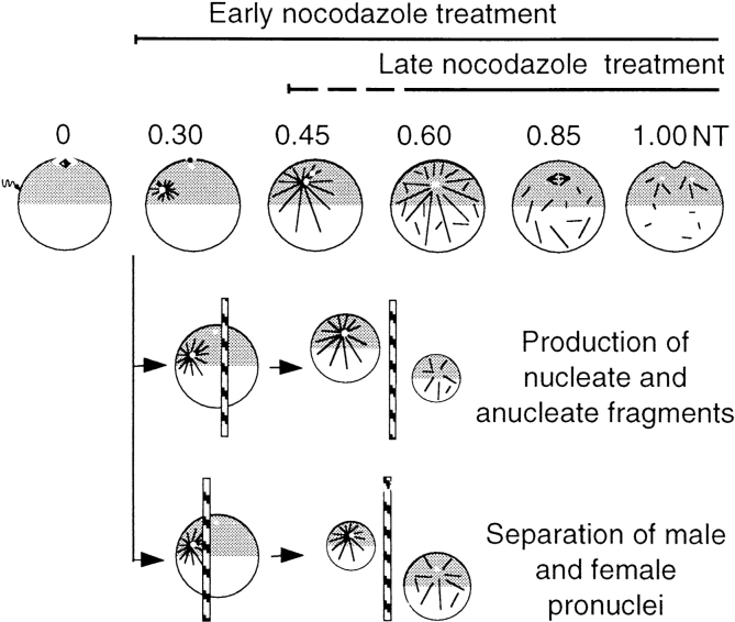
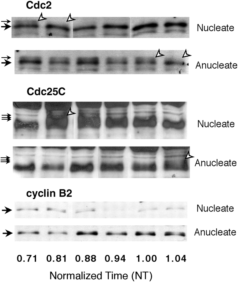
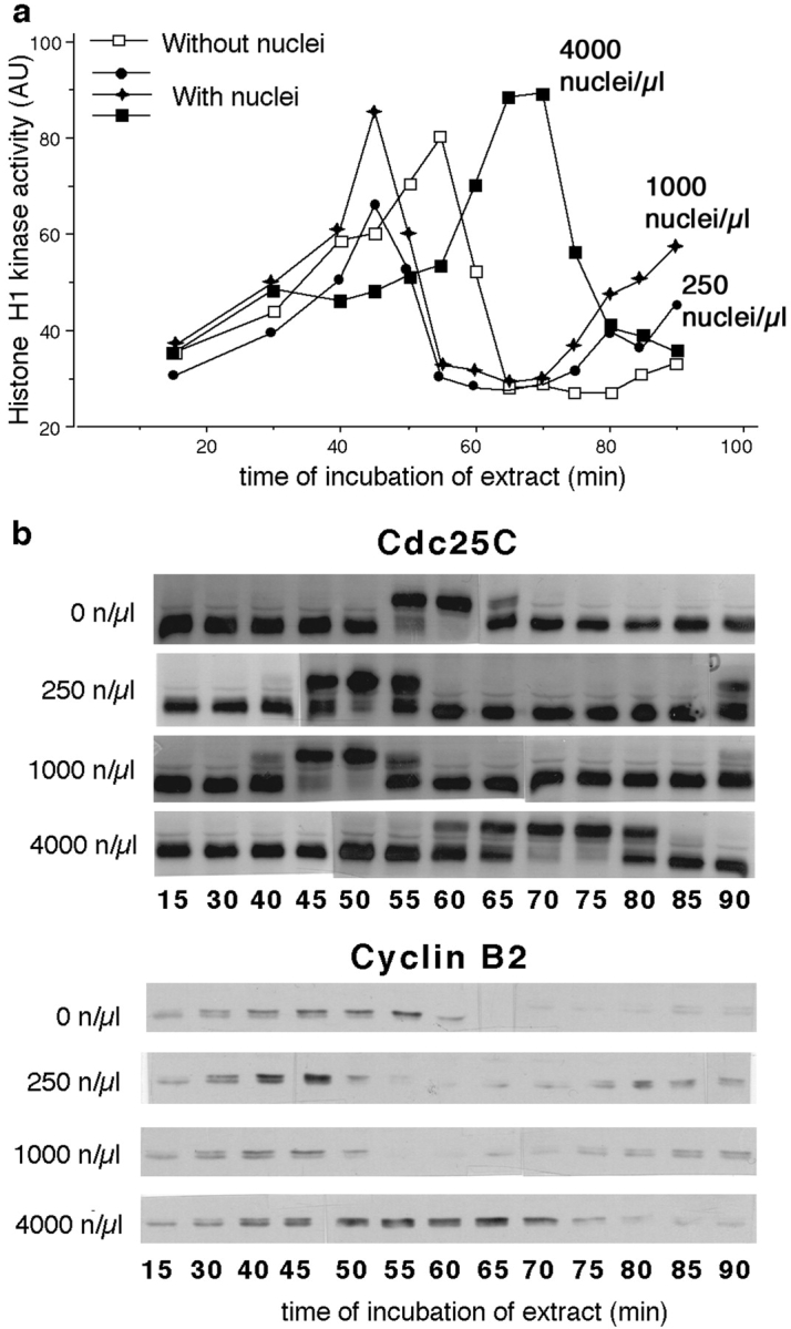
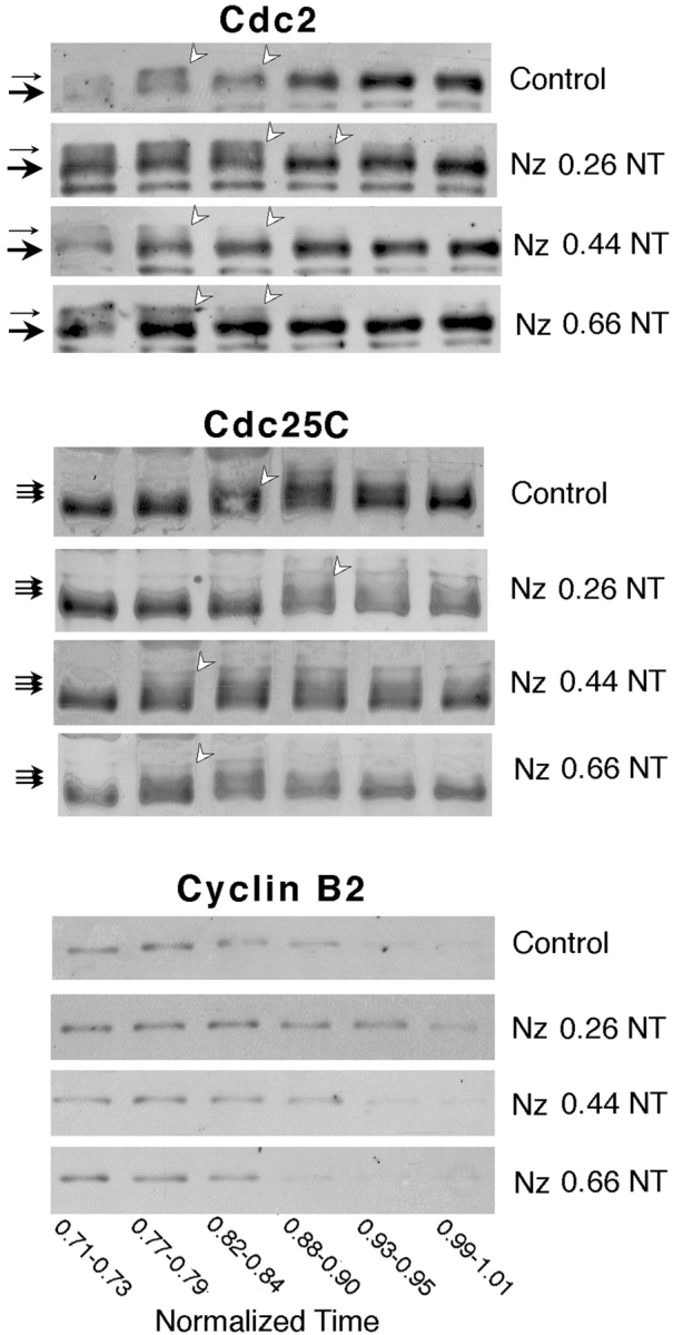
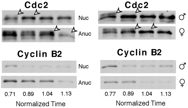
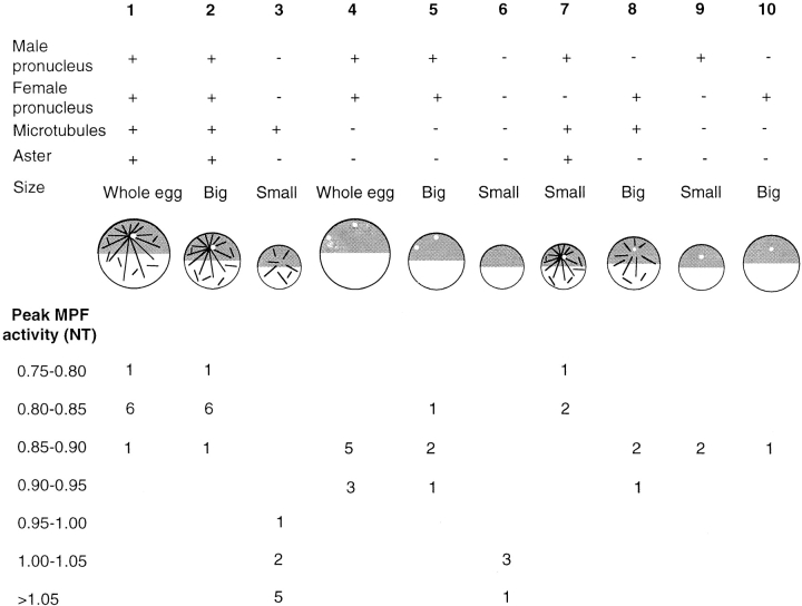
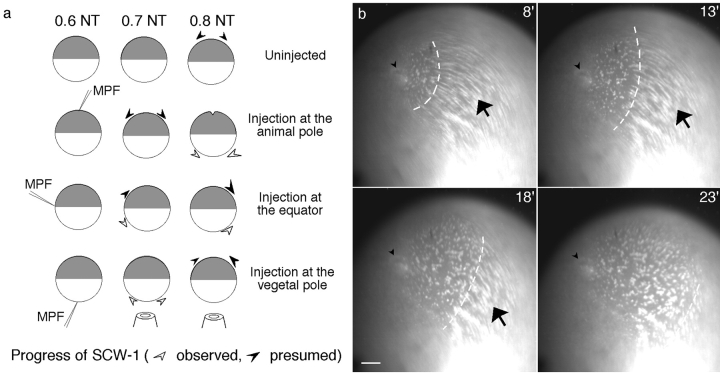
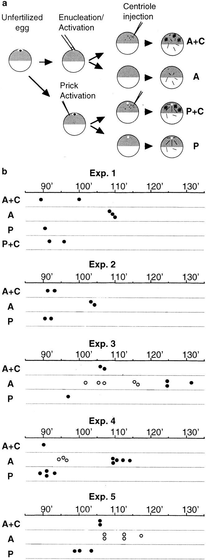
Although maturation/M phase promoting factor (MPF) can activate autonomously in Xenopus egg cytoplasm, indirect evidence suggests that nuclei and centrosomes may focus activation within the cell. We have dissected the contribution of these structures to MPF activation in fertilized eggs and in egg fragments containing different combinations of nuclei, centrosomes, and microtubules by following the behavior of Cdc2 (the kinase component of MPF), the regulatory subunit cyclin B, and the activating phosphatase Cdc25. The absence of the entire nucleus-centrosome complex resulted in a marked delay in MPF activation, whereas the absence of the centrosome alone caused a lesser delay. Nocodazole treatment to depolymerize microtubules through first interphase had an effect equivalent to removing the centrosome. Furthermore, microinjection of isolated centrosomes into anucleate eggs promoted MPF activation and advanced the onset of surface contraction waves, which are close indicators of MPF activation and could be triggered by ectopic MPF injection. Finally, we were able to demonstrate stimulation of MPF activation by the nucleus-centriole complex in vitro, as low concentrations of isolated sperm nuclei advanced MPF activation in cycling cytoplasmic extracts. Together these results indicate that nuclei and microtubule asters can independently stimulate MPF activation and that they cooperate to enhance activation locally.
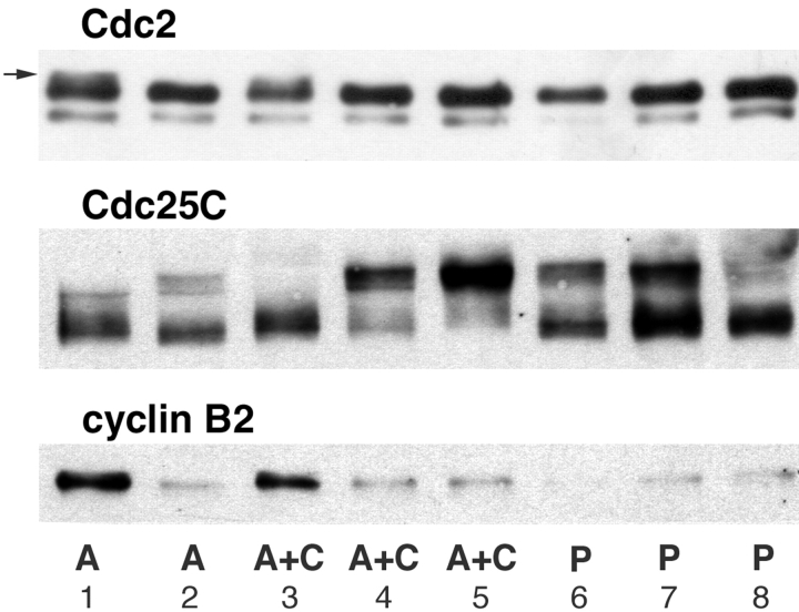

Figures

Figure 1
Figure 1
Dissection of the role of nuclei and microtubules in MPF activation in Xenopus eggs. Diagram showing the different experimental protocols used, in relation to the changing microtubule organization throughout the first cell cycle (Houliston and Elinson 1992). The centriole, brought by the sperm, recruits cytoplasmic components to form an active centrosome that nucleates a giant microtubule aster, which is first manifest by pigment accumulation around the sperm entry point in the animal cortex between 0.2 and 0.3 NT. The sperm aster mediates the migration of both male and female pronuclei to the center of the animal hemisphere where they are found closely apposed at ∼0.5 NT. At this time, general microtubule polymerization occurs throughout the egg. At ∼0.8 NT, the mitotic spindle forms in the animal hemisphere whereas cytoplasmic microtubules depolymerize. At 1.0 NT, the cleavage furrow is initiated at the animal pole. Early nocodazole treatments started between 0.2 and 0.3 NT to prevent sperm aster growth, and late treatments around 0.45 or 0.65 NT to eliminate microtubules at the time of mitosis. Nucleate and anucleate fragments were separated by placing glass rods to one side of the animal pole, thus generating a larger fragment containing the sperm aster and nuclei, and the smaller anucleate fragment. Alternatively, the rod was placed between the animal pole and the sperm entry point to produce one small fragment with the male pronucleus and attached centrosome, and a large fragment containing just the female pronucleus.
Figure 2
Figure 2
MPF activation is delayed in anucleate egg fragments. Small groups of nucleate and anucleate fragments were frozen at different times, processed for Western blotting, and probed with antibodies to detect Cdc2, Cdc25C, and cyclin B2. Approximately 0.2 egg volume equivalents were loaded in each lane. For each blot shown (as well as for those in the figures following), different molecular weight regions of the same blot were probed with the appropriate antibody. Cdc2 was detected with anti-PSTAIR antibody. The small black arrows indicate the phosphorylated (upper, inactive) isoform and the large black arrows the lower isoforms of Cdc2. The lower bands include both the inactive non-T14/Y15–phosphorylated isoform and the active T14/Y15-dephosphorylated isoform. The white arrowheads indicate the maximal detected phosphorylation and the beginning of dephosphorylation of Cdc2 (MPF activation). For Cdc25C, the small arrows mark the phosphorylated isoforms, which appear upon Cdc25C activation (white arrowheads). Activation of both Cdc2 and Cdc25C was delayed by ∼0.25 time units in anucleate fragments. Cyclin B2 accumulated similarly in both fragments until ∼0.8 NT, after which time it was degraded in nucleate but not in anucleate fragments. Reaccumulation of cyclin B2 in the second cycle is first detectable in nucleate fragments at the time of cleavage.
Figure 3
Figure 3
Nuclei stimulate MPF activation in vitro. (a) Histone H1 kinase activity during the first cell cycle of a Xenopus extract made 20 min after egg activation. Increasing volumes (0–8 μl) of nucleus suspension were added to 200 μl aliquots of the extract to give 0, 250, 1,000, and 4,000 nuclei/μl of extract, then divided into 10 μl aliquots, incubated at 21°C, and samples were taken at 5-min intervals over a 90-min period. 250 and 1,000 nuclei/μl advanced the peak of MPF activation by 10 min compared with extracts without nuclei. The delayed MPF activation in the presence of 4,000 nuclei/μl is explained by the activation of the DNA replication checkpoint (Dasso and Newport 1990). (b) Western blot performed with the same samples using anti-Cdc25C and anti-cyclin B2 antibodies. Maximum phosphorylation of Cdc25C and peak cyclin B2 coincided with peak H1 kinase activity.
Figure 4
Figure 4
Early nocodazole treatment delays MPF activation. Western blots (details and annotations as in the legend to Fig. 2) showing the behavior of Cdc2, Cdc25C, and cyclin B2 during first mitosis after treatment at different times with 10 mM nocodazole. Treatments were started at 0.26, 0.44, and 0.66 NT as indicated. The times below each lane show the period of collection and freezing of control and treated eggs. Eggs treated at 0.26 NT showed a delay in Cdc2 and Cdc25C activation of ∼0.1 time units, whereas treatments beginning at 0.44 and 0.66 NT did not. Degradation of cyclin B2 was delayed in the early nocodazole-treated eggs and appeared to degrade more slowly.
Figure 5
Figure 5
MPF activation depends on nuclei and centrosomes. Cdc2 and cyclin B2 proteins followed by Western blotting (see Fig. 2) in nucleate (Nuc) and anucleate (Anuc) fragments compared with fragments containing a single male or female pronucleus. The centrosome associated with the male pronucleus organizes a microtubule aster. Cdc2 dephosphorylation was first detectable in fragments containing both nuclei and asters, then in fragments containing the female pronucleus, and finally in anucleate fragments. Low cyclin B2 levels in the last lane of anucleate samples (left blot) partially reflects reduced loading levels (compare with Cdc2 band intensities).
Figure 6
Figure 6
Additive effects of microtubules, nuclei, and centrosomes in vivo. Combined data from ligation and nocodazole experiments in which the timing of MPF activation was assessed from blots of groups of fragments frozen at successive times with antibodies recognizing Cdc2, cyclin B2, and Cdc25C. The number of cases in which MPF activation was detected during each time interval is indicated for each category.
Figure 7
Figure 7
MPF injection provokes waves of cortical reorganization. (a) Diagram summarizing the effects of precocious injection of active MPF (human recombinant Cdc2–cyclin B) into different sites of fertilized eggs during interphase (0.6 NT). Animal pole injections (n = 2) provoked premature SCWs and cleavage furrow formation. Injection in the equatorial region (n = 2) and vegetal pole (n = 8) produced waves propagating from the injection point that were similar in appearance, although not identical to the first SCW. (b) Superimposed sequences of 10 consecutive images from 1 recording showing the wave of cortical reorganization produced by vegetal injection of MPF. White stained dots are mitochondrial islands labeled with DiOC6(3). These translocate in a coordinate fashion indicating the direction of the cortical rotation relative to the cytoplasm (arrow). The cortical rotation was progressively stopped by the passage of a wave, similar to the first SCW, propagating from the point of injection (arrowhead). The time after injection in minutes is indicated. This ectopic wave is visible as a front of stationary germ plasm islands (dashed line). Bar, 100 μm.
Figure 8
Figure 8
Centrosome injection into anucleate eggs advances SCWs. (a) Experimental design. Definitive assignment of egg categories was made at the end of each experiment based on cleavage pattern (see text). A, anucleate eggs; A+C, anucleate egg with centrosomes; P, prick-activated eggs; P+C, prick-activated eggs with centrosomes. (b) Comparison of timing of the first SCW in different egg categories in five separate experiments. Filled circles indicate the time (minutes after activation) that the first SCW crossed the vegetal hemisphere. Open circles represent the time of exaggerated germ plasm aggregation starting at the end of the cortical rotation seen in anucleate eggs in three experiments, which preceded characteristic SCWs in two of these experiments. Enucleation success for experiments (Exp.) 1, 2, 3, and 4 was 60%, and for experiment 5 was 75% (assessed from groups of 10–20 eggs cultured in parallel).
Figure 9
Figure 9
Centrosome injection into anucleate eggs stimulates MPF activation. Cdc2/Cdc25C/Cyclin B2 blots (details and annotations as in the legend to Fig. 2) of single eggs harvested simultaneously 88 min after activation from an equivalent experiment to that described in the legend to Fig. 8. At this time, the “anucleate” (A) egg in track 1 contained preactive MPF as shown by the presence of inactive Cdc2 isoforms (arrow) and cyclin B2. The “anucleate” centriole-injected (A+C) egg in lane 3 was in an equivalent premitotic state; however, the other two were strongly mitotic, with no detectable inactive, phosphorylated Cdc2, and highly phosphorylated Cdc25C. In contrast, the three prick-activated (P) eggs (lanes 6–8) have all passed through mitosis, as indicated by the partial dephosphorylation of Cdc25C and absence of cyclin B2. The migration of the three proteins in the A egg in lane 2 resembles the activated eggs, suggesting that enucleation was probably unsuccessful.

Similar articles

Cited by

References

    1. Abrieu A., Brassac T., Galas S., Fisher D., Labbé J.C., Dorée M. The Polo-like kinase Plx1 is a component of the MPF amplification loop at the G2/M-phase transition of the cell cycle in Xenopus eggs. J. Cell Sci. 1998;111:1751–1757. - PubMed
    1. Alfa C.E., Ducommun B., Beach D., Hyams J.S. Distinct nuclear and spindle pole body population of cyclin-cdc2 in fission yeast. Nature. 1990;347:680–682. - PubMed
    1. Aligue R., Wu L., Russell P. Regulation of Schizosaccharomyces pombe Wee1 tyrosine kinase. J. Biol. Chem. 1997;272:13320–13325. - PubMed
    1. Ashcroft N.R., Srayko M., Kosinski M.E., Mains P.E., Golden A. RNA-mediated interference of a cdc25 homolog in Caenorhabditis elegans results in defects in the embryonic cortical membrane, meiosis, and mitosis. Dev. Biol. 1999;206:15–32. - PubMed
    1. Bailly E., Doree M., Nurse P., Bornens M. p34cdc2 is located in both nucleus and cytoplasm; part is centrosomally associated at G2/M and enters vesicles at anaphase. EMBO (Eur. Mol. Biol. Organ.) J. 1989;8:3985–3995. - PMC - PubMed

Publication types

MeSH terms