Comparative Study
. 2000 Sep 4;150(5):989-1000.
doi: 10.1083/jcb.150.5.989.

Defects in axonal elongation and neuronal migration in mice with disrupted tau and map1b genes

Affiliations
Comparative Study

Defects in axonal elongation and neuronal migration in mice with disrupted tau and map1b genes

Y Takei et al. J Cell Biol. .

Abstract

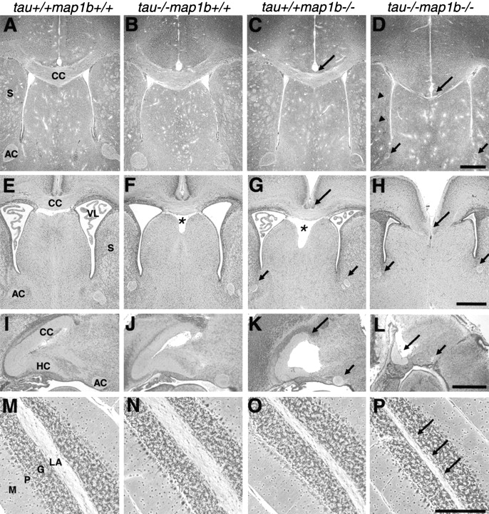
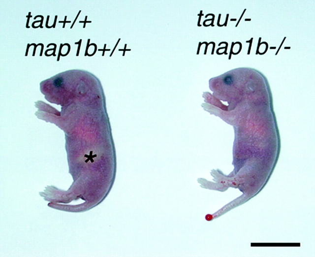
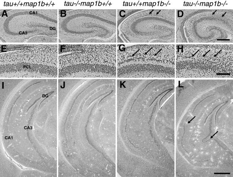
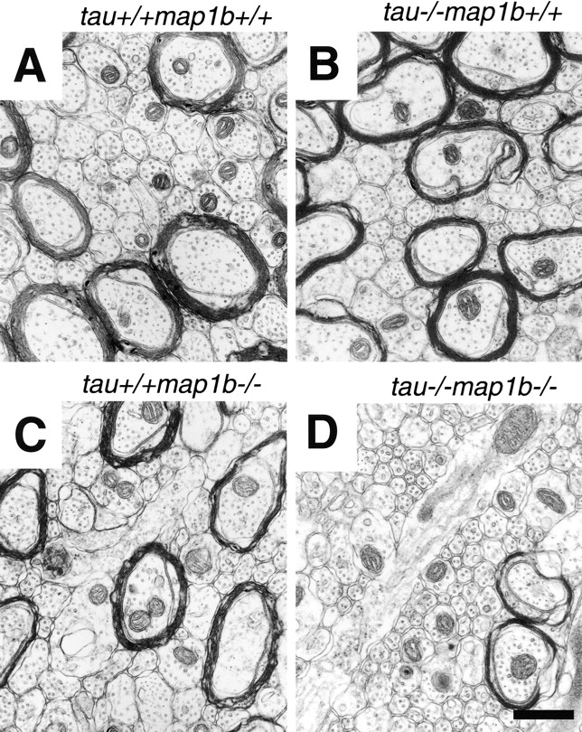
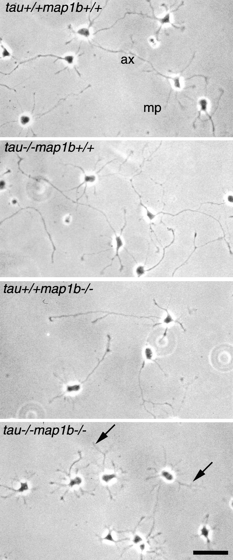
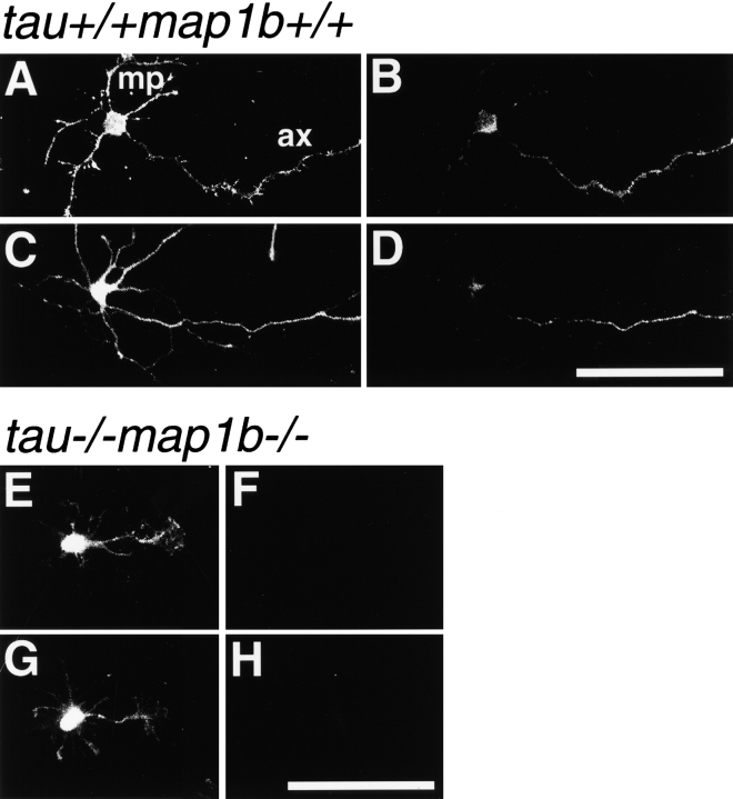
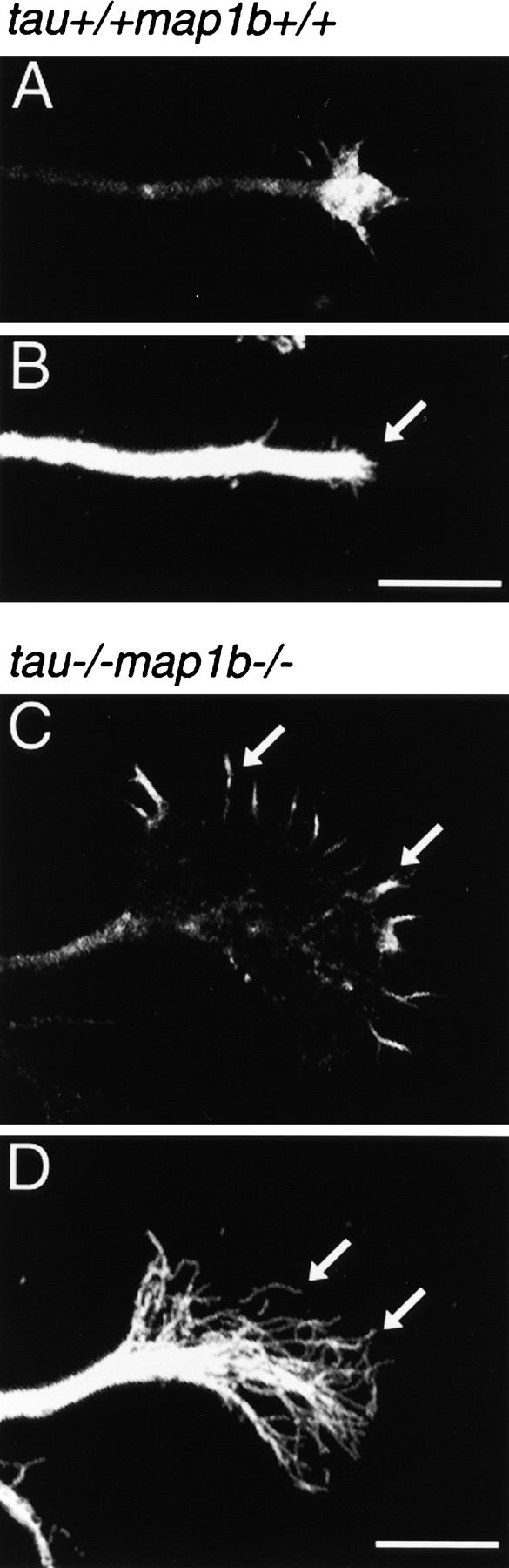
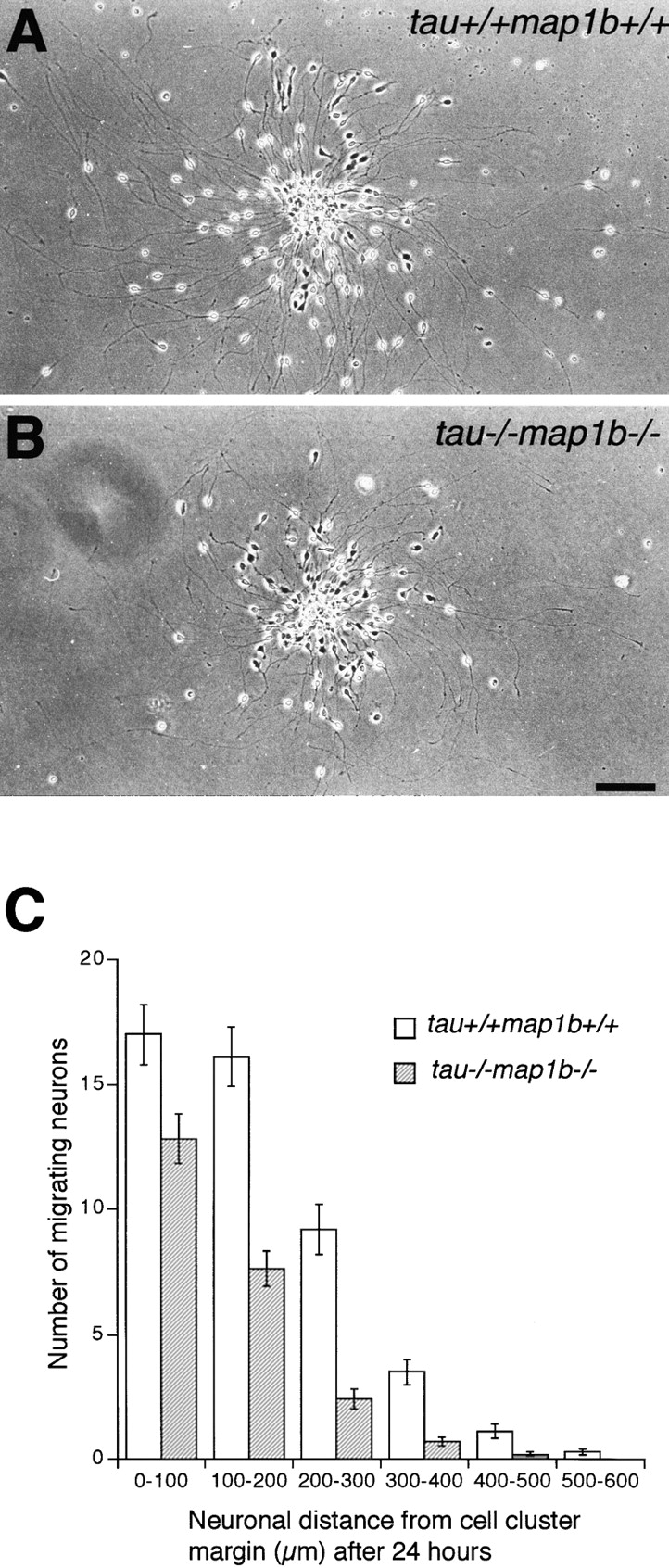
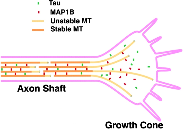
Tau and MAP1B are the main members of neuronal microtubule-associated proteins (MAPs), the functions of which have remained obscure because of a putative functional redundancy (Harada, A., K. Oguchi, S. Okabe, J. Kuno, S. Terada, T. Ohshima, R. Sato-Yoshitake, Y. Takei, T. Noda, and N. Hirokawa. 1994. Nature. 369:488-491; Takei, Y., S. Kondo, A. Harada, S. Inomata, T. Noda, and N. Hirokawa. 1997. J. Cell Biol. 137:1615-1626). To unmask the role of these proteins, we generated double-knockout mice with disrupted tau and map1b genes and compared their phenotypes with those of single-knockout mice. In the analysis of mice with a genetic background of predominantly C57Bl/6J, a hypoplastic commissural axon tract and disorganized neuronal layering were observed in the brains of the tau+/+map1b-/- mice. These phenotypes are markedly more severe in tau-/-map1b-/- double mutants, indicating that tau and MAP1B act in a synergistic fashion. Primary cultures of hippocampal neurons from tau-/-map1b-/- mice showed inhibited axonal elongation. In these cells, a generation of new axons via bundling of microtubules at the neck of the growth cones appeared to be disturbed. Cultured cerebellar neurons from tau-/-map1b-/- mice showed delayed neuronal migration concomitant with suppressed neurite elongation. These findings indicate the cooperative functions of tau and MAP1B in vivo in axonal elongation and neuronal migration as regulators of microtubule organization.


Figures

Figure 2
Figure 2
Defects of axon tract formation in mutant mice. Sections of paraffin-embedded brains of tau+/+map1b++, tau−/−map1b+/+, tau+/+map1b−/−, and tau−/−map1b−/− mice killed at 4 weeks (A–D, M–P) or 0.5 day (E–L) of age. Bodian silver staining (A–H, M–P) or HE staining (I–L). (A–L) Micrographs showing representative areas in frontal (A–H) or mid-sagittal (I–L) sections of the cerebrum. Note the decreased size of the corpus callosum (long arrows) and the anterior commissure (short arrows) in the brains of tau+/+ map1b−/− mice (C, G, and K) and their severe dysgenesis in the brains of tau−/−map1b−/− mice (D, H, and L). Arrowheads in D point the hypoplastic tracts in the striatum. Asterisks indicate the enlarged cavum septi pellucidi (F and G). (M–P) Sagittal sections of the cerebellum. Note that the size of the linea alba is significantly decreased in the brains of tau−/−map1b−/− mice (arrows in P). AC, anterior commissure; CC, corpus callosum; G, granular cell layer; HC, hippocampal commissure; LA, linea alba; M, molecular layer; P, Purkinje cell layer; S, striatum; VL, ventriculus lateralis. Bars: (D, H, and L) 0.5 mm; (P) 100 μm.
Figure 1
Figure 1
Gross appearance of tau+/+map1b+/+ and tau−/− map1b−/− mice (P0.5). Note the lack of milk in the stomach of the tau−/−map1b−/− animal. The area indicating stomach filled with milk in the tau+/+map1b+/+ mouse is marked with an asterisk. Bar, 10 mm.
Figure 3
Figure 3
Defects of neuronal layer formation in mutant mice. Sections of paraffin-embedded brains of tau+/+map1b++, tau−/− map1b+/+, tau+/+map1b−/−, and tau−/−map1b−/− mice killed at 0.5 d (A–H) and 4 wk (I–L) of age, stained by Bodian method. Note that neuronal cell bodies in the pyramidal cell layer are loosely associated with each other (D and H, arrows), and the pyramidal cell layer is undulated (L, arrows) in the brains of tau−/−map1b−/− mice. Arrows in C and G indicate the split in the pyramidal cell layer in the brains of tau+/+map1b−/− mice. CA, cornu ammonis; DG, dentate gyrus; and PCL, pyramidal cell layer. Bars: (D) 0.25 mm; (H) 0.1 mm; (L) 0.5 mm.
Figure 4
Figure 4
Drastic reduction in area of axons and MT number per axon in neurons of tau−/−map1b−/− mice. (A–D) Electron micrographs showing representative areas in cross sections of the anterior commissure of tau+/+ map1b++, tau−/−map1b+/+, tau+/+ map1b−/−, and tau−/−map1b−/− mice at postnatal week 4. Bar, 0.5 μm.
Figure 5
Figure 5
Phenotypes of hippocampal neurons in culture. Hippocampal neurons obtained from tau+/+map1b++, tau−/− map1b+/+, tau+/+map1b−/−, and tau−/−map1b−/− mice were cultured for 3 d and observed by phase-contrast microscopy. Axonal outgrowth of cells from tau−/−map1b−/− mice was significantly inhibited (arrows). ax, axon; and mp, minor process. Bar, 50 μm.
Figure 6
Figure 6
Expression and cellular localization of tau and MAP1B in hippocampal cells. Neurons from tau+/+ map1b+/+ (A–D) or tau−/− map1b−/− mice (E–H), cultured for 3 d, were double-labeled with phalloidin (A, C, E, and G) and monoclonal antibodies recognizing the tau-1 epitope (B and F) or phosphorylated MAP1B (D and H). Both immunoreactivities are axon-dominant in the cells of tau+/+map1b +/+ mice (B and D), whereas they are negative in the cells from tau−/− map1b−/− mice (F and H). ax, axon; and mp, minor process. Bars, 50 μm.
Figure 7
Figure 7
Cytoskeletal organization in growth cones. Growth cones of hippocampal neurons from tau+/+map1b+/+ (A and B) or tau−/−map1b−/− (C and D) mice cultured for 3 d were double-labeled with phalloidin (A and C) and a monoclonal antibody against tubulin (B and D). Note that the growth cone of hippocampal neurons from tau−/−map1b−/− mice is extensive (C), throughout which MTs are flared out (D, arrows), instead of being bundled as seen in the neurons from tau+/+map1b+/+ mice (B, arrow). Bars, 10 μm.
Figure 8
Figure 8
In vitro analysis of neuronal migration. Migration of tatau+/+map1b+/+ (A) and tau−/−map1b−/− (B) cerebellar granule cells from reaggregate cell clusters after 24 h of culture. (C) Mean ± SEM of the number of migrating cerebellar granule cells and their distance of migration from the reaggregate cell cluster in tau+/+map1b+/+ and tau−/−map1b−/− mice after 24 h. Three animals of each genotype were used, and 35 tau+/+ map1b+/+ and 33 tau−/−map1b−/− clusters were examined. Bar, 100 μm.
Figure 9
Figure 9
Schematic diagram of growth cones showing the role of tau and MAP1B in organizing MTs in growth cones. Tau and MAP1B are considered to zip MTs in the advancing growth cones, and convert the dynamic MTs into highly organized parallel arrays of axonal MTs.

References

    1. Bix G.J., Clark G.D. Platelet-activating factor receptor stimulation disrupts neuronal migration in vivo. J. Neurosci. 1998;18:307–318. - PMC - PubMed
    1. Black M.M., Slaughter T., Fischer I. Microtubule-associated protein 1b (MAP1b) is concentrated in the distal region of growing axons. J. Neurosci. 1994;14:857–870. - PMC - PubMed
    1. Black M.M., Slaughter T., Moshiach S., Obrocka M., Fischer I. Tau is enriched on dynamic microtubules in the distal region of growing axons. J. Neurosci. 1996;16:3601–3619. - PMC - PubMed
    1. Bloom G.S., Luca F.C., Vallee R.B. Microtubule-associated protein 1Bidentification of a major component of the neuronal cytoskeleton. Proc. Natl. Acad. Sci. USA. 1985;82:5404–5408. - PMC - PubMed
    1. Bodian P.A. A new method for staining nerve fibers and nerve endings in mounted paraffin sections. Anat. Rec. 1936;65:89–96.

Publication types

MeSH terms

Substances