. 2000 Sep 4;150(5):1013-26.
doi: 10.1083/jcb.150.5.1013.

A myosin I is involved in membrane recycling from early endosomes

Affiliations

A myosin I is involved in membrane recycling from early endosomes

E M Neuhaus et al. J Cell Biol. .

Abstract

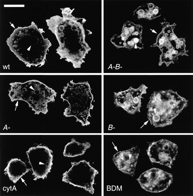
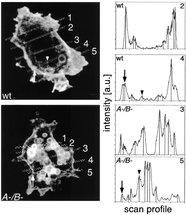
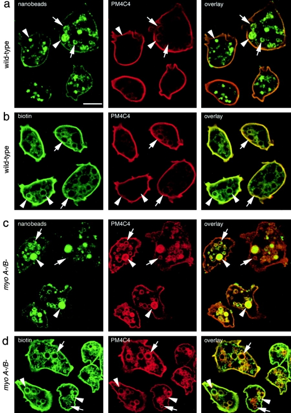
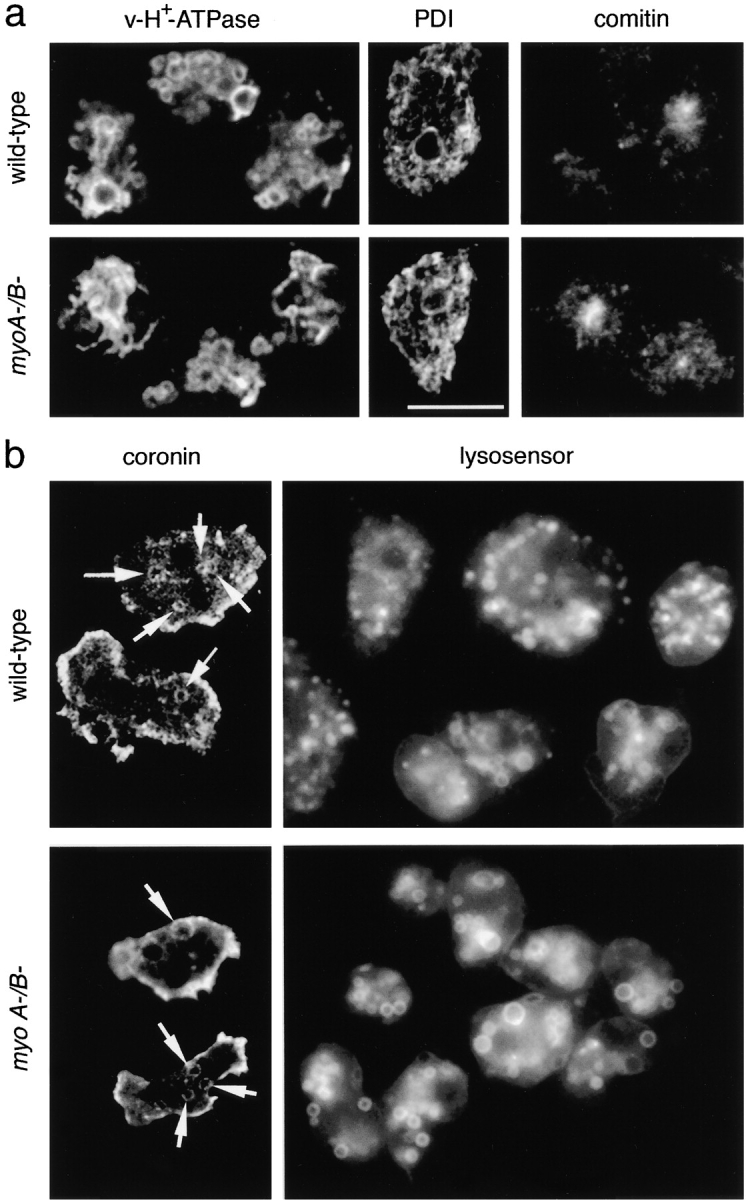
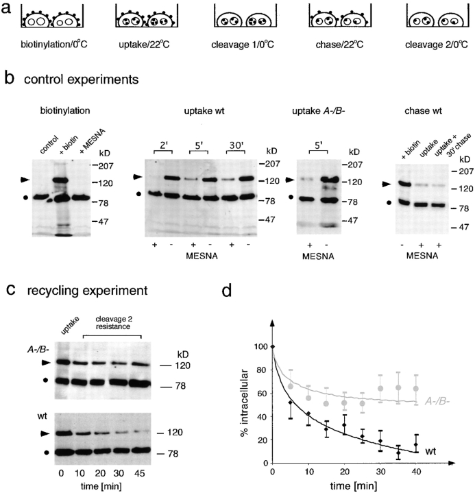
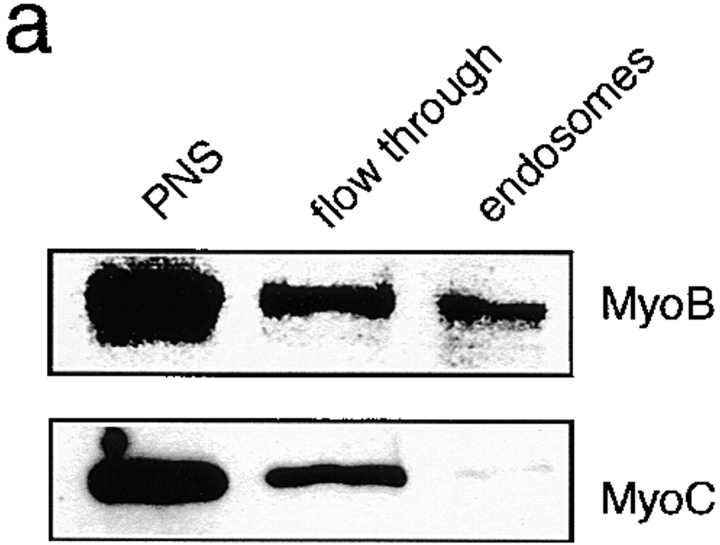
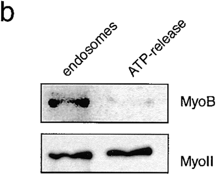
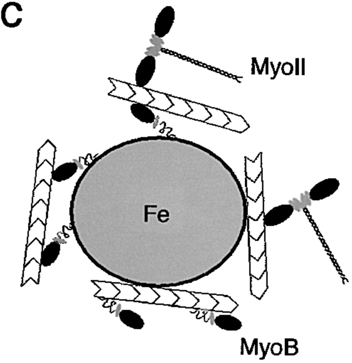
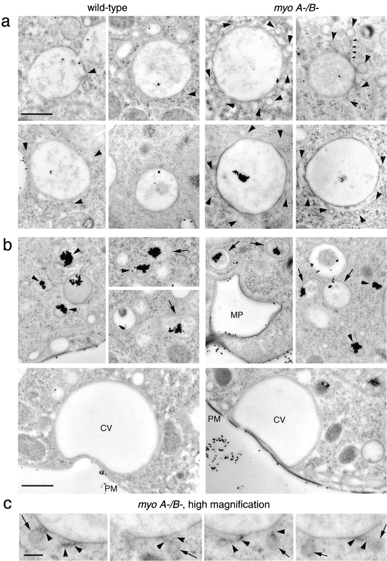
Geometry-based mechanisms have been proposed to account for the sorting of membranes and fluid phase in the endocytic pathway, yet little is known about the involvement of the actin-myosin cytoskeleton. Here, we demonstrate that Dictyostelium discoideum myosin IB functions in the recycling of plasma membrane components from endosomes back to the cell surface. Cells lacking MyoB (myoA(-)/B(-), and myoB(-) cells) and wild-type cells treated with the myosin inhibitor butanedione monoxime accumulated a plasma membrane marker and biotinylated surface proteins on intracellular endocytic vacuoles. An assay based on reversible biotinylation of plasma membrane proteins demonstrated that recycling of membrane components is severely impaired in myoA/B null cells. In addition, MyoB was specifically found on magnetically purified early pinosomes. Using a rapid-freezing cryoelectron microscopy method, we observed an increased number of small vesicles tethered to relatively early endocytic vacuoles in myoA(-)/B(-) cells, but not to later endosomes and lysosomes. This accumulation of vesicles suggests that the defects in membrane recycling result from a disordered morphology of the sorting compartment.


Figures

Figure 1
Figure 1
Distribution of a plasma membrane marker. An antibody directed against the plasma membrane marker PM4C4 strongly labeled the plasma membrane (arrows) and to a lesser extent intracellular endosomal vacuoles (arrowheads) in wild-type D. discoideum cells (wt). myoA/B (A-B-) cells displayed higher labeling intensities on intracellular endosomal vacuoles than at the plasma membrane. myoA cells (A-) and cytA-treated wild-type cells (cytA) showed a staining pattern resembling the wild-type situation. myoB cells (B-) and BDM-treated wild-type cells (BDM) looked similar to myoA/B cells. Single confocal sections. Bar, 10 μm.
Figure 2
Figure 2
Quantification of the membrane concentration of the marker PM4C4. The steady state concentration of PM4C4 on the cell surface (arrows) compared with endomembranes (arrowheads) of wild-type (wt) and myoA/B mutant cells A-/B- was determined. Representative confocal sections of both cell types with two examples of scan profiles are shown. Peaks corresponding to plasma membrane (arrows) and endosomes (arrowheads) are marked as in the corresponding confocal section. Analysis of peak intensities of 78 line scans through wild-type cells and 53 line scans through myoA/B cells resulted in ratios of plasma membrane versus endosomal membranes stainings of 2.80 ± 1.43 for wild-type and 0.34 ± 0.25 for myoA/B cells.
Figure 3
Figure 3
Colocalization of endocytic markers with PM4C4-positive vacuoles. (a and c) Intracellular PM4C4-positive vacuoles (PM4C4) contained endocytosed yellow-green fluorescent nanobeads as fluid phase marker (nanobeads) in both wild-type (a) and myoA/B cells (c). Some of the PM4C4 vacuoles contained high concentrations of fluid phase marker (a and c, arrowheads); others contained only low concentrations (a and c, arrows). (b and d) The PM4C4 vacuoles also contained plasma membrane proteins that had been biotinylated at 0°C and internalized via endocytosis after warming to 22°C (biotin) in wild-type (b) and in myoA/B cells (d). The relative labeling intensities of the vacuoles varied between both channels. Some vacuoles contained higher biotin signals (b and d, arrowheads) and appeared green in the overlay, whereas others contained higher PM4C4 signals (b and d, arrows) and appeared red in the overlay. Altogether, myoA/B cells (d, biotin) contained more biotinylated protein on internal vacuoles than wild-type cells (b, biotin), similar to what was observed for the plasma membrane marker PM4C4 (b and d). Bar, 10 μm.
Figure 4
Figure 4
(a) Other major endomembrane compartments are similar in wild-type and myoA/B cells. The contractile vacuole system was visualized with an antibody against the vacuolar H+-ATPase (v-H+-ATPase); the ER was labeled with an antibody against PDI; the Golgi apparatus was visualized with an antibody against the actin-binding protein comitin. (b) Endosomes in myoA/B null cells are acidic. myoA/B and wild-type cells showed similar number of coronin-positive vacuoles (coronin). Staining of wild-type D. discoideum cells with LysoSensor Green visualized the membrane of acidic organelles with a broad size distribution (lysosensor). In myoA/B cells, the labeled acidic compartments were more homogeneous in size. Bar, 10 μm.
Figure 5
Figure 5
Establishment of a recycling assay using reversible biotinylation of plasma membrane proteins. (a) Schematic representation of the optimized recycling assay. Cell surface proteins were biotinylated (biotinylation) and internalization was allowed for 5 min (uptake). Afterwards, remaining biotin in the plasma membrane was cleaved off (cleavage 1) and further trafficking of internalized membrane was allowed (chase). Biotin was cleaved off of reexposed proteins (cleavage 2) and the remaining amount of intracellular labeling was followed over time. (b) Control experiments. Biotinylation: Western blot of D. discoideum cell lysates. An endogenous biotin-containing protein of 80 kD (•) was used as internal standard (first lane). After labeling of cell surface proteins at 0°C with a biotin-coupled cross-linker containing a disulfide bridge, a strong signal at 120 kD (arrowhead) and several minor bands were visible (second lane). This biotin labeling (arrowhead) could be cleaved off quantitatively by the reducing agent MESNA (third lane). Uptake wt: Due to internalization of labeled plasma membrane at 22°C, increasing amounts of the 120-kD biotin signal (arrowhead) became protected from cleavage 1 with MESNA (+MESNA lanes). The total amount of biotin in the samples before cleavage 1 (−MESNA lanes) was constant over time. Uptake A/B: myoA/B cells also internalized biotinylated plasma membrane proteins, and a cleavage 1 (+MESNA lane) resistant 120-kD biotin signal (arrowhead) was detected after 5 min uptake at 22°C. Chase wt: After internalization, the cleavage 1–resistant signal (arrowhead, uptake) stayed constant even after 30 min of chase (arrowhead, uptake + 30′ chase). (c) Recycling experiment. Representative Western blots from recycling experiments. After an initial uptake of biotinylated plasma membrane proteins (first lane, uptake) the cleavage 2–resistant 120-kD signal (arrowhead) decreased with increasing duration of chase (recycling) in wild-type cells. In contrast, in myoA/B cells the internalized 120-kD signal (arrowhead) stayed constant from 10 min on. (d) Quantification. Curves resulting from quantification of Western blots from three to five independent recycling experiments carried out as described in c. Wild-type cells are represented by black diamonds and myoA/B cells by gray circles. Error bars represent the SEM.
Figure 6
Figure 6
MyoB is found in an early endosomal fraction. (a) Wild-type D. discoideum cells were fed with colloidal iron for 15 min. Early endosomes were isolated by magnetic fractionation and probed for the presence of class I myosins by Western blotting using anti-MyoB and anti-MyoC antibodies. MyoB was found in the isolated endosome fraction, whereas MyoC was not significantly present. (b) ATP-release experiment. Isolated endosomes were incubated with ATP and pellets (endosomes) and supernatants (after ATP release) were analyzed by Western blotting with anti-MyoB and anti–myosin II antibodies. Myosin II was quantitatively released by ATP, most likely indicating that it was bound to endosome-attached actin filaments via its head domain. MyoB was not released, indicating that it was likely specifically bound to endosomes via its tail domain. (c) Schematic illustration of the observed binding modes.
Figure 6
Figure 6
MyoB is found in an early endosomal fraction. (a) Wild-type D. discoideum cells were fed with colloidal iron for 15 min. Early endosomes were isolated by magnetic fractionation and probed for the presence of class I myosins by Western blotting using anti-MyoB and anti-MyoC antibodies. MyoB was found in the isolated endosome fraction, whereas MyoC was not significantly present. (b) ATP-release experiment. Isolated endosomes were incubated with ATP and pellets (endosomes) and supernatants (after ATP release) were analyzed by Western blotting with anti-MyoB and anti–myosin II antibodies. Myosin II was quantitatively released by ATP, most likely indicating that it was bound to endosome-attached actin filaments via its head domain. MyoB was not released, indicating that it was likely specifically bound to endosomes via its tail domain. (c) Schematic illustration of the observed binding modes.
Figure 7
Figure 7
(a) Ultrastructure of early endocytic vacuoles in myoA/B cells. Thin sections of rapidly frozen myoA/B-deficient D. discoideum cells (myoA/B) presented an accumulation of small (110–120 nm) vesicles (arrowheads) around big endolysosomal vacuoles containing internalized BSA-gold. These vacuoles contained only few dispersed gold particles, indicating that they were relatively early endosomes. Some of the vesicles were tethered to the vacuoles (small arrowheads). Vacuoles in wild-type cells (wild-type) did not show this accumulation of vesicles (arrowheads). (b) Ultrastructure of late endosomes and of the contractile vacuole. Later endocytic compartments containing higher concentrations of gold particles (aggregated because the BSA coat is digested by lysosomal enzymes) were identified by the presence of intralumenal membranous structures (arrows) or a relatively electron-dense content (arrowheads). Newly formed macropinosomes (MP) contained few dispersed gold particles and were surrounded by an F-actin layer. The contractile vacuole (CV) was identified by the complete absence of ingested gold particles, its characteristic shape, and direct apposition to the plasma membrane (PM). None of these structures showed an accumulation of small vesicles, neither in wild-type, nor in myoA/B cells. (c) Higher magnification of tethered vesicles in myoA/B cells. Some of the small vesicles accumulated around early endocytic vacuoles in myoA/B cells had a relatively electron-dense periphery (arrows), and often seemed to be connected to the vacuole by thin tethers (arrowheads). Bars: (a and b) 1 μm; (c) 0.2 μm.

Similar articles

Cited by

References

    1. Abercrombie M., Heaysman J.E.M., Pegrum S.M. The locomotion of fibroblasts in culture. 3. Movement of particles on the dorsal surface of the leading lamella. Exp. Cell Res. 1970;62:389–398. - PubMed
    1. Adessi C., Chapel A., Vincon M., Rabilloud T., Klein G., Satre M., Garin J. Identification of major proteins associated with Dictyostelium discoideum endocytic vesicles. J. Cell Sci. 1995;108:3331–3337. - PubMed
    1. Aguado-Velasco C., Bretscher M. Circulation of plasma membrane in Dictyostelium . Mol. Biol. Cell. 1999;10:4419–4427. - PMC - PubMed
    1. Aubry L., Klein G., Martiel J.L., Satre M. Kinetics of endosomal pH evolution in Dictyostelium discoideum amoebae. Study by fluorescence spectroscopy. J. Cell Sci. 1993;105:861–866. - PubMed
    1. Aubry L., Klein G., Martiel J.L., Satre M. Fluid-phase endocytosis in the amoebae of the cellular slime mould Dictyostelium discoideummathematical modelling of kinetics and pH evolution. J. Theor. Biol. 1997;184:89–98.

Publication types