. 2000 Sep 4;150(5):1027-36.
doi: 10.1083/jcb.150.5.1027.

Preservation of mitochondrial structure and function after Bid- or Bax-mediated cytochrome c release

Affiliations

Preservation of mitochondrial structure and function after Bid- or Bax-mediated cytochrome c release

O von Ahsen et al. J Cell Biol. .

Abstract

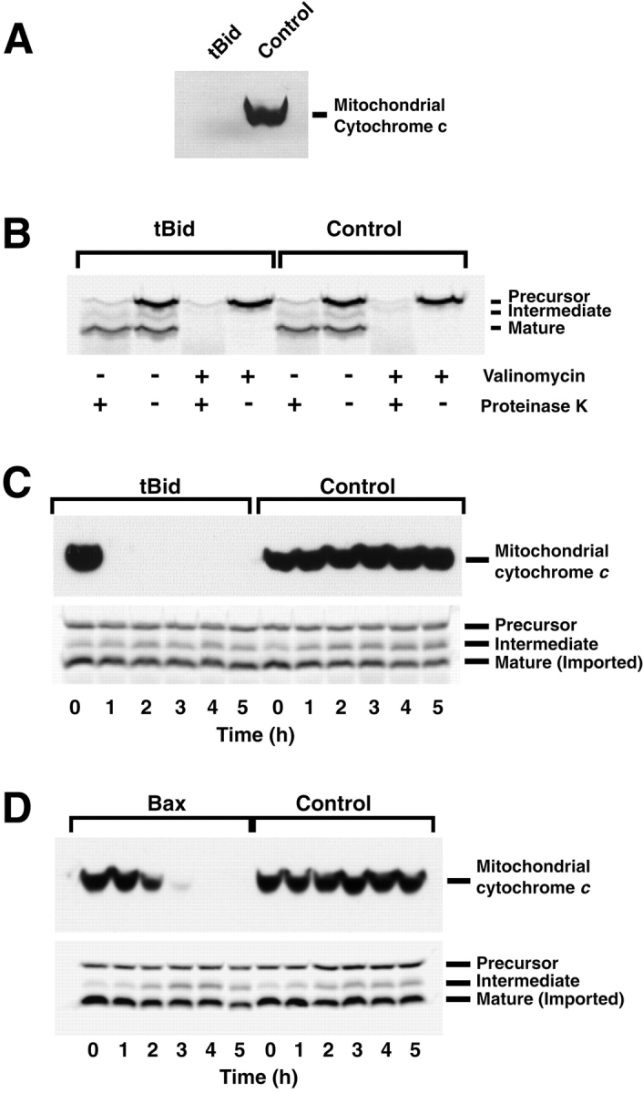
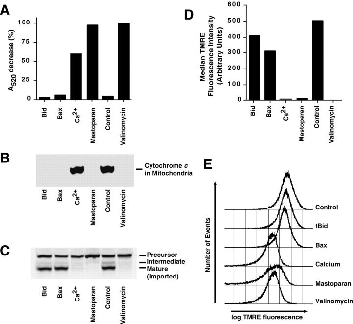
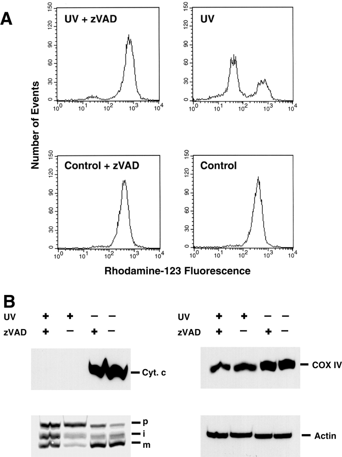
Proapoptotic members of the Bcl-2 protein family, including Bid and Bax, can activate apoptosis by directly interacting with mitochondria to cause cytochrome c translocation from the intermembrane space into the cytoplasm, thereby triggering Apaf-1-mediated caspase activation. Under some circumstances, when caspase activation is blocked, cells can recover from cytochrome c translocation; this suggests that apoptotic mitochondria may not always suffer catastrophic damage arising from the process of cytochrome c release. We now show that recombinant Bid and Bax cause complete cytochrome c loss from isolated mitochondria in vitro, but preserve the ultrastructure and protein import function of mitochondria, which depend on inner membrane polarization. We also demonstrate that, if caspases are inhibited, mitochondrial protein import function is retained in UV-irradiated or staurosporine-treated cells, despite the complete translocation of cytochrome c. Thus, Bid and Bax act only on the outer membrane, and lesions in the inner membrane occurring during apoptosis are shown to be secondary caspase-dependent events.


Figures

Figure 1
Figure 1
Protein import function is maintained by isolated mitochondria even after complete cytochrome c release. Mitochondria (1.5 mg/ml protein) were incubated in MIB containing 80 mM KCl, with or without 10 μg/ml tBid for 30 min at 22°C. (A) An aliquot was analyzed for mitochondrial cytochrome c content, showing a complete loss of cytochrome c from mitochondria incubated with tBid. (B) Mitochondria were reisolated and resuspended in import buffer containing 2 mM ATP, and the sample was divided into four aliquots. After a 5-min incubation in the presence or absence of valinomycin (a K+ ionophore) to dissipate membrane potential, the precursor protein was added. After 30 min, the import reaction was stopped by the addition of valinomycin and cooling the samples on ice. Where indicated, proteinase K was added for 10 min to digest nonimported precursor. After addition of PMSF and a 5-min incubation on ice, mitochondria were reisolated and analyzed by SDS-PAGE and autoradiography. Inhibition of processing by valinomycin proves that protein import was dependent on membrane potential. Imported protein was processed to a mature size and protected against externally added protease. (C and D) Mitochondria remain import-competent for several hours after the complete loss of cytochrome c induced by tBid or Bax. Mitochondria (1.5 mg/ml) were incubated in PT buffer with or without the addition of 10 μg/ml tBid (C) or 50 μg/ml Bax (D). At the times indicated, aliquots were removed and analyzed for mitochondrial cytochrome c content (top) or import competence (bottom), as indicated by the processing of the precursor to a mature size by the mitochondrial matrix processing peptidase.
Figure 2
Figure 2
Bid-induced cytochrome c release does not depend on mitochondrial PT. Mitochondria (0.3 mg/ml as protein) were incubated in PT buffer, with the addition (as indicated) of 10 μg/ml tBid, 50 μg/ml Bax, 1 mM CaCl2, 20 μg/ml mastoparan, or 1 μM valinomycin for 30 min at 22°C. (A) Mitochondrial swelling was assessed by absorbance at 520 nm measured before and 25 min after the indicated additions were made. The A520 decrease produced by valinomycin was taken as 100%. 30 min after additions, aliquots were removed and analyzed for mitochondrial cytochrome c content (B) or import competence (C), 50 nM TMRE was added to the remaining samples, and (D and E) dye uptake was measured by flow cytometry after a further 10 min at 22°C. After this, valinomycin (1 μM) was added to all samples and a second measurement of background fluorescence was made. D shows the median fluorescence with this background subtracted (linear scale), while E shows histograms (log scale). Data are representative of five independent experiments.
Figure 3
Figure 3
Cyclosporin A blocks PT in Xenopus mitochondria, but has no effect on tBid-induced cytochrome c release. (A) Xenopus mitochondria were preincubated in PT buffer with or without 10 μM cyclosporin A (A) for 15 min at 22°C, and CaCl2 was added at the indicated concentrations. After 30 min, TMRE retention was measured by flow cytometry. (B) The lack of effect of cyclosporin A on tBid-induced cytochrome c release. Xenopus mitochondria were preincubated in PT buffer with or without 10 μM cyclosporin A for 15 min at 22°C, and tBid was added at the indicated concentrations. After 30 min at 22°C, the mitochondria were reisolated and their cytochrome c content was analyzed by Western blotting.
Figure 4
Figure 4
tBid produces no observable changes in mitochondrial ultrastructure, whereas Ca2+ causes swelling of the matrix. Shown are representative images from thin (∼70 nm) sections using conventional electron microscopy. Before fixation, Xenopus mitochondria (0.3 mg/ml protein) were incubated for 30 min at 22°C in import buffer with (A) 10 μg/ml tBid, (B) buffer only, or (C) 1 mM CaCl2. No significant difference between tBid-treated and control mitochondria could be observed (A and B). Only the calcium treatment led to matrix swelling (C). No breaks in outer membranes could be seen; even after calcium-induced swelling, the outer membranes apparently stay intact. Electron tomography also shows that tBid produces no significant structural alterations in mitochondria, whereas Ca2+ causes matrix swelling. D–F show cross-sections (2.3-nm thickness) through the middle of electron tomographic reconstructions of semi-thick (∼500 nm) sections of mitochondria treated with 10 μg/ml tBid (D), buffer (E), or 1 mM CaCl2 (F). Surface-rendered volumes of the same three mitochondria reconstructed for D–F are shown in G–I. Here, the outer membrane is shown in dark blue, the inner boundary membrane in light blue, and the cristal membranes in yellow. Bars, 250 nm.
Figure 5
Figure 5
Mitochondria isolated from HL-60 cells retain protein import function and intact ΔΨm after cytochrome c release. Mitochondria (5 mg/ml as protein) were incubated in PT buffer, with the addition (as indicated) of 10 μg/ml tBid, 1 mM CaCl2, or 20 μg/ml mastoparan for 30 min at 22°C. (A) Mitochondrial swelling was assessed by comparing absorbance at 520 nm before and 25 min after the indicated additions were made. The A520 decrease produced by mastoparan was taken as 100%. 30 min after additions, aliquots were removed and analyzed for mitochondrial cytochrome c content (B) or import competence (C), 50 nM TMRE was added to the remaining samples, and (D) dye uptake was measured by flow cytometry after a further 10 min at 22°C. Finally, a second measurement of background fluorescence was made after valinomycin (1 μM) was added to all samples. B shows the median fluorescence with this background subtracted. (E) HL-60 mitochondria remain import-competent for at least 3 h after complete cytochrome c release. HL-60 cell mitochondria (5 mg/ml protein) were incubated in PT buffer in the presence or absence of tBid (10 μg/ml) as indicated. Aliquots of the reaction were taken at the indicated times and analyzed for cytochrome c content and import competence. As a control showing import dependence on ΔΨm, 1 μM valinomycin was added where indicated. Data are representative of three independent experiments.
Figure 6
Figure 6
Bid releases cytochrome c from mitochondria in permeabilized HeLa cells without disruption of membrane potential or protein import. Permeabilized HeLa cells (2 × 10⁵) were incubated in 100 μl of import buffer with the indicated amounts of Bid for 30 min at 22°C. An aliquot was spun down to analyze cytochrome c content, and the remaining sample was used to assess import competence. Cell pellets were extracted with NP-40 lysis buffer and analyzed as described in Fig. 7.
Figure 8
Figure 8
Mitochondrial import function remains partially intact in cells undergoing staurosporine-induced apoptosis, provided that caspases are inhibited. HeLa cells were cultured for 20 h in medium containing 1 μM staurosporine and/or 100 μM zVAD-fmk as indicated. Approximately 70% of staurosporine-treated cells were apoptotic at this time, whereas, in the presence of zVAD-fmk, 60% were intact and 40% necrotic. (A) Cells were harvested and TMRE uptake was assessed by flow cytometry (5,000 events shown without gating). Staurosporine-treated cells show a loss of ΔΨm, which is largely prevented by caspase inhibition. (B) Cytochrome c release and import competence were assessed after digitonin permeabilization as in Fig. 7. Quantitation by phosphorimaging revealed 38 and 13% imported protein in staurosporine-treated samples with or without zVAD-fmk. (Control samples gave 100% and 84%; import in the control sample with zVAD was defined as 100%.) The cytochrome c blot was stripped and reprobed with anti–COX-IV to demonstrate equal gel loading.
Figure 8
Figure 8
Mitochondrial import function remains partially intact in cells undergoing staurosporine-induced apoptosis, provided that caspases are inhibited. HeLa cells were cultured for 20 h in medium containing 1 μM staurosporine and/or 100 μM zVAD-fmk as indicated. Approximately 70% of staurosporine-treated cells were apoptotic at this time, whereas, in the presence of zVAD-fmk, 60% were intact and 40% necrotic. (A) Cells were harvested and TMRE uptake was assessed by flow cytometry (5,000 events shown without gating). Staurosporine-treated cells show a loss of ΔΨm, which is largely prevented by caspase inhibition. (B) Cytochrome c release and import competence were assessed after digitonin permeabilization as in Fig. 7. Quantitation by phosphorimaging revealed 38 and 13% imported protein in staurosporine-treated samples with or without zVAD-fmk. (Control samples gave 100% and 84%; import in the control sample with zVAD was defined as 100%.) The cytochrome c blot was stripped and reprobed with anti–COX-IV to demonstrate equal gel loading.
Figure 7
Figure 7
When caspases are inhibited, UV-irradiated HeLa cells display a complete loss of mitochondrial cytochrome c, but retain mitochondrial import function. (A) Loss of ΔΨm in apoptotic cells is blocked by caspase inhibition. HeLa cells were exposed to 180 mJ/cm2 UVC light and further cultured for 8 h in medium with or without 100 μM zVAD-fmk as indicated. After this time, >50% of UV-treated cells showed an apoptotic phenotype in the absence of zVAD-fmk. Cells were harvested, and rhodamine-123 retention was measured by flow cytometry (10,000 events shown without gating). In the presence of zVAD-fmk, apoptotic cells retain ΔΨm. (B) Mitochondrial import competence is retained in the presence of zVAD-fmk. After flow cytometric analysis (A), the cells were permeabilized with digitonin and analyzed for cytochrome c content and import-competence. Cell pellets were extracted with NP-40 lysis buffer (150 mM NaCl, 1% NP-40, and 50 mM Tris-Cl, pH 8.0), and the protein content of the extract was determined by the Bradford assay. Finally, 50 μg of protein from each sample was subjected to SDS-PAGE and Western blotting. Quantitative analysis by phosphorimaging revealed 82 and 17% imported protein in the UV-treated samples with or without zVAD, respectively. Control samples gave 100 and 89% import, respectively (the control sample with zVAD was taken as 100%). Blots were stripped and reprobed with antibodies to COX-IV and actin to demonstrate equal gel loading.

References

    1. Bossy-Wetzel E., Newmeyer D.D., Green D.R. Mitochondrial cytochrome c release in apoptosis occurs upstream of DEVD-specific caspase activation and independently of mitochondrial transmembrane depolarization. EMBO (Eur. Mol. Biol. Organ.) J. 1998;17:37–49. - PMC - PubMed
    1. Cecconi F., Alvarez-Bolado G., Meyer B.I., Roth K.A., Gruss P. Apaf1 (CED-4 homolog) regulates programmed cell death in mammalian development. Cell. 1998;94:727–737. - PubMed
    1. Chautan M., Chazal G., Cecconi F., Gruss P., Golstein P. Interdigital cell death can occur through a necrotic and caspase-independent pathway. Curr. Biol. 1999;9:967–970. - PubMed
    1. Desagher S., Osen-Sand A., Nichols A., Eskes R., Montessuit S., Lauper S., Maundrell K., Antonsson B., Martinou J.C. Bid-induced conformational change of Bax is responsible for mitochondrial cytochrome c release during apoptosis. J. Cell Biol. 1999;144:891–901. - PMC - PubMed
    1. Deshmukh M., Johnson E.M., Jr. Evidence of a novel event during neuronal deathdevelopment of competence-to-die in response to cytoplasmic cytochrome c. Neuron. 1998;21:695–705. - PubMed

Publication types

MeSH terms