. 2000 Sep 4;150(5):1057-70.
doi: 10.1083/jcb.150.5.1057.

Endothelial cell-surface gp60 activates vesicle formation and trafficking via G(i)-coupled Src kinase signaling pathway

Affiliations

Endothelial cell-surface gp60 activates vesicle formation and trafficking via G(i)-coupled Src kinase signaling pathway

R D Minshall et al. J Cell Biol. .

Abstract

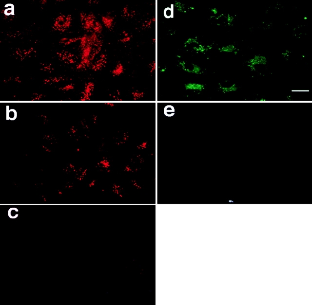
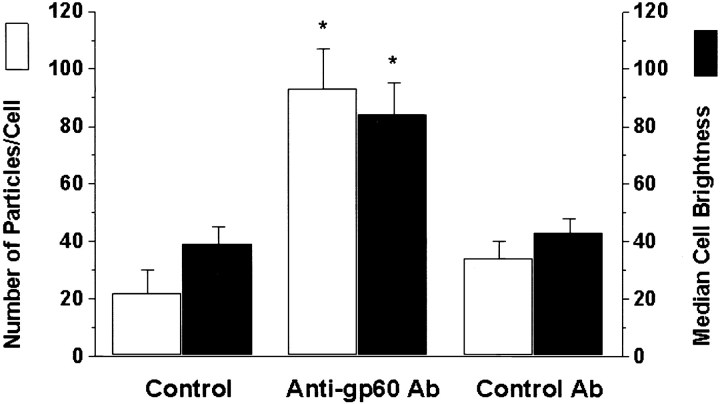
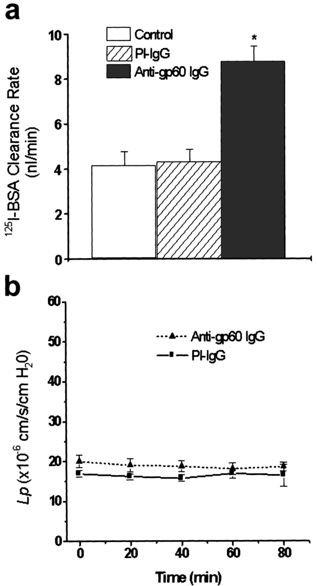
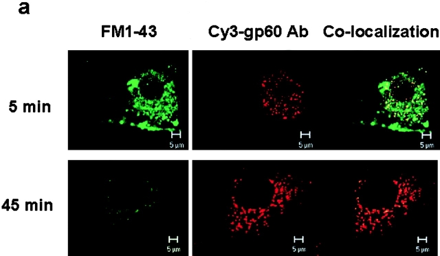
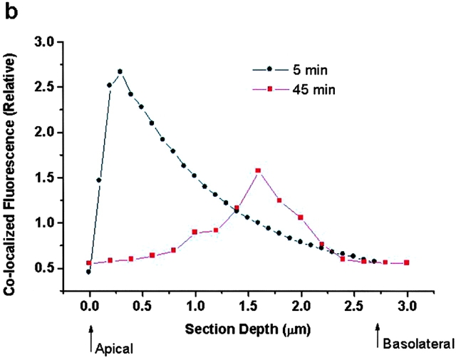
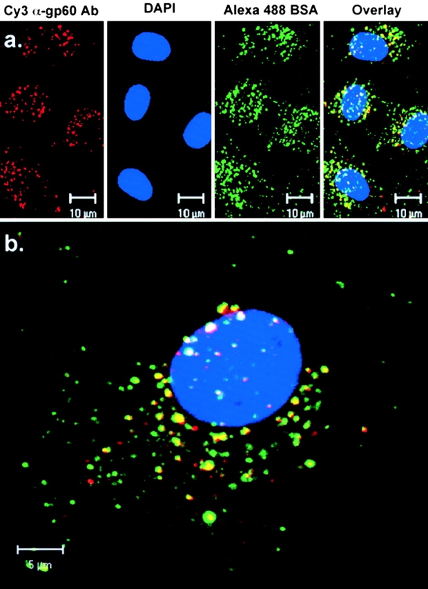
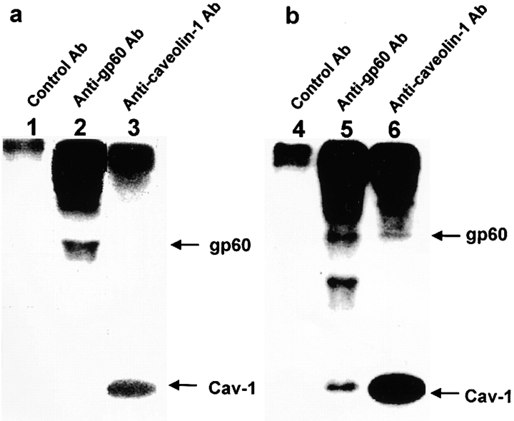
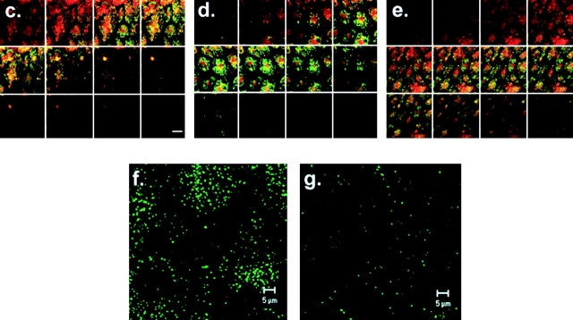
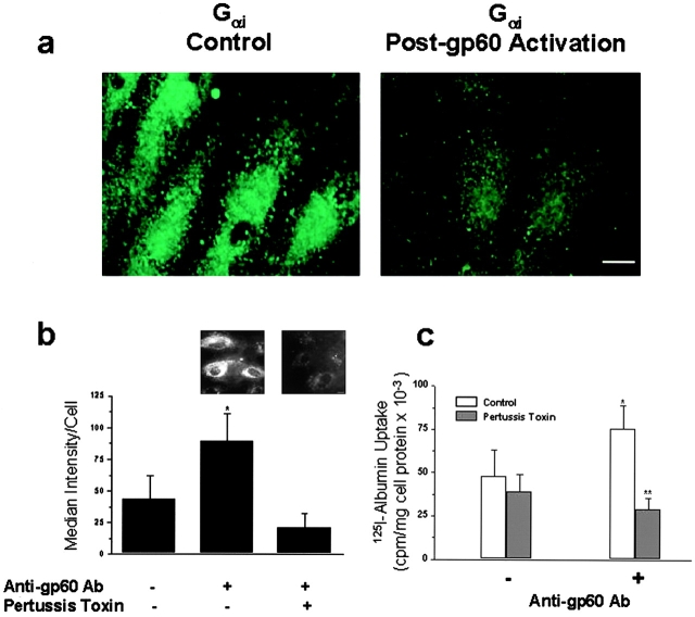
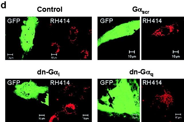
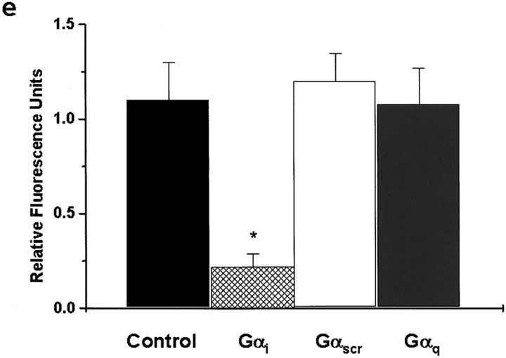
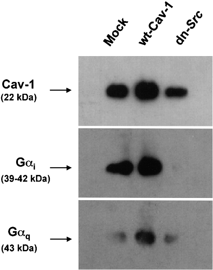
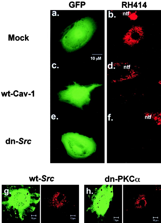
We tested the hypothesis that the albumin-docking protein gp60, which is localized in caveolae, couples to the heterotrimeric GTP binding protein G(i), and thereby activates plasmalemmal vesicle formation and the directed migration of vesicles in endothelial cells (ECs). We used the water-soluble styryl pyridinium dye N-(3-triethylaminopropyl)-4-(p-dibutylaminostyryl) pyridinium dibromide (FM 1-43) to quantify vesicle trafficking by confocal and digital fluorescence microscopy. FM 1-43 and fluorescently labeled anti-gp60 antibody (Ab) were colocalized in endocytic vesicles within 5 min of gp60 activation. Vesicles migrated to the basolateral surface where they released FM 1-43, the fluid phase styryl probe. FM 1-43 fluorescence disappeared from the basolateral EC surface without the loss of anti-gp60 Ab fluorescence. Activation of cell-surface gp60 by cross-linking (using anti-gp60 Ab and secondary Ab) in EC grown on microporous filters increased transendothelial (125)I-albumin permeability without altering liquid permeability (hydraulic conductivity), thus, indicating the dissociation of hydraulic conductivity from the albumin permeability pathway. The findings that the sterol-binding agent, filipin, prevented gp60-activated vesicle formation and that caveolin-1 and gp60 were colocalized in vesicles suggest the caveolar origin of endocytic vesicles. Pertussis toxin pretreatment and expression of the dominant negative construct encoding an 11-amino acid G(alphai) carboxyl-terminal peptide inhibited endothelial (125)I-albumin endocytosis and vesicle formation induced by gp60 activation. Expression of dominant negative Src (dn-Src) and overexpression of wild-type caveolin-1 also prevented gp60-activated endocytosis. Caveolin-1 overexpression resulted in the sequestration of G(alphai) with the caveolin-1, whereas dn-Src inhibited G(alphai) binding to caveolin-1. Thus, vesicle formation induced by gp60 and migration of vesicles to the basolateral membrane requires the interaction of gp60 with caveolin-1, followed by the activation of the downstream G(i)-coupled Src kinase signaling pathway.


Figures

Figure 1
Figure 1
Labeling of gp60 and endocytic vesicles in live endothelial cells. (a) BLMVEC were incubated with Cy3-labeled anti-gp60 Ab at 37°C for 15 min and washed at a low temperature (4°C) to label gp60; red fluorescent images reflect cell surface and internalized fluorescence. (b) Cells were treated identically to a, except that cells were washed with a low pH (3.0) fluid to show internalized gp60. (c) Control photomicrograph only shows background staining when incubation was carried out at 4°C and washed with pH 3.0 buffer. (d) Cells were incubated for 15 min at 37°C with FM 1-43 and washed at normal pH with dye-free buffer to label endosomes; green fluorescence indicates FM 1-43 incorporated in endosomes during the incubation period. (e) 15-min incubation with FM 1-43 at 4°C (to inhibit endocytosis) prevents fluorescent dye internalization. Other details are described in Materials and Methods. Identical results were observed with the chemically similar styryl dye, RH 414 (as shown in Fig. 8 and Fig. 9); thus, FM 1-43 and RH 414 dyes were used interchangeably to label endocytic vesicles. Results are representative of five experiments. Bar, 20 μm.
Figure 2
Figure 2
Activation of gp60 using cross-linking Ab increases vesicle formation in endothelial cells. BPMVEC were incubated with either anti-gp60 Ab (plus secondary Ab) or control Ab (preimmune IgG; 5 μg/ml) for 30 min at 4°C. Cells were incubated at 37°C for 15 min with 5 μg/ml FM 1-43 plus 10 mg/ml BSA. Median brightness values of cells and the number of fluorescent particles per cell were determined after three washes with buffer. The asterisk indicates increased particle density and median cell brightness (P < 0.05) from corresponding control values.
Figure 4
Figure 4
Gp60 activation increases transendothelial flux of 125I-albumin without increasing barrier hydraulic conductivity. (a) BLMVEC monolayers on filters were washed twice with Hepes-DME and incubated with either anti-gp60 Ab or preimmune (PI) IgG (10 μg/ml) for 30 min at 22°C, followed by treatment with 10 μg/ml goat anti–rabbit secondary Ab for 30 min. Monolayers were used for transendothelial 125I-albumin permeability measurements at 37°C. Both luminal and albuminal compartments contained 30 mg/ml of unlabeled albumin. Asterisk indicates the difference from control (P < 0.001). (b) Lack of effect of gp60 cross-linking on endothelial monolayer hydraulic conductivity (Lp). A system consisting of BLMVEC cultured on filters using the two compartment system was used to measure Lp (Qiao et al. 1993); medium albumin concentration was 5 mg/ml. Monolayers were washed and incubated with either anti-gp60 Ab (to induce gp60 cross-linking) or preimmune IgG (10 μg/ml) for 10 min, followed by secondary Ab treatment for 5 min. Values are the mean ± SEM (n = 5).
Figure 3
Figure 3
Colocalization and migration of gp60 and plasmalemma-derived vesicles. (a) Cy3-labeled anti-gp60 Ab was used to fluorescently tag gp60 in BLMVEC incubated in 10 mg/ml BSA. Endosomes were labeled at the same time with 5 μg/ml FM 1-43 to show colocalization of vesicles with gp60. After coincubation with both probes for the indicated times, cell-surface fluorescence was removed by extensive rinsing with pH 5.0 buffer at 4°C. Top row shows confocal images (63× objective), near the luminal cell surface, of early (5 min) intracellular fluorescence because of vesicle marker FM 1-43 (green, left), cy3-labeled anti-gp60 Ab (red, middle), and colocalization image (yellow, right). Bottom row shows albumin cell surface after exocytosis. Note the relative absence of colocalization at 45 min. (b) Plot of migration of gp60-containing vesicles in an endothelial cell. Plot gives relative colocalized fluorescence intensity versus depth of the optical section through the endothelial cell. Peak fluorescent intensity occurred near the luminal cell surface at 5 min after colabeling, whereas the peak is shifted towards the basolateral surface at 45 min. The peak colocalized fluorescence intensity decreased at 45 min compared with 5 min because of exocytosis of FM 1-43 and dilution of its fluorescence in extracellular fluid. Results are representative of five experiments.
Figure 3
Figure 3
Colocalization and migration of gp60 and plasmalemma-derived vesicles. (a) Cy3-labeled anti-gp60 Ab was used to fluorescently tag gp60 in BLMVEC incubated in 10 mg/ml BSA. Endosomes were labeled at the same time with 5 μg/ml FM 1-43 to show colocalization of vesicles with gp60. After coincubation with both probes for the indicated times, cell-surface fluorescence was removed by extensive rinsing with pH 5.0 buffer at 4°C. Top row shows confocal images (63× objective), near the luminal cell surface, of early (5 min) intracellular fluorescence because of vesicle marker FM 1-43 (green, left), cy3-labeled anti-gp60 Ab (red, middle), and colocalization image (yellow, right). Bottom row shows albumin cell surface after exocytosis. Note the relative absence of colocalization at 45 min. (b) Plot of migration of gp60-containing vesicles in an endothelial cell. Plot gives relative colocalized fluorescence intensity versus depth of the optical section through the endothelial cell. Peak fluorescent intensity occurred near the luminal cell surface at 5 min after colabeling, whereas the peak is shifted towards the basolateral surface at 45 min. The peak colocalized fluorescence intensity decreased at 45 min compared with 5 min because of exocytosis of FM 1-43 and dilution of its fluorescence in extracellular fluid. Results are representative of five experiments.
Figure 5
Figure 5
Colocalization of gp60 with albumin. Confocal images of BLMVEC grown to confluence on gelatin-coated glass coverslips showing fluorescent staining of gp60, DAPI, and Alexa 488-albumin. (a) Single section (<1.0 μm thick; four frame average) near the apical membrane surface showing gp60 (red), DAPI (blue), albumin (green), and the overlay of gp60, albumin, and DAPI immunostaining in BLMVEC. (b) Merged images of gp60, DAPI, and albumin in confocal z-axis optical sections (0.2-μm step size; two frame averages) through a single BLMVEC. Projection image (22 sections stacked) en face (b) show the distribution of gp60 and albumin in the cell.
Figure 6
Figure 6
Coimmunoprecipitation and migration of caveolin-1 and gp60. BLMVEC were grown to confluence, serum-deprived overnight, and metabolically labeled for 4 h with 200 μCi/ml 32P-orthophosphate. Cells were either stimulated for 20 min with 6 mg/ml BSA (b) or not stimulated with BSA (a) and lysed. Total cell lysate was immunoprecipitated with preimmune rabbit IgG (lanes 1 and 4), rabbit anti-gp60 IgG (lanes 2 and 5), or rabbit anti–caveolin-1 IgG (lanes 3 and 6), separated by SDS-PAGE, transferred to a nitrocellulose membrane, and visualized by autoradiography. As shown in lane 4 (b), the cell lysate that immunoprecipitated with the anti-gp60 Ab also contained a 22-kD protein, which migrated similarly to that immunoprecipitated with anti–caveolin-1 Ab (b, lane 6). (b) Lane 6 shows that the cell lysate that immunoprecipitated with anti–caveolin-1 Ab also pulled down a protein similar to that immunoprecipitated with the anti-gp60 Ab (b, lane 5). Control Ab did not immunoprecipitate either 32P-labeled protein. In the absence of albumin stimulation, gp60 was not coimmunoprecipitated with the anti–caveolin-1 Ab (a, lane 2 and 3). Results are representative of three experiments. (c–e) Merged images of gp60 (red) and caveolin-1 (green) immunostaining of BLMVEC monolayers after albumin exposure for 0 (c), 3 (d), or 30 min (e). Gp60 and caveolin-1 staining appeared near the apical surface in the absence of added albumin (c, top panel). With the addition of albumin, gp60 migrated towards the basolateral aspect of the cell monolayer (d, at 3 min, middle panel, and e, at 30 min, bottom panel of images). The green fluorescence did not redistribute with time to the same extent as gp60. (f and g) Effects of filipin on fluorescent albumin uptake. BLMVEC were pretreated with vehicle (f, control) or 50 nM filipin (g) for 30 min at 37°C, and then incubated with medium containing 5 mg/ml BSA and 50 μg/ml Alexa 488 albumin for 30 min at 37°C. The cells were washed and imaged for albumin internalization. Results are representative of five experiments. Bar, 20 μm.
Figure 6
Figure 6
Coimmunoprecipitation and migration of caveolin-1 and gp60. BLMVEC were grown to confluence, serum-deprived overnight, and metabolically labeled for 4 h with 200 μCi/ml 32P-orthophosphate. Cells were either stimulated for 20 min with 6 mg/ml BSA (b) or not stimulated with BSA (a) and lysed. Total cell lysate was immunoprecipitated with preimmune rabbit IgG (lanes 1 and 4), rabbit anti-gp60 IgG (lanes 2 and 5), or rabbit anti–caveolin-1 IgG (lanes 3 and 6), separated by SDS-PAGE, transferred to a nitrocellulose membrane, and visualized by autoradiography. As shown in lane 4 (b), the cell lysate that immunoprecipitated with the anti-gp60 Ab also contained a 22-kD protein, which migrated similarly to that immunoprecipitated with anti–caveolin-1 Ab (b, lane 6). (b) Lane 6 shows that the cell lysate that immunoprecipitated with anti–caveolin-1 Ab also pulled down a protein similar to that immunoprecipitated with the anti-gp60 Ab (b, lane 5). Control Ab did not immunoprecipitate either 32P-labeled protein. In the absence of albumin stimulation, gp60 was not coimmunoprecipitated with the anti–caveolin-1 Ab (a, lane 2 and 3). Results are representative of three experiments. (c–e) Merged images of gp60 (red) and caveolin-1 (green) immunostaining of BLMVEC monolayers after albumin exposure for 0 (c), 3 (d), or 30 min (e). Gp60 and caveolin-1 staining appeared near the apical surface in the absence of added albumin (c, top panel). With the addition of albumin, gp60 migrated towards the basolateral aspect of the cell monolayer (d, at 3 min, middle panel, and e, at 30 min, bottom panel of images). The green fluorescence did not redistribute with time to the same extent as gp60. (f and g) Effects of filipin on fluorescent albumin uptake. BLMVEC were pretreated with vehicle (f, control) or 50 nM filipin (g) for 30 min at 37°C, and then incubated with medium containing 5 mg/ml BSA and 50 μg/ml Alexa 488 albumin for 30 min at 37°C. The cells were washed and imaged for albumin internalization. Results are representative of five experiments. Bar, 20 μm.
Figure 7
Figure 7
Role of Gi signaling in gp60-induced vesicle formation. (a) Cell-surface Gαi immunostaining decreases after gp60 activation. BLMVEC exposed to vehicle (left) or anti-gp60 Ab (right) for 10 min at 37°C were labeled with polyclonal anti-Gαi Ab plus Alexa 488–conjugated goat anti–rabbit secondary Ab. Confocal images (eight frame average, <1.0-μm thick optical sections near the apical plasma membrane) were acquired with a Zeiss LSM 210. (b) Pertussis toxin inhibits gp60-activated vesicle formation. BLMVEC were preincubated for 6 h at 37°C in medium containing 100 ng/ml pertussis toxin. At end of the preincubation period, endocytosis was stimulated by 30 min of incubation in 5 μg/ml anti-gp60 Ab to cross-link gp60. FM 1-43 (5 μg/ml) was added to cells during the final 15 min of Ab incubation; excess styryl dye was washed away with three changes of ice-cold buffer. Control cells received no toxin during 6-h preincubation period. Median cellular fluorescence intensity is shown. Pertussis toxin blocked FM 1-43 endocytic marker dye uptake that was induced by the activation of gp60 with anti-gp60 Ab (gp60 cross-linking). The asterisk indicates an increase (P < 0.05) of median fluorescence intensity because of gp60 cross-linking (in the absence of pertussis toxin). (b, inset) Fluorescent images of cells: (left) no pertussis toxin; (right) pertussis toxin treatment. Results are representative of three experiments. (c) Pertussis toxin (100 ng/ml for 6 h) inhibits 125I-albumin uptake in endothelial cells induced by gp60 cross-linking. Cells were incubated in 125I-labeled tracer albumin for 25 min after gp60 cross-linking, washed three times in pH 2.5 buffer, lysed, and counted for 125I-labeled albumin. Bars indicate SD (n = 3). The single asterisk indicates a difference from no anti-gp60 Ab (−) control group (P < 0.05); and the double asterisk indicates a decrease relative to the anti-gp60 Ab–activated (+) group (P < 0.05). (d and e) Expression of Gαi carboxyl-terminal peptide in endothelial cells inhibits gp60-activated endocytosis. (d) BLMVEC were cotransfected with GFP in pcDNA3.1 and a pcDNA3.1 expression vector containing the minigene construct encoding an 11–amino acid carboxyl-terminal sequence of the guanine nucleotide binding protein Gαi (dn-Gαi), Gαq, (dn-Gαq), or a randomized sequence (Gα-scr). The cells were serum-deprived for 24 h and incubated for 15 min with 6 mg/ml BSA and 5 μg/ml RH 414. Images of RH 414 dye uptake were acquired in transfected cells (GFP-positive) by confocal microscopy. (e) Summary of RH 414 fluorescence intensity in G-protein minigene–transfected cells. Expression of Gαi-carboxyl-terminal peptide inhibited gp60-activated endocytosis in BLMVEC, whereas BLMVEC expressing Gαq peptide or Gα-randomized sequence peptide showed the characteristic endocytic response secondary to gp60 activation. The asterisk indicates decrease from corresponding control values (cells transfected with dn-Gαq or Gα-randomized sequence peptide; P < 0.05). Bar: (a) 10 μm; (b) 2 μm.
Figure 7
Figure 7
Role of Gi signaling in gp60-induced vesicle formation. (a) Cell-surface Gαi immunostaining decreases after gp60 activation. BLMVEC exposed to vehicle (left) or anti-gp60 Ab (right) for 10 min at 37°C were labeled with polyclonal anti-Gαi Ab plus Alexa 488–conjugated goat anti–rabbit secondary Ab. Confocal images (eight frame average, <1.0-μm thick optical sections near the apical plasma membrane) were acquired with a Zeiss LSM 210. (b) Pertussis toxin inhibits gp60-activated vesicle formation. BLMVEC were preincubated for 6 h at 37°C in medium containing 100 ng/ml pertussis toxin. At end of the preincubation period, endocytosis was stimulated by 30 min of incubation in 5 μg/ml anti-gp60 Ab to cross-link gp60. FM 1-43 (5 μg/ml) was added to cells during the final 15 min of Ab incubation; excess styryl dye was washed away with three changes of ice-cold buffer. Control cells received no toxin during 6-h preincubation period. Median cellular fluorescence intensity is shown. Pertussis toxin blocked FM 1-43 endocytic marker dye uptake that was induced by the activation of gp60 with anti-gp60 Ab (gp60 cross-linking). The asterisk indicates an increase (P < 0.05) of median fluorescence intensity because of gp60 cross-linking (in the absence of pertussis toxin). (b, inset) Fluorescent images of cells: (left) no pertussis toxin; (right) pertussis toxin treatment. Results are representative of three experiments. (c) Pertussis toxin (100 ng/ml for 6 h) inhibits 125I-albumin uptake in endothelial cells induced by gp60 cross-linking. Cells were incubated in 125I-labeled tracer albumin for 25 min after gp60 cross-linking, washed three times in pH 2.5 buffer, lysed, and counted for 125I-labeled albumin. Bars indicate SD (n = 3). The single asterisk indicates a difference from no anti-gp60 Ab (−) control group (P < 0.05); and the double asterisk indicates a decrease relative to the anti-gp60 Ab–activated (+) group (P < 0.05). (d and e) Expression of Gαi carboxyl-terminal peptide in endothelial cells inhibits gp60-activated endocytosis. (d) BLMVEC were cotransfected with GFP in pcDNA3.1 and a pcDNA3.1 expression vector containing the minigene construct encoding an 11–amino acid carboxyl-terminal sequence of the guanine nucleotide binding protein Gαi (dn-Gαi), Gαq, (dn-Gαq), or a randomized sequence (Gα-scr). The cells were serum-deprived for 24 h and incubated for 15 min with 6 mg/ml BSA and 5 μg/ml RH 414. Images of RH 414 dye uptake were acquired in transfected cells (GFP-positive) by confocal microscopy. (e) Summary of RH 414 fluorescence intensity in G-protein minigene–transfected cells. Expression of Gαi-carboxyl-terminal peptide inhibited gp60-activated endocytosis in BLMVEC, whereas BLMVEC expressing Gαq peptide or Gα-randomized sequence peptide showed the characteristic endocytic response secondary to gp60 activation. The asterisk indicates decrease from corresponding control values (cells transfected with dn-Gαq or Gα-randomized sequence peptide; P < 0.05). Bar: (a) 10 μm; (b) 2 μm.
Figure 8
Figure 8
Effects of caveolin-1 overexpression and dominant negative Src (dn-Src) expression on the association of Gαi and Gαq with caveolin-1. BLMVEC transfected with pcDNA3.1 alone (Mock); and wt-caveolin-1 (cav-1) or dn-Src was grown to confluence (48 h posttransfection). Cells were washed and lysed (see details in Materials and Methods), and the lysate was immunoprecipitated using 2.5 μg/ml anti–caveolin-1 Ab. Precipitated proteins were separated by SDS-PAGE and transferred to the Duralose membrane. The membrane was blotted with anti–caveolin-1 Ab (top), anti-Gαi Ab (middle), and anti-Gαq Ab (bottom). Representative data from three experiments are shown.
Figure 9
Figure 9
Effects of caveolin-1 overexpression and dominant negative Src (dn-Src) expression on gp60-induced vesicle formation. BLMVEC cotransfected with GFP and either 0.25 μg/ml vector alone, wt-caveolin-1, or dn-Src were plated in 4-well Lab-tek chambers and, at 48 h after transfection, were assayed for RH 414 uptake induced by albumin. Cells were incubated with 5 μg/ml RH 414 for 15 min at 37°C, rinsed three times in HBSS (4°C), and whole cell fluorescent images of RH 414 were acquired in GFP-positive cells. (a and b) BLMVEC transfected with vector alone; (c and d) cell transfected with wild type caveolin-1; (e and f) cell transfected with dn-Src. (a, c, and e) Fluorescein filter set showing a selection of a GFP-positive (and cotransfected) cell. (b, d, and f) Rhodamine filter set showing whole cell fluorescence of RH 414 from the same field shown in a, c, or e. Activation of gp60 failed to induce the formation of vesicles in the cells overexpressing caveolin-1 or expressing dn-Src. (g and h) Expression of wt-Src or dn-PKCα had no effect on RH 414 uptake. Results are representative of three to five experiments.

Similar articles

Cited by

References

    1. Anderson R.G. The caveolae membrane system. Annu. Rev. Biochem. 1998;67:199–225. - PubMed
    1. Betz W.J., Mao F., Bewick G.S. Activity-dependent staining and destaining of vertebrate motor nerve terminals. J. Neurosci. 1992;12:363–375. - PMC - PubMed
    1. Bocci V. Efficient labeling of serum proteins with 131I using chloramine-T. Int. J. Appl. Radiat. Isot. 1964;15:449–456. - PubMed
    1. Chun M., Liyanage U.K., Lisanti M.P., Lodish H.F. Signal transduction of a G protein-coupled receptor in caveolicolocalization of endothelin and its receptor with caveolin. Proc. Natl. Acad. Sci. USA. 1994;91:11728–11732. - PMC - PubMed
    1. Del Vecchio P.J., Siflinger-Birnboim A., Shepard J.M., Bizios R., Cooper J.A., Malik A.B. Endothelial monolayer permeability to macromolecules. Fed. Proc. 1987;46:2511–2515. - PubMed

Publication types

MeSH terms