. 2000 Sep 4;150(5):1161-76.
doi: 10.1083/jcb.150.5.1161.

Two cell adhesion molecules, nectin and cadherin, interact through their cytoplasmic domain-associated proteins

Affiliations

Two cell adhesion molecules, nectin and cadherin, interact through their cytoplasmic domain-associated proteins

K Tachibana et al. J Cell Biol. .

Abstract

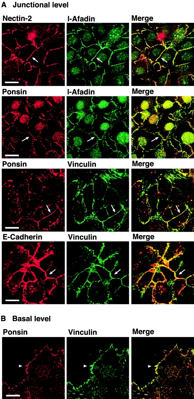
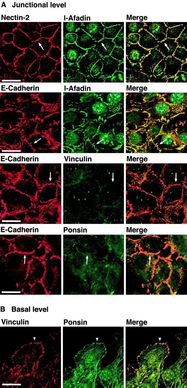
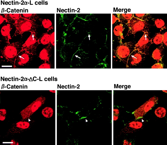
We have found a new cell-cell adhesion system at cadherin-based cell-cell adherens junctions (AJs) consisting of at least nectin and l-afadin. Nectin is a Ca(2+)-independent homophilic immunoglobulin-like adhesion molecule, and l-afadin is an actin filament-binding protein that connects the cytoplasmic region of nectin to the actin cytoskeleton. Both the trans-interaction of nectin and the interaction of nectin with l-afadin are necessary for their colocalization with E-cadherin and catenins at AJs. Here, we examined the mechanism of interaction between these two cell-cell adhesion systems at AJs by the use of alpha-catenin-deficient F9 cell lines and cadherin-deficient L cell lines stably expressing their various components. We showed here that nectin and E-cadherin were colocalized through l-afadin and the COOH-terminal half of alpha-catenin at AJs. Nectin trans-interacted independently of E-cadherin, and the complex of E-cadherin and alpha- and beta-catenins was recruited to nectin-based cell-cell adhesion sites through l-afadin without the trans-interaction of E-cadherin. Our results indicate that nectin and cadherin interact through their cytoplasmic domain-associated proteins and suggest that these two cell-cell adhesion systems cooperatively organize cell-cell AJs.


Figures

Figure 1
Figure 1
Structures of various constructs of nectin, l-afadin, ponsin, E-cadherin, and α-catenin. CTM, COOH-terminal motif of four aa residues; GAL4AD, GAL4 activation domain; and CBD, catenin-binding domain.
Figure 2
Figure 2
α-Catenin–dependent colocalization of nectin and l-afadin with E-cadherin. αF9Dα(−/−) and F9Dα(−/−) cells were double stained with various combinations of the anti–E-cadherin, anti–l-afadin, and anti–nectin-2 Abs. (arrows) Cell–cell adhesion sites between two cells; and (arrowheads) cell–cell adhesion sites where >2 cells adhere to each other. The results shown are representative of three independent experiments. Bars, 10 μm.
Figure 3
Figure 3
β-Catenin–independent colocalization of nectin, l-afadin, ponsin, and vinculin with E-cadherin. nEα-L cells were double stained with various combinations of the anti–nectin-2, anti–l-afadin, antiponsin, antivinculin, and anti–E-cadherin Abs. There was nuclear staining with the anti–l-afadin or antiponsin Ab, but the nuclear staining was not an artifact of second antibodies, although its significance is not known. (arrows) Cell–cell adhesion sites; and (arrowhead) focal contacts. The results shown are representative of three independent experiments. Bars, 10 μm.
Figure 4
Figure 4
Nectin- and l-afadin–independent colocalization of ponsin and vinculin with E-cadherin. nEαN-L cells were double stained with various combinations of the anti–nectin-2, anti–l-afadin, antiponsin, antivinculin, and anti–E-cadherin Abs. Alternatively, nEαN-L cells were transfected with pEGFP-ponsin and stained with the anti–E-cadherin Ab. (arrows) Cell–cell adhesion sites; and (arrowhead) focal contacts. The results shown are representative of three independent experiments. Bars, 10 μm.
Figure 5
Figure 5
Ponsin- and vinculin-independent colocalization of nectin and l-afadin with E-cadherin. nEαC-L cells were double stained with various combinations of the anti–nectin-2, anti–l-afadin, antiponsin, antivinculin, and anti–E-cadherin Abs. (arrows) Cell–cell adhesion sites; and (arrowhead) focal contacts. The results shown are representative of three independent experiments. Bars, 10 μm.
Figure 6
Figure 6
Interaction of l-afadin with α-catenin on affinity chromatography. GST-α-catenin-C was applied to metal affinity beads on which Myc-His6-l-afadin was immobilized. After the beads were extensively washed, elution was performed with 100 mM imidazole chloride, pH 7.5. Each fraction was subjected to SDS-PAGE (10% polyacrylamide gel), followed by protein staining with Coomassie brilliant blue. The results shown are representative of three independent experiments.
Figure 7
Figure 7
Localization of nectin and l-afadin in nectin-2α-L and -2α-ΔC-L cells. Nectin-2α-L and -2α-ΔC-L cells were double stained with the anti–nectin-2 and anti–l-afadin Abs. (arrows) Nectin-2α–based cell–cell adhesion sites; and (arrowheads) nectin-2α-ΔC–based cell–cell adhesion sites. The results shown are representative of three independent experiments. Bar, 10 μm.
Figure 8
Figure 8
l-Afadin–dependent recruitment of α-catenin to nectin-based cell–cell adhesion sites in the absence of E-cadherin. Nectin-2α-L and -2α-ΔC-L cells were transfected with pPGKIZ-HA-α-catenin and double stained with the anti–nectin-2 and anti-HA Abs. (arrow) Nectin-2α–based cell–cell adhesion sites; and (arrowhead) nectin-2α-ΔC–based cell–cell adhesion sites. The results shown are representative of three independent experiments. Bars, 10 μm.
Figure 9
Figure 9
l-Afadin–dependent recruitment of β-catenin to nectin-based cell–cell adhesion sites without the trans-interaction of E-cadherin. Nectin-2α-L and -2α-ΔC-L cells were transfected with pEF-tEC and double stained with the anti–nectin-2 and anti–β-catenin Abs. (arrows) Nectin-2α–based cell–cell adhesion sites; and (arrowhead) nectin-2α-ΔC–based cell–cell adhesion sites. The results shown are representative of three independent experiments. Bars, 10 μm.
Figure 10
Figure 10
l-Afadin–dependent recruitment of E-cadherin to nectin-based cell–cell adhesion sites without the trans-interaction of E-cadherin. Nectin-1α-L cells were cocultured with nectin-1α-EL or -1α-ΔC-EL cells in the presence or absence of gD and double stained with the anti–nectin-1α Ab1 and the anti–E-cadherin Ab. In the absence of gD, E-cadherin was concentrated at ∼40% of the adhesion sites between nectin-1α-L and -1α-EL cells. The recruitment of E-cadherin was observed in ∼20% of the adhesion sites between nectin-1α-L and -1α-ΔC-EL cells. In the presence of gD, the recruitment of E-cadherin was observed in ∼8% of the adhesion sites between nectin-1α-L and -1α-EL cells. (arrows) Cell–cell adhesion sites between two nectin-1α-L cells or between two nectin-1α-ΔC-L cells; (arrowheads) cell–cell adhesion sites between nectin-1α-L and -1α-EL or -1α-ΔC-EL cells; and (double arrowheads) cell–cell adhesion sites between two nectin-1α-EL cells or between two nectin-1α-ΔC-EL cells. The results shown are representative of three independent experiments.

References

    1. Adra C.N., Boer P.H., McBurney M.W. Cloning and expression of the mouse pgk-1 gene and the nucleotide sequence of its promoter. Gene. 1987;60:65–74. - PubMed
    1. Aoki J., Koike S., Ise I., Sato-Yoshida Y., Nomoto A. Amino acid residues on human poliovirus receptor involved in interaction with poliovirus. J. Biol. Chem. 1994;269:8431–8438. - PubMed
    1. Aoki J., Koike S., Asou H., Ise I., Suwa H., Tanaka T., Miyasaka M., Nomoto A. Mouse homolog of poliovirus receptor-related gene 2 product, mPRR2, mediates homophilic cell aggregation. Exp. Cell Res. 1997;235:374–384. - PubMed
    1. Asakura T., Nakanishi H., Sakisaka T., Takahashi K., Mandai K., Nishimura M., Sasaki T., Takai Y. Similar and differential behavior between the nectin-afadin-ponsin and cadherin-catenin systems during the formation and disruption of the polarized junctional alignment in epithelial cells. Genes Cells. 1999;4:573–581. - PubMed
    1. Bradford M.M. A rapid and sensitive method for the quantitation of microgram quantities of protein utilizing the principle of protein-dye binding. Anal. Biochem. 1976;72:248–254. - PubMed

Publication types

MeSH terms